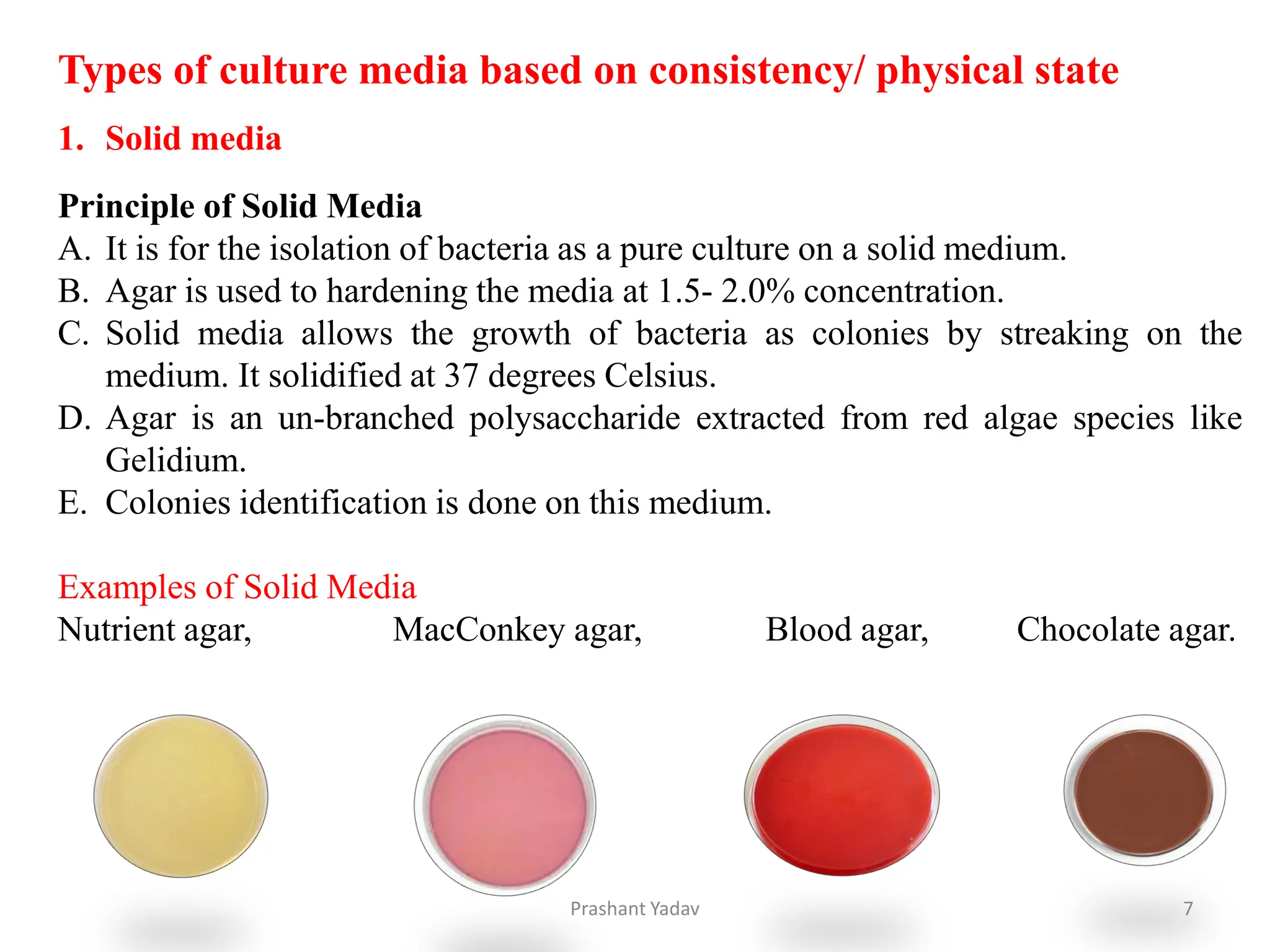
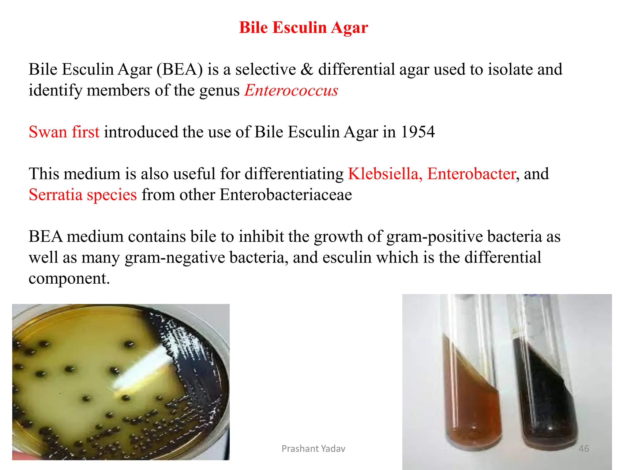
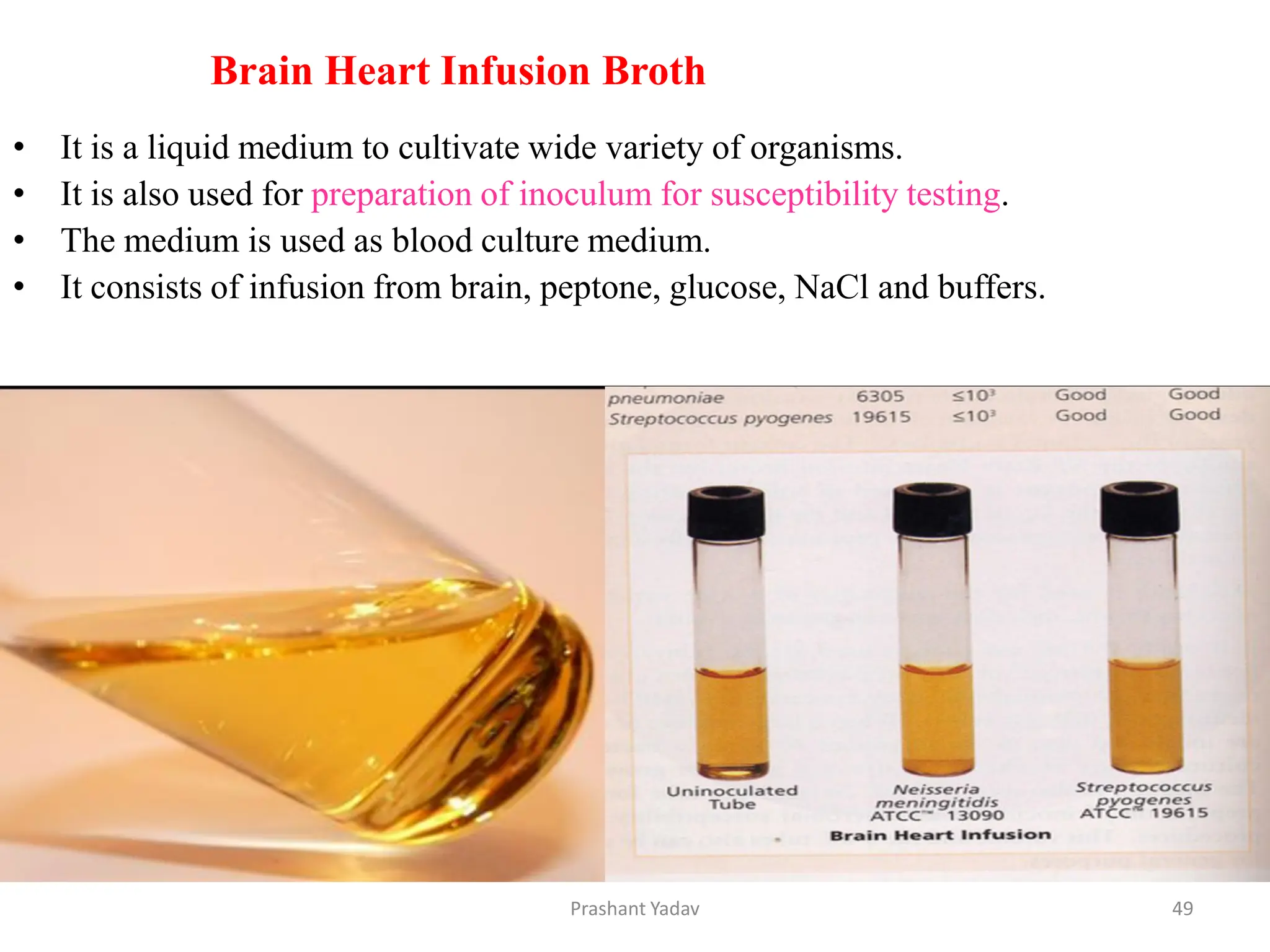
Culture media in microbiology refers to the solid or liquid substances used to cultivate and grow microorganisms such as bacteria, fungi, and viruses in a laboratory setting. These media provide the necessary nutrients, environment, and conditions for microorganisms to thrive and reproduce. The choice of culture media depends on the specific requirements of the microorganisms being studied, as different organisms have varied nutritional needs and environmental preferences.
2. Culture Media
Culture media, also known as growth media, are specific mixtures of
nutrients and other substances that support the growth of
microorganisms such as bacteria and fungi (yeasts and molds).
History:
Louis Pasteur used simple broths made up of urine or meat
extracts. Robert Koch realized the importance of solid media and
used potato pieces to grow bacteria. It was on the suggestion of
Fannie Eilshemius, wife of Walther Hesse (who was an assistant
to Robert Koch) that agar was used to solidify culture media.
Before the use of agar, attempts were made to use gelatin as
solidifying agent. Gelatin had some inherent problems; it existed
as liquid at normal incubating temperatures (35-37o C) and was
digested by certain bacteria.
2
Prashant Yadav
3. Common ingredients of culture media
Peptone- Source of carbon and nitrogen. : It is a complex mixture of partially
digested proteins, obtained from various sources such as heart muscle, casein or
fibrin, or soya
Electrolytes- Sodium chloride or other electrolytes.
Beef extract- Source of amino acid, vitamins, minerals.
Yeast extract- Source of vitamin, carbon, nitrogen.
Distilled water- Source of hydrogen and oxygen.
Agar- Solidifying agent. : It is prepared from the cell wall of seaweeds and
available commercially in powder form. It is used in concentration of 1–2% for
solid medium, 0.5% for semisolid agar and 6% to inhibit Proteus swarming.
Blood and serum: They are important components of enriched media; provide
extra nutrition to fastidious bacteria. Usually 5–10% of sheep blood is used.
Alternatively, horse, ox, or human blood can also be used.
3
Prashant Yadav
4. How to prepare culture media?
1. Weight the amount of ingredients powder on weighing machine.
2. Dissolve the ingredients in distilled water.
3. Pour the media into flask.
4. Add agar and boiled it to dissolve.
5. Sterilization is done in autoclave to prevent from contamination,
at 121ºC for 15 min at 15lbs.
6. After the autoclave place the media flask in laminar air flow.
7. Sterilize the laminar air flow with 70% alcohol.
8. A bit cools down the media and pours into sterile Petri-plates for
solidification.
9. Then sample is ready to spread(spreader) / streak
4
Prashant Yadav
6. Types of culture media based on consistency/ physical state
1. Solid medium
2. Semi-solid medium
3. Liquid medium
Types of culture media based on constituents / Nutritional
There are seven routine laboratory media.
1. Basal media/ Simple media
2. Enriched media
3. Selective media
4. Enrichment media
5. Indicator media or differential media
6. Transport media
7. Storage media
Types of culture media based on oxygen requirement
1. Aerobic media
2. Anaerobic media
6
Prashant Yadav
7. Types of culture media based on consistency/ physical state
1. Solid media
Principle of Solid Media
A. It is for the isolation of bacteria as a pure culture on a solid medium.
B. Agar is used to hardening the media at 1.5- 2.0% concentration.
C. Solid media allows the growth of bacteria as colonies by streaking on the
medium. It solidified at 37 degrees Celsius.
D. Agar is an un-branched polysaccharide extracted from red algae species like
Gelidium.
E. Colonies identification is done on this medium.
Examples of Solid Media
Nutrient agar, MacConkey agar, Blood agar, Chocolate agar.
7
Prashant Yadav
8. Types of culture media based on consistency/ physical state
Semi-solid media
Principle of Semi-solid media
This media shows the motility of bacteria and the cultivation of
microaerophilic bacteria.
This media has agar at a concentration of 0.5% or less,
It has a jelly consistency.
Semi Solid Media are used for the determination of bacterial motility.
Examples of Semi-solid media
Stuart’s and Amies media, Hugh and Leifson’s oxidation fermentation
medium, and Mannitol motility media.
The growth of bacteria in semi-solid appears as a thick line in the medium.
8
Prashant Yadav
9. Liquid media
Principle of Liquid media
This media shows the growth of a large number of bacteria.
It is called Broth that allows bacteria to grow uniformly with turbidity.
The growth occurs at 37ºC in an incubator for 24hrs.
Liquid media don’t have the addition of agar; it is for fermentation studies.
Examples of Liquid media
1. Nutrient broth,
2. Tryptic soy broth,
3. MR-VP broth,
4. phenol red carbohydrate broth.
Growth of bacteria in liquid media- Turbidity is
seen at the end of the broth.
9
Prashant Yadav
10. Types of culture media based on constituents / Nutritional
1. Basal media/ Simple media
This media allows the growth of non- fastidious bacteria without any
enrichment source; used for sub-culturing. It’s a non-selective medium.
having Carbon and Nitrogen
Staphylococcus and Enterobacteriaceae grow in this media.
Example- Nutrient Agar, Peptone water, Nutrient broth
10
Prashant Yadav
11. Peptone Water
Ingredients Gms / L
Peptone 10.0
Sodium chloride 5.0
Composition of Peptone Water
Final pH (at 25°C) 7.2±0.2
Result on Peptone Water
A positive result (inoculated medium): Growth, turbidity seen
A negative result (uninoculated medium): No growth
Uses of Peptone Water
Peptone Water is used as a growth medium and as a base for carbohydrate
fermentation media.
It is a broth medium used for the detection of indole.
Peptone Water with pH adjusted to 8.4 (alkaline condition) is suitable for the
cultivation and enrichment of Vibrio species.
11
Prashant Yadav
12. Nutrient Broth
Nutrient Broth is used for the general cultivation of less fastidious
microorganisms, can be enriched with blood or other biological
fluids.
Ingredients
Ingredient 1 L 500 mL
beef extract 1 g 0.5 g
Yeast extract 2 g 1 g
Peptone 5 g 2.5 g
Sodium
chloride
(NaCl)
5 g 2.5 g
12
Prashant Yadav
13. Composition of nutrient agar
Ingredients gram / liter
Peptone 5.0g
Meat extract 1.0g
Yeast extract 2.0 g
Sodium chloride(NaCl) 5.0 g
Bacteriological Agar 12.0 g
Nutrient agar
Nutrient agar is a general purpose medium suitable for the cultivation
of a wide variety of nonfastidious microorganisms.
Use to isolate separate colonies for :
i. Colony morphology
ii. Pigmentation
iii. Biochemical identification tests
iv. Serotyping
v. Antibiotic susceptibility testing
13
Prashant Yadav
14. 2. Enriched media
Enriched media contain the nutrients required to support the growth of a wide
variety of organisms, including some of the more fastidious ones. They are
commonly used to harvest as many different types of microbes as are present in
the specimen.
Example-:
Blood agar
Chocolate agar
Loeffler's serum
MacConkey agar
Lowenstein-Jensen media
14
Prashant Yadav
15. It is an enriched medium that supports the growth of fastidious bacteria and
inhibits the growth of some bacteria like Neisseria and Haemophilus.
The blood also aids in visualizing hemolytic reactions of different bacteria.
The hemolytic reactions, however, depend on the type of animal blood used.
Sheep blood is mostly used for Group A Streptococci as it provides the best results, but
it fails to support the growth of Haemophilus haemolyticus.
It is because sheep blood is deficient in pyridine nucleotides.
Blood agar
Composition of Blood Agar
0.5% Peptone
0.3% beef extract/yeast extract
1.5% agar
0.5% NaCl
Distilled water
(Since Blood Agar is made from Nutrient Agar, above is the composition of Nutrient
Agar)
5% Sheep Blood
pH should be from 7.2 to 7.6 (7.4)
15
Prashant Yadav
16. Hemolysis
Certain bacterial species produce extracellular enzymes that lyse red blood cells
in the blood agar (hemolysis).
1. Alpha hemolysis
Alpha hemolysis is defined by a greenish-grey or brownish discoloration
around the colony as a result of the partial lysis of the red blood cells.
During α-hemolysis, H2O2 produced by the bacteria causes hemoglobin present
in the RBC of the medium is converted into methemoglobin.
Some of the α-hemolytic species are a part of the human normal flora, but some
species like Streptococcus pneumonia cause pneumonia and other such severe
infections.
16
Prashant Yadav
17. 2. Beta hemolysis
Beta hemolysis is defined by a clear zone of hemolysis under and around the colonies
when grown on blood agar.
The clear zone appears as a result of the complete lysis of the red blood cells present in
the medium, causing denaturation of hemoglobin to form colorless products.
β-hemolytic bacteria include group A streptococci like S. pyogenes and group B
streptococcus like S. agalactiae, both of which are associated with severe infections in
humans.
3. Gamma hemolysis
Gamma hemolysis is also called non- hemolysis as no lysis of red blood cells occurs.
As a result, no change of coloration or no zone of hemolysis is observed under or
around the colonies.
Species like Neisseria meningiditis are non-hemolytic or gamma-hemolytic.
17
Prashant Yadav
18. S.N Organism Colony Morphology Hemolysis
1. Neisseria meningiditis
Grey and unpigmented colonies that appear round,
smooth, moist, glistening, and convex, with a clearly
defined edge.
Non-hemolytic or γ-
hemolytic.
2. Salmonella Typhi
Smooth colorless colonies that are smooth, moist, and
flat with a diameter range of 2-4 mm.
Non-hemolytic or γ-
hemolytic.
3. Staphylococcus aureus
Golden yellow colored circular, convex and smooth
colonies of the diameter range of 2-4 mm; opaque
colonies with a zone of hemolysis.
β-hemolytic.
4.
Staphylococcus
epidermidis
Circular, colonies of the size 1-4 mm in diameter;
grey to white-colored with low convex elevation;
moist, glistening colonies.
Non-hemolytic or γ-
hemolytic.
5. Streptococcus pyogenes
White-greyish-colored colonies with a diameter of >
0.5 mm; the colonies are surrounded by a zone of β-
hemolysis that is often two to four times as large as
the colony diameter.
β-hemolytic.
6.
Streptococcus
pneumonia
small, grey, moist (sometimes mucoidal in
encapsulated virulent strains), colonies with the
characteristic zone of alpha-hemolysis (green); due to
autolysis, often produces a dimple-like zone of
hemolysis than the typical crater-like appearance.
α-hemolytic.
7.
Pseudomonas
aeruginosa
Large colonies of the size 2-5mm in diameter; flat
colonies that are grey to white-colored with an
undulate margin with a zone of β-hemolysis.
β-hemolytic.
18
Prashant Yadav
20. Chocolate agar
Chocolate Agar (CHOC) is a non-selective, enriched growth medium that
is the lysed blood agar.
The agar is named for its color when the red blood cells (RBCs) lysis
gives the medium a chocolate-brown color without having chocolate
products.
It is used for the isolation of fastidious bacteria, such as Haemophilus
influenzae,
The red blood cells are lysed changing the medium color chocolate
brown.
The lysis of RBC during the heating process releases intracellular co-
enzyme nicotinamide adenine dinucleotide (Factor V or NAD) into the
agar for utilization by fastidious bacteria (the heating process also
inactivates growth inhibitors).
20
Prashant Yadav
21. Ingredients Gms /L
Casein/Animal Tissue Digest 15.0g
Cornstarch 1.0g
Sodium chloride 5.0g
Dipotassium Phosphate 4.0g
Monopotassium Phosphate 1.0g
Hemoglobin solution 2%
Koenzyme enrichment 10.0ml
Agar 10.0g
Composition of Chocolate Agar
Final pH 7.2 ± 0.2 (at 25°C)
Organism Colony morphology
Neisseria gonorrhoeae pinkish-brown and translucent, and are typically 0.5-1 mm in
diameter
N. meningitidis grayish, non-hemolytic, round, convex, smooth, moist, larger in
size as compared to N. gonorrhoeae
Haemophilus influenzae Nonhemolytic, colorless, moist colonies with a characteristic
“mousy” odor.
21
Prashant Yadav
22. Loeffler's serum
Loeffler medium enhances primary and secondary isolation and cultivation of
fastidious pathogenic microorganisms, especially from the nose and throat .
Loeffler Serum slope is used for the cultivation
of Corynebacterium diphtheriae.
Due to its serum content, Loeffler medium can be used for the determination of
proteolytic activities of microorganisms.
Gram-positive microorganisms other than Corynebacterium may produce
metachromatic granules when grown on Loeffler Medium.
Loeffler serum slope cannot confirm the organisms and thus further
biochemical, immunological, molecular, or mass spectrometry testing be
performed on colonies from pure culture for complete identification.
22
Prashant Yadav
23. Ingredients Gms/liter
Proteose Peptone 2.5gm
Dextrose 2.5gm
Sodium Chloride 1.25gm
Beef Extract 2.5gm
Horse Serum 750.0 ml
Composition of Loeffler Medium
Organisms Growth
Corynebacterium diphtheriae
Growth; minute, and cream-colored colonies
with slightly raised centers; metachromatic
granules seen in methylene blue stain
Corynebacterium pseudodiptheriticum Growth; minute, and cream-colored colonies
Pseudomonas aeruginosa Good growth; green colonies with proteolysis
Staphylococcus aureus Good growth; yellow to gold colonies
Streptococcus pyogenes Fair to good growth; non-proteolytic
23
Prashant Yadav
24. MacConkey agar
MacConkey agar is a selective and differential media used for the isolation and
differentiation of non-fastidious gram-negative rods, particularly members of the
family Enterobacteriaceae and the genus Pseudomonas.
It is used in the differentiation of lactose fermenting from lactose non-fermenting
gram-negative bacteria.
It is used for the isolation of coliforms and intestinal pathogens in water, dairy
products and biological specimens.
Lactose fermenting strains grow as red or pink and may be surrounded by a zone of
acid precipitated bile. The red color is due to production of acid from lactose,
absorption of neutral red and a subsequent color change of the dye when the pH of
medium falls below 6.8.
Lactose non-fermenting strains
Such as Shigella and Salmonella are colorless and transparent and typically do not
alter appearance of the medium. Yersinia enterocolitica may appear as small, non-
lactose fermenting colonies after incubation at room temperature.
24
Prashant Yadav
25. Ingredients Amount
Peptone (Pancreatic digest of
gelatin)
17 gm
Proteose peptone (meat and casein) 3 gm
Lactose monohydrate 10 gm
Bile salts 1.5 gm
Sodium chloride 5 gm
Neutral red 0.03 gm
Crystal Violet 0.001 g
Agar 13.5 gm
Distilled Water
Add to make
1 Liter
Composition of MacConkey Agar
Organism Color Remarks
Escherichia coli red/pink non-mucoid
Aerobacter aerogenes pink mucoid
Enterococcus species red minute, round
Staphylococcus species pale pink opaque
Pseudomonas aeruginosa green-brown fluorescent growth
25
Prashant Yadav
27. Lowenstein-Jensen media
Lowenstein Jensen (LJ) Media is a selective medium that is commonly used for
the cultivation and isolation of Mycobacterium, specifically Mycobacterium
tuberculosis from clinical specimens.
Incorporated congo red and malachite green to inhibit unwanted bacteria.
Ingredients Amount
Potato Flour 30.0gm
L- Asparagine 3.6gm
Monopotassium Phosphate 2.4gm
Magnesium Citrate 0.6gm
Malachite Green 0.4gm
Magnesium Sulfate 0.24gm
Glycerol 12ml
Egg Base 1000ml
Composition of Lowenstein Jensen (LJ) Media
27
Prashant Yadav
28. It is used for the diagnosis of Mycobacterial infections.
It is used for testing antibiotic susceptibility of isolates.
It is also used for differentiating different species of mycobacterium (by
colony morphology, growth rate, biochemical characteristics and microscopy).
The media should be protected from all sources of light, as malachite green is
very photosensitive.
Cultures should be read within 5–to 7 days and once a week thereafter for up
to 8 weeks.
The green color of the medium is due to the presence of malachite green
which is one of the selective agents to prevent growth of most other
contaminants.
The typical colonies are non pigmented, rough, dry on LJ medium.
Rapid growers have mature colonies within 7 days; slow growers require more
than 7 days for mature colony forms.
White, cream or buff = Nonchromogenic (NC)
Lemon, yellow, orange, red = Chromogenic (Ch)
28
Prashant Yadav
29. Selective media are used to select for the growth of a particular
"selected" microorganism.
Selective media generally selects for the growth of a desired organism,
stopping the growth of or altogether killing non-desired organisms.
They are solid media containing inhibitory substances that
inhibit the normal flora present in the specimen and allow
the pathogens to grow
Lowenstein–Jensen (LJ) medium
Thiosulfate citrate bile salt sucrose (TCBS) agar
DCA (deoxycholate citrate agar and
XLD (xylose lysine deoxycholate) agar
Potassium tellurite agar (PTA):
3. Selective media
29
Prashant Yadav
30. Thiosulfate citrate bile salt sucrose (TCBS) agar
Thiosulfate citrate bile salts sucrose (TCBS) agar is a selective as well as
differential culture medium used for selective isolation of Vibrio spp from a
variety of clinical specimens.
TCBS has a very high pH 8.5-9.0 (alkaline pH)
Vibrio spp produce either yellow or green colonies on TCBS, depending on
whether they are able to ferment sucrose.
If the sucrose is fermented, yellow colonies are produced Vibrio
cholerae produces yellow colonies in TCBS whereas Vibrio
parahaemolyticus produces green colonies
Preparation TCBS Agar
Suspend 89.08 gm of dehydrated medium in 1000ml of distilled or deionized
water.
Heat to boiling to dissolve the medium completely.
Do not autoclave.
Cool to 45-50°C.
Mix well and pour into sterile Petri plates.
30
Prashant Yadav
31. Microorganisms Characteristics
Vibrio cholera
Flat yellow colonies, 2-3 mm in
diameter
Vibrio alginolyticus Large yellow colonies
Vibrio fluvialis, Vibrio
vulnificus
Yellow or translucent colonies
Vibrio parahaemolyticus
Colorless colonies with a green
center
Pseudomonas, Aeromonas Blue colonies
Enterobacteria or others Tiny transparent colonies
Vibrio cholera
Vibrio parahaemolyticus
TCBS
31
Prashant Yadav
32. DCA (deoxycholate citrate agar)
They are used for the isolation of enteric pathogens, such as Salmonella and Shigella
from stool. pH 7.3 to 7.5 is inhibitory for Gram-Positive Bacteria
DO NOT AUTOCLAVE.
Organisms Growth
Escherichia coli
Poor growth; pink with bile precipitate negative
reaction for H2S
Salmonella Enterit
idis
Good-luxuriant growth; colorless; positive
reaction for H2S, black centered colonies
Salmonella Typhi
murium
Good-luxuriant growth; colorless; positive
reaction for H2S, black centered colonies
Shigella flexneri Good growth; colorless
Salmonella Abony
Good-luxuriant growth; colorless; positive
reaction for H2S, black centered colonies
Shigella sonnei
Colonies are smooth and initially colorless,
becoming pale pink on further incubation due
to late lactose fermentation
Enterobacter/Kleb
siella spp.
Large, pale mucoid colonies with the pink
center
32
Prashant Yadav
33. XLD (xylose lysine deoxycholate) agar
•(XLD) agar is a selective growth medium used for the isolation
of Salmonella and Shigella species from clinical samples and from food
• XLD Agar was developed by Taylor for the differentiation, isolation, and
identification of enteric pathogens, and to support the growth of more fastidious
enteric organisms.
Preparation
•Suspend 56.68 grams in 1000 ml distilled water.
•Heat with frequent agitation until the medium boils.
DO NOT AUTOCLAVE OR OVERHEAT.
•Transfer immediately to a water bath at 50°C.
•After cooling, pour into sterile Petri plates.
Organisms Colony characteristics
Salmonella H2S positive Red colonies with black centers
Shigella spp.
and Salmonella H2S negative
Red colonies
E. coli Large, flat, yellow colonies
Proteus spp. Red to Yellow colonies
Enterobacter / Klebsiella Mucoid, yellow colonies
33
Prashant Yadav
34. Potassium tellurite agar (PTA)
• It is used for isolation of Corynebacterium diphtheriae.
• It is also a selective medium for the cultivation and isolation
of Corynebacterium species.
• It is applicable for both clinical and non-clinical specimens to
recover Corynebacterium species.
Colony Morphology
Organism Growth and Colour of the colony
Corynebacterium diphtheriae ATCC 11913: Good, Grey-black
Escherichia coli ATCC 25922 : Inhibited
Throat or nasal swab is directly inoculated and
streaked on this agar medium.
34
Prashant Yadav
35. Enrichment media are a type of highly selective media which allows the growth
of only a particular type of microorganism in the medium.
The liquid media inhibits the growth of the unwanted bacteria , allow the
growth of a particular types of microorganism in the medium, contain added
antibiotics ,dye , chemicals or altered pH.
EX. Selenite F broth, APW, tetrathionate broth
4. Enrichment media
APW
Selenite F broth
Tetrathionate broth
35
Prashant Yadav
36. These media differentiate between two groups of bacteria
by using an indicator, which changes the color of the
colonies of a particular group of bacteria but not the other
group.
•MacConkey agar-
•CLED agar (cysteine lactose electrolyte-deficient agar)
5.Differential Media
36
Prashant Yadav
37. CLED agar, which stands for Cystine-Lactose-Electrolyte-Deficient agar, is a
selective and differential culture medium used in microbiology for the isolation
and differentiation of urinary tract pathogens. It is specifically designed for the
cultivation of bacteria from urine samples.
CLED agar composition
Ingredients gram/liter Ingredients gram/liter
Balanced
Peptone
4.0 g Tryptone 4.0 g
Beef extract 3.0 g Lactose 10.0 g
L-Cystine 0.128 g Bromothymol
Blue
0.02 g
Agar 15.0 g Final pH 7.3 +/- 0.2
CLED agar (cysteine lactose electrolyte-deficient agar)
37
Prashant Yadav
38. Organism Colony Morphology
Escherichia coli
Large, elevated, yellow, opaque colonies with a center more intense
yellow; yellowish medium
Klebsiella species
extremely mucoid colonies varying in color from yellow to whitish-
blue. yellowish medium.
Proteus species
translucent blue colonies are usually smaller than Escherichia coli.
blue-green to blue medium.
Salmonella species flat blue colonies
Pseudomonas aeruginosa
green colonies with a typical matte surface and rough periphery.
“Sweet” odor. Blue-green agar
Enterococcus faecalis yellow colonies about 0.5 mm in diameter. yellow medium
Staphylococcus aureus
deep yellow colonies about 0.75 mm diameter, uniform in color. yellow
medium
Corynebacteria very small grey colonies
Lactobacilli similar to corynebacteria but with a rougher surface
38
Prashant Yadav
39. 6.Transport media
They are used for the transport of the clinical specimens suspected to contain
delicate organism or when delay is expected while transporting the
specimens from the site of collection to the laboratory Bacteria do not
multiply in the transport media, they only remain viable.
• Cary and Blair Medium
• Amies medium
• Stuarts medium
• Venkatraman Ramakrishnan (VR) medium
• Alkaline Peptone Water (APW)
• Sach’s buffered glycerol saline
• Viral Transport Medium
39
Prashant Yadav
40. Cary Blair Transport Medium is a semi-solid medium recommended for use
in the transportation and preservation of clinical specimens, primarily stool
and rectal swabs.
Ingredients gm/litre
Disodium hydrogen phosphate 1.1
Sodium thioglycollate 1.5
Sodium chloride 5.0
Calcium chloride 0.1
Agar 5.0
pH 8.4 ± 0.2@ 25°C
Cary and Blair Medium
Appearance of Cary Blair Transport
Medium
Dehydrated medium: Off-white color,
free-flowing powder.
Prepared medium: Light amber color,
semi-solid gel.
Positive controls Expected results
Shigella flexneri ATCC 12022 Good growth on subculture
Salmonella Typhimurium ATCC
14028
Good growth on subculture
Negative control
Uninoculated medium No change
40
Prashant Yadav
41. Amies Transport Medium is an improved transport medium, containing
charcoal to prolong the viability of pathogenic organisms.
It is a transport medium used to preserve the viability of anaerobes such
as Neisseria gonorrhea and other pathogens from swabs.
It is recommended for the throat, vaginal, and wound samples.
It may not be suitable for the transport of fastidious organisms
Ingredients Gms/liter
Sodium chloride 3.000
Potassium chloride 0.200
Calcium chloride 0.100
Magnesium chloride 0.100
Monopotassium phosphate 0.200
Disodium phosphate 1.150
Sodium thioglycollate 1.000
Charcoal 10.000
Agar 4.000
Amies Transport Medium
41
Prashant Yadav
42. Organisms Growth
Escherichia coli Good recovery on subculture
Klebsiella pneumoniae Good recovery on subculture
Neisseria meningitidis Good recovery on subculture
Pseudomonas aeruginosa Good recovery on subculture
Salmonella Typhi Good recovery on subculture
Shigella flexneri Good recovery on subculture
Staphylococcus aureus Good recovery on subculture
Vibrio cholerae Good recovery on subculture
Bacteroides fragilis Good recovery on subculture
Haemophilus influenzae Good recovery on subculture
Neisseria gonorrhoeae Good recovery on subculture
Streptococcus pyogenes Good recovery on subculture
42
Prashant Yadav
43. Venkatraman-Ramakrishnan medium is used to transport fecal samples that are
suspected of containing Vibrio cholerae. This medium allows the survival of
the bacteria as it is transferred to the lab.
Venkatraman Ramakrishnan (VR) medium
It is prepared by mixing
20gm of crude sea salt
5gm of peptone
1l of distilled water
Alkaline Peptone Water (APW)
Alkaline Peptone Water (APW) is one of the commonly used enrichment
liquid media which is highly recommended for the isolation of Vibrio cholerae from
clinical specimens like feces as well as Non-clinical specimens like Food & Water.
This Alkaline pH of APW the growth of Vibrio species and inhibits the growth of
commensal intestinal bacteria which are commonly present in the Clinical
Specimens.
43
Prashant Yadav
44. Observations & Identification of Vibrio cholerae
Vibrio cholerae grows rapidly in the Alkaline Peptone Water produces
turbidity on and below the surface specifically within 3-6 hours.
You can follow the further steps. the cultured organism is Vibrio or not:
Hanging drop Preparation for the motility of bacteria.
Gram staining of the culture (Vibrio cholerae appear as Gram-
negative curved bacilli)
Culture on TCBS medium.
Components Quantity (in grams)
Peptone 5.0
Sodium chloride 5.0
Distilled water 500 ml
Final pH at Room
Temperature
8.6±0.2
44
Prashant Yadav
45. Viral Transport Medium
A viral transport media (VTM) is used to carry and maintain the viability of
specimens (viruses) to a laboratory for the identification and further processing
of the sample
Viral Transport Medium components,
Hank’s Balanced Salts
Bovine Serum Albumin
Sucrose
Gelatin
Gelatin Peptone
Veal Infusion
Vancomycin
Amphotericin B
Polymyxin B
Phenol Red
pH 7.3 ± 0.3 at 25 °C *
Adjusted to suit performance parameter.
45
Prashant Yadav
46. Bile Esculin Agar
Bile Esculin Agar (BEA) is a selective & differential agar used to isolate and
identify members of the genus Enterococcus
Swan first introduced the use of Bile Esculin Agar in 1954
This medium is also useful for differentiating Klebsiella, Enterobacter, and
Serratia species from other Enterobacteriaceae
BEA medium contains bile to inhibit the growth of gram-positive bacteria as
well as many gram-negative bacteria, and esculin which is the differential
component.
46
Prashant Yadav
47. Ingredients Gms/liter
Peptic digest of animal
tissue
5.00
Beef extract 3.00
Esculin 1.00
Bile salts 40.0
Ferric citrate 0.50
Agar 15.00
It is recommended for use as a differential medium in the isolation and presumptive
identification of enterococci/group D streptococci.
To identify and isolate bacteria capable of hydrolyzing the esculin, in the presence of
Bile
Bile esculin media contains peptone and esculin for nutrition, as well as bile for the
purpose of inhibiting Gram-positive bacteria
Composition of Bile Esculin Agar
47
Prashant Yadav
48. Organism Growth Bile esculin hydrolysis
Enterococcus faecalis Good
Positive reaction;
Blackening of medium
Escherichia coli Good Negative reaction
Enterococcus faecium Luxuriant
Positive reaction;
Blackening of the medium
around the growth.
Yersinia enterocolitica Good-luxuriant
Positive reaction;
Blackening of the medium.
Quality Control of Bile Esculin Test
Positive control of Bile Esculin Test: Enterococcus species (e.g. E. faecalis)
Negative control of Bile Esculin Test: Viridans streptococcus, not group D
48
Prashant Yadav
49. Brain Heart Infusion Broth
• It is a liquid medium to cultivate wide variety of organisms.
• It is also used for preparation of inoculum for susceptibility testing.
• The medium is used as blood culture medium.
• It consists of infusion from brain, peptone, glucose, NaCl and buffers.
49
Prashant Yadav
50. Mueller–Hinton agar is a microbiological growth medium that is
commonly used for antibiotic susceptibility testing, specifically disk
diffusion tests.
It is also used to isolate and maintain Neisseria and Moraxella species.
It is a non-selective, non-differential medium, This means that almost all
organisms plated on here will grow.
Antibiotic susceptibility testing using the Kirby-Bauer disc diffusion
method.
Ingredients Gms/Litre
Beef extract 2.0
Acid hydrolysate of casein 17.5
Starch 1.5
Agar 17.0
Composition of MHA
Mueller Hinton Agar (MHA)
50
Prashant Yadav
51. Sabouraud Dextrose Agar(SDA)
Sabouraud Dextrose Agar or SDA was formulaed by Raymond Sabouraud in 1892
Sabouraud Dextrose Agar is used for the cultivation of fungi (yeasts, molds)
This medium is also employed to determine microbial contamination in food,
cosmetics, and clinical specimens
The medium has a low pH (about 5.0), which inhibits the growth of most bacteria
It has a high concentration of either glucose or maltose and also contains mycological
peptone.
Ingredients Gms/Liter
Dextrose 40.0
Peptone 10.0
Agar 15.0
Composition of Sabouraud Dextrose Agar
Sabouraud dextrose broth
SDB has the same formulation as
above, without agar added.
51
Prashant Yadav
52. Fungi Colony morphology
Candida albicans
Pasty opaque slightly domed, smooth, and cream
or white colonies
Aspergillus flavus
Yellow-green powdery on front and pale
yellowish on reverse
Aspergillus niger
The initial growth is white, becoming black later
on giving a “salt and pepper appearance” which
results from darkly pigmented conidia borne in
large numbers on conidiophores and reverse
turning pale yellow
Aspergillus fumigatus
Bluish-green powdery colonies on front and pale
yellow on the reverse.
Trichosporon mucoides White to cream, yellowish, wrinkled
Geotrichum candidum
White to cream-colored, flat with aerial
mycelium
52
Prashant Yadav
53. Bird Seed Agar
Bird Seed Agar is used for the selective isolation of Cryptococcus
neoformans and Cryptococcus gattii. from clinical specimens
Cryptococcus neoformans usually grows as a yeast (unicellular) and replicates by
budding.
Cryptococcus neoformans is an encapsulated yeast that produces the enzyme
phenoloxidase, an enzyme necessary in melanin synthesis
It is also known as Caffeic Acid Agar or Niger Seed Agar or Staib Agar.
Ingredients Grams/Litre
Guizotia abyssinica (niger seed) 50
Glucose 1
KH2PO4 (potassium dihydrogen
orthophosphate)
1
Creatinine 1
Agar 15
Composition of Bird Seed Agar
Additives: to each 500 ml bottle.
•Penicillin G (20 units/ml) 1 mL
•Gentamicin (40 mg/ml) 1 mL
Result-:
The presence of golden brown to black pigmented smooth colonies is indicative
of Cryptococcus neoformans. Other species like Cryptococcus laurentii, Saccharomyces
cerevisiae, etc produce non-pigmented colonies. Candida appears as white colonies. 53
Prashant Yadav
55. CHROMagar Candida (CAC) is a selective medium for the isolation of fungi that
simultaneously provides direct differentiation and identification of several
Candida species. The yeasts produce enzymes that react with chromogenic
substrates in the CAC medium, producing colonies of different colors
55
Prashant Yadav