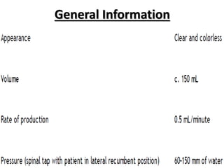
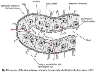
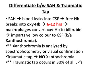
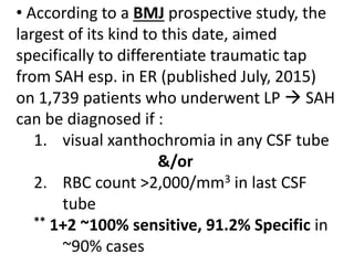
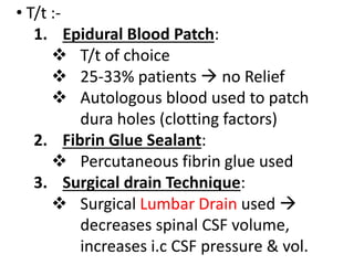
CSF
Vaibhav Shriya
Intern
General Information
Functions
Fig: Microscopy of the choroid plexus showing the path taken by fluids in the formation of CSF
Fig: Flow of CSF (dashed lines show CSF movement inside CNS cavities)
Composition
Differentiate b/w SAH & Traumatic
Tap
• SAH  blood leaks into CSF  free Hb
breaks into oxy-Hb  6-12 hrs 
macrophages convert oxy-Hb to bilirubin
 imparts yellow colour to CSF (k/a
Xanthochromia).
•** Xanthochromia is analyzed by
spectrophotometry or visual confirmation
•Traumatic tap  NO Xanthochromia
•** Traumatic tap occurs in 30% of all LP’s
• According to a BMJ prospective study, the
largest of its kind to this date, aimed
specifically to differentiate traumatic tap
from SAH esp. in ER (published July, 2015)
on 1,739 patients who underwent LP  SAH
can be diagnosed if :
1. visual xanthochromia in any CSF tube
&/or
2. RBC count >2,000/mm3 in last CSF
tube
** 1+2 ~100% sensitive, 91.2% Specific in
~90% cases
Artificial CSF (aCSF)
• Experimentally used Buffer solution
• Isolated brains, brain slices are
immersed in it
• Exposed brain parts are also
immersed in it to maintain O2 supply
• also used to buffer pH at biological
levels and maintain osmolarity
Composition of aCSF
127 mM NaCl
1.0 mM KCl
1.2 mM KH2PO4
26 mM NaHCO3
10 mM D-glucose
2.4 mM CaCl2
1.3 mM MgCl2
Spontaneous Intracranial
Hypotension (SIH)
• aka Spontaneous Spinal CSF Leak (SCSFL)
• occurs d/t holes in dura mater  CSF
leaks out  orthostatic headache (acute,
severe, worsens in sitting posture,
improves on lying down), N/V, neck pain
&/or stiffness
• Lack of CSF pressure & volume  may
cause Coning through Foramen Magnum
• Lower brain portion is believed to impact
one or more CN’s :-
• Average time from onset of symptoms until
definitive diagnosis is 13 months
• Diagnosis :-
1. CT Myelogram (CTM): measurement
of CSF pressure from LP  CTM
2. MRI:
 heavily T2-weighted MR
myelography
 Intrathecal contrast  MR
myelography
3. Assay: Beta-2 Transferrin assay
4. CSF Analysis:
 very low or even negative opening
pressures during LP
 18-46%  CSF opening pressure
within Normal range
 May show lymphocytic
pleocytosis, xanthochromia,
elevated protein levels
5. C/F: as above
• T/t :-
1. Epidural Blood Patch:
 T/t of choice
 25-33% patients  no Relief
 Autologous blood used to patch
dura holes (clotting factors)
2. Fibrin Glue Sealant:
 Percutaneous fibrin glue used
3. Surgical drain Technique:
 Surgical Lumbar Drain used 
decreases spinal CSF volume,
increases i.c CSF pressure & vol.
4. Neurosurgical repair:
 For patients unresponsive to
Epidural Blood Patch or Fibrin Glue
 Area of dura leak is Ligated &
clipped
 Alternatively, small compress k/a
Pledget placed over dura leak 
sealed with gel foam & fibrin glue
• Long term Prognosis = Poor
• Complications = Coma, Temporary
paralysis, infection, BP issues, reduced
ICP, brain herniation
THANK YOU

CSF (Cerebrospinal Fluid)

  • 1.
  • 2.
  • 3.
  • 4.
    Fig: Microscopy ofthe choroid plexus showing the path taken by fluids in the formation of CSF
  • 5.
    Fig: Flow ofCSF (dashed lines show CSF movement inside CNS cavities)
  • 6.
  • 7.
    Differentiate b/w SAH& Traumatic Tap • SAH  blood leaks into CSF  free Hb breaks into oxy-Hb  6-12 hrs  macrophages convert oxy-Hb to bilirubin  imparts yellow colour to CSF (k/a Xanthochromia). •** Xanthochromia is analyzed by spectrophotometry or visual confirmation •Traumatic tap  NO Xanthochromia •** Traumatic tap occurs in 30% of all LP’s
  • 8.
    • According toa BMJ prospective study, the largest of its kind to this date, aimed specifically to differentiate traumatic tap from SAH esp. in ER (published July, 2015) on 1,739 patients who underwent LP  SAH can be diagnosed if : 1. visual xanthochromia in any CSF tube &/or 2. RBC count >2,000/mm3 in last CSF tube ** 1+2 ~100% sensitive, 91.2% Specific in ~90% cases
  • 9.
    Artificial CSF (aCSF) •Experimentally used Buffer solution • Isolated brains, brain slices are immersed in it • Exposed brain parts are also immersed in it to maintain O2 supply • also used to buffer pH at biological levels and maintain osmolarity
  • 10.
    Composition of aCSF 127mM NaCl 1.0 mM KCl 1.2 mM KH2PO4 26 mM NaHCO3 10 mM D-glucose 2.4 mM CaCl2 1.3 mM MgCl2
  • 11.
    Spontaneous Intracranial Hypotension (SIH) •aka Spontaneous Spinal CSF Leak (SCSFL) • occurs d/t holes in dura mater  CSF leaks out  orthostatic headache (acute, severe, worsens in sitting posture, improves on lying down), N/V, neck pain &/or stiffness • Lack of CSF pressure & volume  may cause Coning through Foramen Magnum • Lower brain portion is believed to impact one or more CN’s :-
  • 13.
    • Average timefrom onset of symptoms until definitive diagnosis is 13 months • Diagnosis :- 1. CT Myelogram (CTM): measurement of CSF pressure from LP  CTM 2. MRI:  heavily T2-weighted MR myelography  Intrathecal contrast  MR myelography 3. Assay: Beta-2 Transferrin assay
  • 14.
    4. CSF Analysis: very low or even negative opening pressures during LP  18-46%  CSF opening pressure within Normal range  May show lymphocytic pleocytosis, xanthochromia, elevated protein levels 5. C/F: as above
  • 15.
    • T/t :- 1.Epidural Blood Patch:  T/t of choice  25-33% patients  no Relief  Autologous blood used to patch dura holes (clotting factors) 2. Fibrin Glue Sealant:  Percutaneous fibrin glue used 3. Surgical drain Technique:  Surgical Lumbar Drain used  decreases spinal CSF volume, increases i.c CSF pressure & vol.
  • 16.
    4. Neurosurgical repair: For patients unresponsive to Epidural Blood Patch or Fibrin Glue  Area of dura leak is Ligated & clipped  Alternatively, small compress k/a Pledget placed over dura leak  sealed with gel foam & fibrin glue • Long term Prognosis = Poor • Complications = Coma, Temporary paralysis, infection, BP issues, reduced ICP, brain herniation
  • 17.