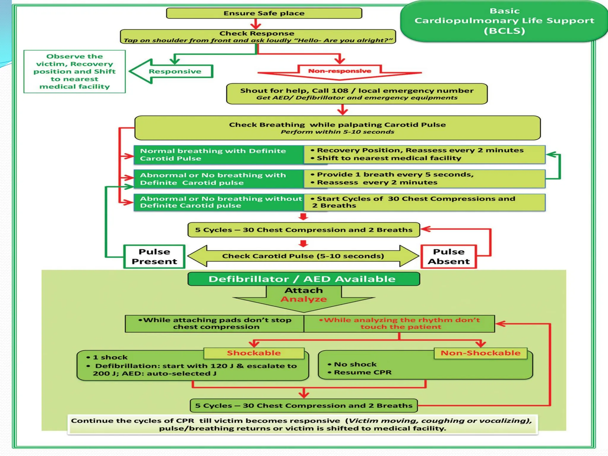
BASIC CARDIO-PULMONARY
RESUSCITATION
Sadhana
Chattopadhyay
Abrupt cessation of spontaneous
and effective ventilation and
systemic perfusion
What Is Cardiac Arrest ?
How Do you Recognize a Cardiac Arrest?
EARLY
Unresponsiveness
In case of adults absence of carotid pulse
and in case of infants Brachial pulse
Absence of normal respiration( the victim
is not breathing or only gasping)
LATE SIGN
 Cyanosis
 Cold clammy skin
 Dilated pupils
 ECG –Asystole / PEA/ pulse less VT/ VF
REVERSABLE CAUSES
‘HIT THE TARGET’
H – Hypoxia
I – Increased H Ions [Acidosis],
T – Tension Pneumothorax,
T – Toxins/Poisons,
H – Hypovolaemia,
E – Electrolyte Imbalance [Hypo-/Hyperkalaemia],
T – TamponadeCardiac,
A – Acute Coronary Syndrome,
R – Raised Intracranial Pressure [SubarachnoidHaemorrhage]
G – Glucose [Hypo-/hyperglycaemia],
E – Embolism (Pulmonary Thrombosis),
T – Temperature [Hypothermia]).
CARDIO-PULMONARY RESUSCITATION
A basic life support for the purpose of
oxygenating the brain, heart and other
vital organs until the appropriate
definite medical treatment can restore
the normal heart and lung function.
Core Links In BCLS
Compression rate is 120/min
Compression depth is now 2-2.4 inches in adults
Allow complete chest recoil after each compression
Minimize interruption during chest compression(<
10secs)
Avoid hyperventilation.
Rotate compressor every 2 mins
High quality CPR criteria
1. Assessment and Activate ERS & get an AED
Make sure that scene is safe
Check for response --Tap & shout "Are you all right?"
Check pulse & breathing for absent or abnormal breathing
simultaneously
Activate code blue (7201) & get an AED/Defibrilator
STEPS OF BASIC LIFE SUPPORT
2. Chest compression
Check for pulse for 10 sec.
If no pulse within 10 sec. start CPR with chest
compressions first followed by 2 breaths at 5
sec interval over 1 sec
STEPS OF BASIC LIFE SUPPORT
Two-finger chest compression technique in infant (1 rescuer).
Two thumb-encircling hands chest compression in infant
(2 rescuers)‫‏‬
STEPS OF BASIC LIFE SUPPORT
>Position self correctly (i.e. close to and adequately above
patient, kneel on the bed close to patient),
>Locate correct hand position. (2 finger above the xyphoid
sternum)
>Push hard at 2-2.4 inches & Push fast at the rate of
120/minute
STEPS OF BASIC LIFE SUPPORT
3. Opening Airway
 Clean the airway by finger sweep in case of visible
foreign body, or oral suction
 Tilt the head back and lift the chin((using head –
tilt/chin – lift), Double maneuver – SNIFFING
POSITION
STEPS OF BASIC LIFE SUPPORT
4. Giving mouth- to-mouth breaths
 The nostrils of the victim are pinched closed to assist with an airtight
seal
 The provider puts his mouth completely over the patient’s mouth
 If victim is not breathing or only gasping, GIVE 2 RESCUE BREATHS &
Observe for visible chest rise
 If victim is not breathing or only gasping, GIVE 2 RESCUE BREATHS &
Observe for visible chest rise
STEPS OF BASIC LIFE SUPPORT
4. Using bag –mask device
 The provider ensures a tight seal between the mask and the
patient’s face.
 The bag is squeezed with one hand for 1 second, forcing at least
500 mL of air into the patient’s lungs.
DEFIBRILLATION
Defibrillation is the
application of
electrical shock to
help restore the
heart’s regular
rhythm
Shockable (VT)
 Monomorphic VT
– Broad complex rythm
– Rapif rate
– Constant QRS morphology
 Polymorphic VT
– Torsade de pointes
VENTRICULAR TACHYCARDIA
Fast heart rhythm which does not allow the
heart to fill properly and cardiac output is
compromised and reduced
Shockable (VF)
 Uncoordinated electrical
activity
 Coarse/fine
 Exclude artefact
– Movement
– Electrical interference
 Bizarre irregular
waveform
 No recognisable QRS
complexes
 Random frequency and
amplitude
Non-shockable (Asystole)
 Absent ventricular (QRS) activity
 Atrial activity (P waves) may persist
 Rarely a straight line trace
 Adrenaline 1 mg IV then every 3-5 min
Non-shockable (Pulseless Electrical Activity)
 Clinical features of cardiac arrest
 ECG normally associated with an output
 Adrenaline 1 mg IV then every 3-5 min
Non-shockable (Pulseless Electrical Activity)
 Clinical features of cardiac arrest
 ECG normally associated with an output
 Adrenaline 1 mg IV then every 3-5 min
Placement of Defibrillator's Paddles
There are two accepted positions to optimize current
delivery to the heart:
(1) Anteroapical – Sternal paddle is placed to the right of
the sternum just below the clavicle on mid-clavicular line,
and the Apex paddle is centred lateral to the normal cardiac
apex in the mid-axillary line on 4th to 5th intercostal space
(2)Anteroposterior – the anterior pad/paddle is placed over
the praecordium or apex, and the posterior pad/paddle is
placed on the back in the left or right infrascapular region.
Mid
Amount of Jule
 Adult – 120-J , 120J ,120J ,200J,200J
 Paediatric- 2-4J /kg of BW
Be ready with a defibrillator
Assess shock-able rhythm
Remove metallic items from patient's body
Maintain PAAS(P-power cord, A-attach
defibrillator lead, A-analyze shock, S-shock)
Apply jelly properly
Apply 25lb pressure on paddle for fixation
Be clear before shock(I clear, you clear , all
clear)
DO’S & DON’TS
DO'S
DON'T s
 Do not -
Defibrillate on ECG lead
Defibrillate on hairy or wet chest
Defibrillate over a pacemaker generator
box(permanent pace maker)
Defibrillate until temporary pacemaker is turned off
Have any direct or indirect contact with the patient
Have the patient in contact with the metal fixtures
Use loose or extension cord
Charge or discharge paddles in the air
Pass charged paddles to another members of the
staff
Discharge over medication patch
Touch beds
Drug interventions
Common complications due to CPR
Complications are
rib fracture,
sternal fractures,
bleeding in the anterior mediastinum,
heart contusion,
hemopericardium
upper airway Complications,
damage to the abdominal viscera - lacerations of the liver and spleen,
fat emboli,
pulmonary complications - pneumothorax, hemothorax, lung contusions.
??????????????????????
Thank You

CPR DEMONSTRATION for nursing students and stafff

  • 1.
  • 2.
    Abrupt cessation ofspontaneous and effective ventilation and systemic perfusion What Is Cardiac Arrest ?
  • 3.
    How Do youRecognize a Cardiac Arrest? EARLY Unresponsiveness In case of adults absence of carotid pulse and in case of infants Brachial pulse Absence of normal respiration( the victim is not breathing or only gasping)
  • 4.
    LATE SIGN  Cyanosis Cold clammy skin  Dilated pupils  ECG –Asystole / PEA/ pulse less VT/ VF
  • 5.
    REVERSABLE CAUSES ‘HIT THETARGET’ H – Hypoxia I – Increased H Ions [Acidosis], T – Tension Pneumothorax, T – Toxins/Poisons, H – Hypovolaemia, E – Electrolyte Imbalance [Hypo-/Hyperkalaemia], T – TamponadeCardiac, A – Acute Coronary Syndrome, R – Raised Intracranial Pressure [SubarachnoidHaemorrhage] G – Glucose [Hypo-/hyperglycaemia], E – Embolism (Pulmonary Thrombosis), T – Temperature [Hypothermia]).
  • 6.
    CARDIO-PULMONARY RESUSCITATION A basiclife support for the purpose of oxygenating the brain, heart and other vital organs until the appropriate definite medical treatment can restore the normal heart and lung function.
  • 8.
  • 9.
    Compression rate is120/min Compression depth is now 2-2.4 inches in adults Allow complete chest recoil after each compression Minimize interruption during chest compression(< 10secs) Avoid hyperventilation. Rotate compressor every 2 mins High quality CPR criteria
  • 10.
    1. Assessment andActivate ERS & get an AED Make sure that scene is safe Check for response --Tap & shout "Are you all right?" Check pulse & breathing for absent or abnormal breathing simultaneously Activate code blue (7201) & get an AED/Defibrilator STEPS OF BASIC LIFE SUPPORT
  • 11.
    2. Chest compression Checkfor pulse for 10 sec. If no pulse within 10 sec. start CPR with chest compressions first followed by 2 breaths at 5 sec interval over 1 sec STEPS OF BASIC LIFE SUPPORT
  • 12.
    Two-finger chest compressiontechnique in infant (1 rescuer).
  • 13.
    Two thumb-encircling handschest compression in infant (2 rescuers)‫‏‬
  • 14.
    STEPS OF BASICLIFE SUPPORT >Position self correctly (i.e. close to and adequately above patient, kneel on the bed close to patient), >Locate correct hand position. (2 finger above the xyphoid sternum) >Push hard at 2-2.4 inches & Push fast at the rate of 120/minute
  • 15.
    STEPS OF BASICLIFE SUPPORT 3. Opening Airway  Clean the airway by finger sweep in case of visible foreign body, or oral suction  Tilt the head back and lift the chin((using head – tilt/chin – lift), Double maneuver – SNIFFING POSITION
  • 16.
    STEPS OF BASICLIFE SUPPORT 4. Giving mouth- to-mouth breaths  The nostrils of the victim are pinched closed to assist with an airtight seal  The provider puts his mouth completely over the patient’s mouth  If victim is not breathing or only gasping, GIVE 2 RESCUE BREATHS & Observe for visible chest rise  If victim is not breathing or only gasping, GIVE 2 RESCUE BREATHS & Observe for visible chest rise
  • 17.
    STEPS OF BASICLIFE SUPPORT 4. Using bag –mask device  The provider ensures a tight seal between the mask and the patient’s face.  The bag is squeezed with one hand for 1 second, forcing at least 500 mL of air into the patient’s lungs.
  • 18.
    DEFIBRILLATION Defibrillation is the applicationof electrical shock to help restore the heart’s regular rhythm
  • 19.
    Shockable (VT)  MonomorphicVT – Broad complex rythm – Rapif rate – Constant QRS morphology  Polymorphic VT – Torsade de pointes
  • 20.
    VENTRICULAR TACHYCARDIA Fast heartrhythm which does not allow the heart to fill properly and cardiac output is compromised and reduced
  • 21.
    Shockable (VF)  Uncoordinatedelectrical activity  Coarse/fine  Exclude artefact – Movement – Electrical interference  Bizarre irregular waveform  No recognisable QRS complexes  Random frequency and amplitude
  • 22.
    Non-shockable (Asystole)  Absentventricular (QRS) activity  Atrial activity (P waves) may persist  Rarely a straight line trace  Adrenaline 1 mg IV then every 3-5 min
  • 23.
    Non-shockable (Pulseless ElectricalActivity)  Clinical features of cardiac arrest  ECG normally associated with an output  Adrenaline 1 mg IV then every 3-5 min
  • 24.
    Non-shockable (Pulseless ElectricalActivity)  Clinical features of cardiac arrest  ECG normally associated with an output  Adrenaline 1 mg IV then every 3-5 min
  • 27.
    Placement of Defibrillator'sPaddles There are two accepted positions to optimize current delivery to the heart: (1) Anteroapical – Sternal paddle is placed to the right of the sternum just below the clavicle on mid-clavicular line, and the Apex paddle is centred lateral to the normal cardiac apex in the mid-axillary line on 4th to 5th intercostal space (2)Anteroposterior – the anterior pad/paddle is placed over the praecordium or apex, and the posterior pad/paddle is placed on the back in the left or right infrascapular region.
  • 28.
  • 29.
    Amount of Jule Adult – 120-J , 120J ,120J ,200J,200J  Paediatric- 2-4J /kg of BW
  • 30.
    Be ready witha defibrillator Assess shock-able rhythm Remove metallic items from patient's body Maintain PAAS(P-power cord, A-attach defibrillator lead, A-analyze shock, S-shock) Apply jelly properly Apply 25lb pressure on paddle for fixation Be clear before shock(I clear, you clear , all clear) DO’S & DON’TS DO'S
  • 31.
    DON'T s  Donot - Defibrillate on ECG lead Defibrillate on hairy or wet chest Defibrillate over a pacemaker generator box(permanent pace maker) Defibrillate until temporary pacemaker is turned off Have any direct or indirect contact with the patient Have the patient in contact with the metal fixtures Use loose or extension cord Charge or discharge paddles in the air Pass charged paddles to another members of the staff Discharge over medication patch Touch beds
  • 34.
  • 37.
    Common complications dueto CPR Complications are rib fracture, sternal fractures, bleeding in the anterior mediastinum, heart contusion, hemopericardium upper airway Complications, damage to the abdominal viscera - lacerations of the liver and spleen, fat emboli, pulmonary complications - pneumothorax, hemothorax, lung contusions.
  • 38.