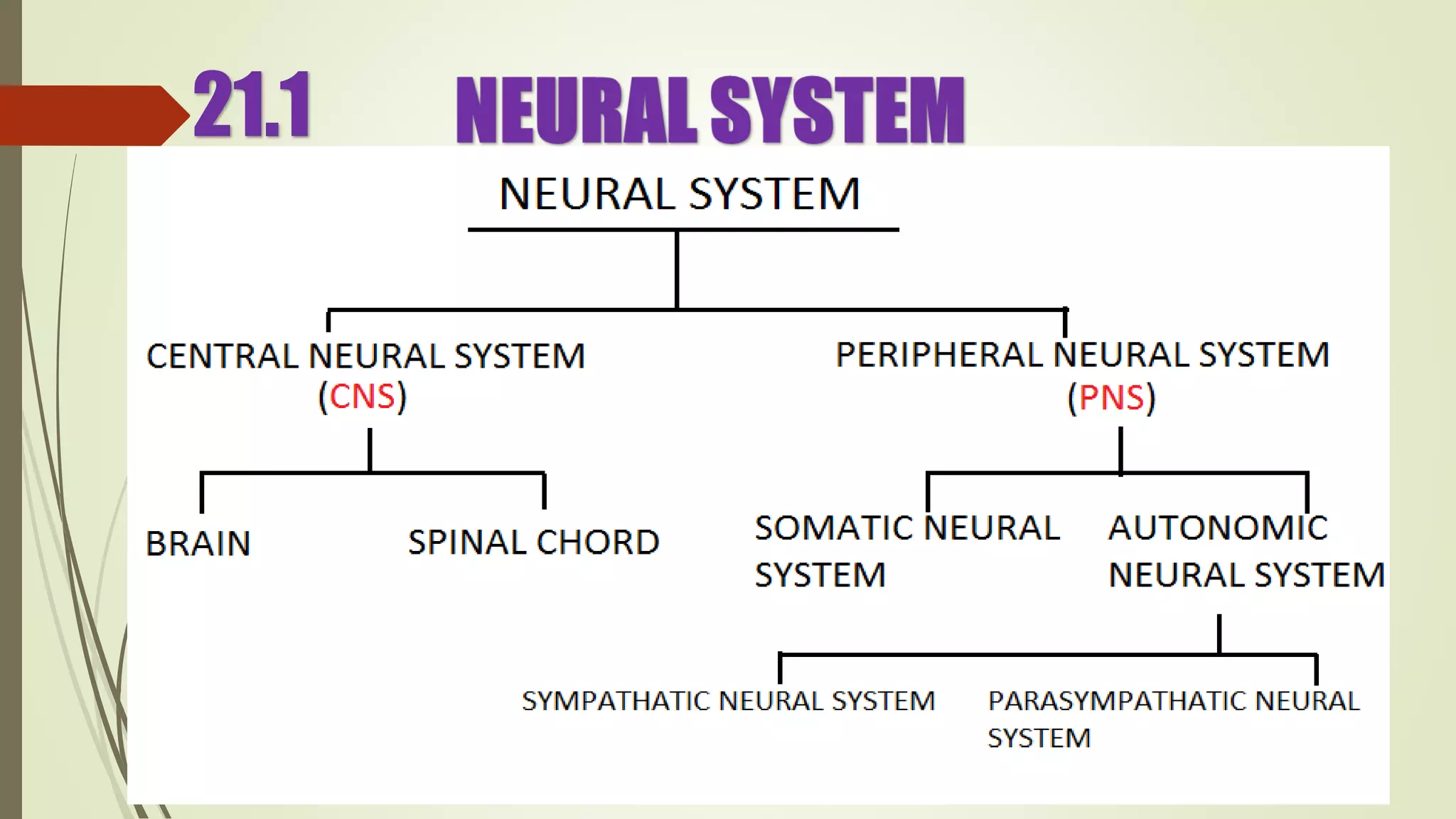
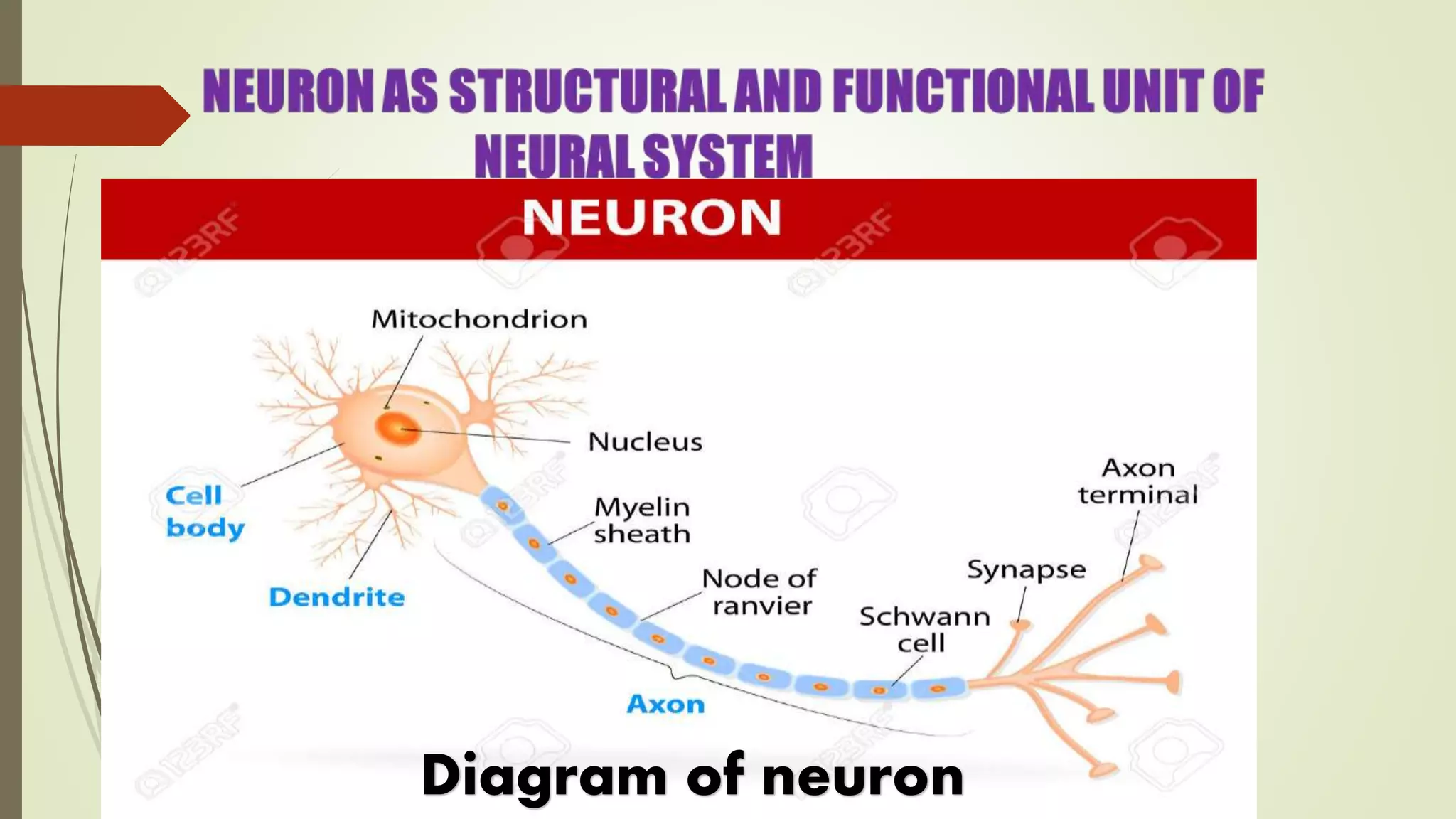
1. The document provides information about neural control and coordination from the NCERT biology textbook. It describes the structure and function of the nervous system, including neurons, nerve impulses, and reflex actions.
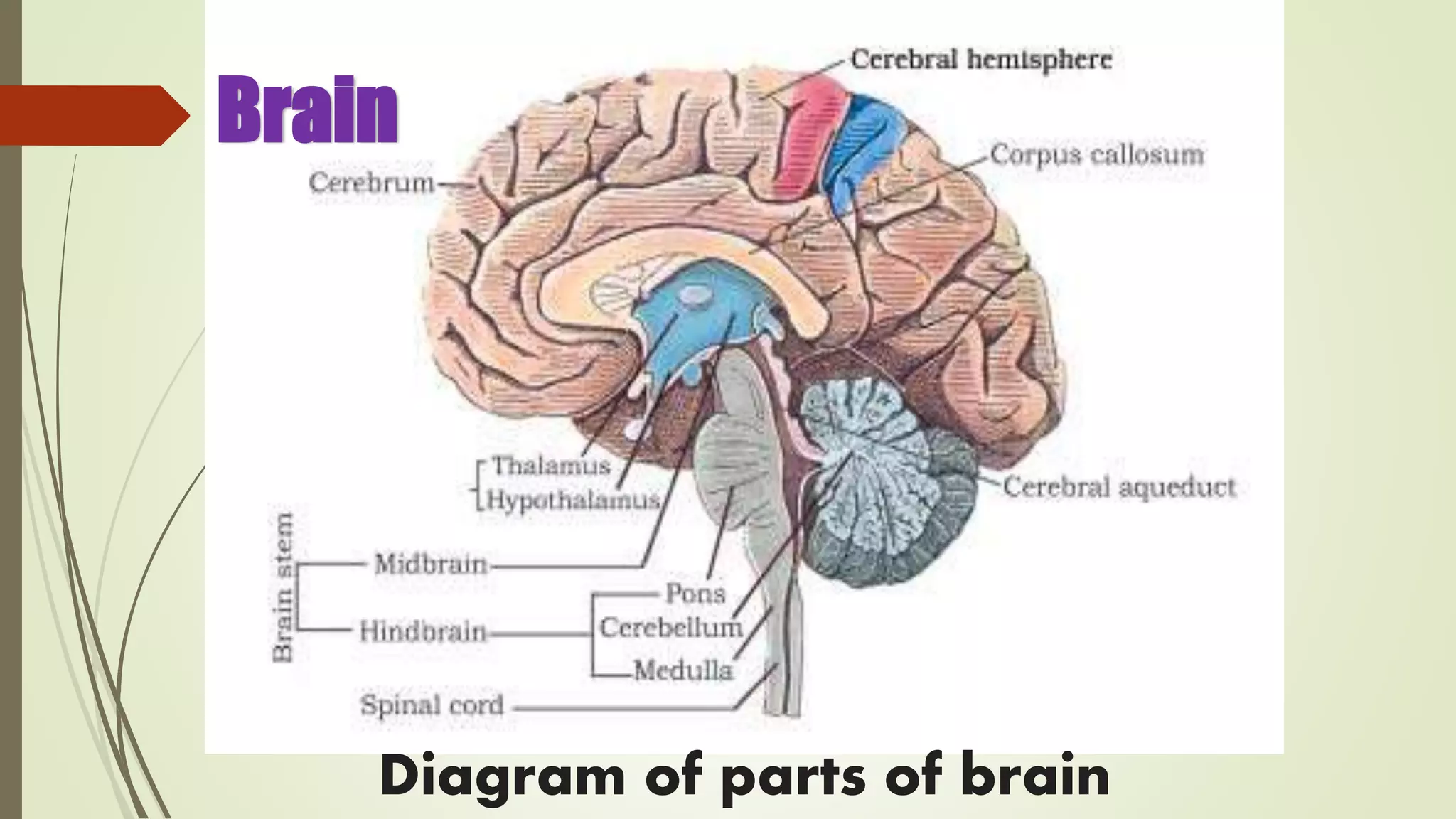
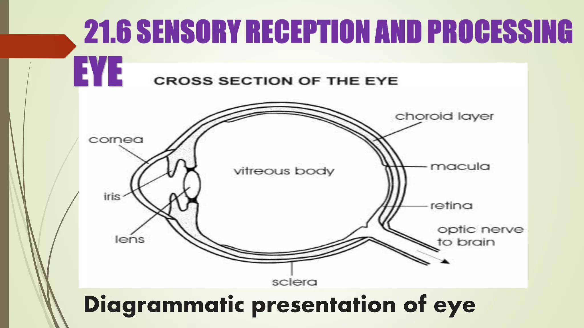
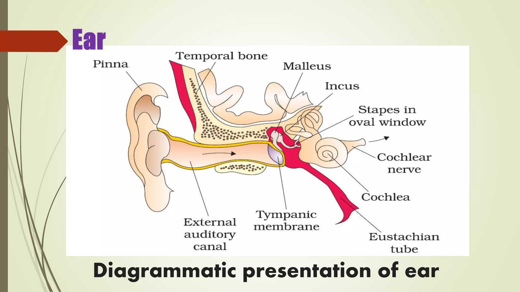
2. Key parts of the central nervous system like the brain, spinal cord, and various brain regions are defined. The processes of vision and hearing are also summarized.
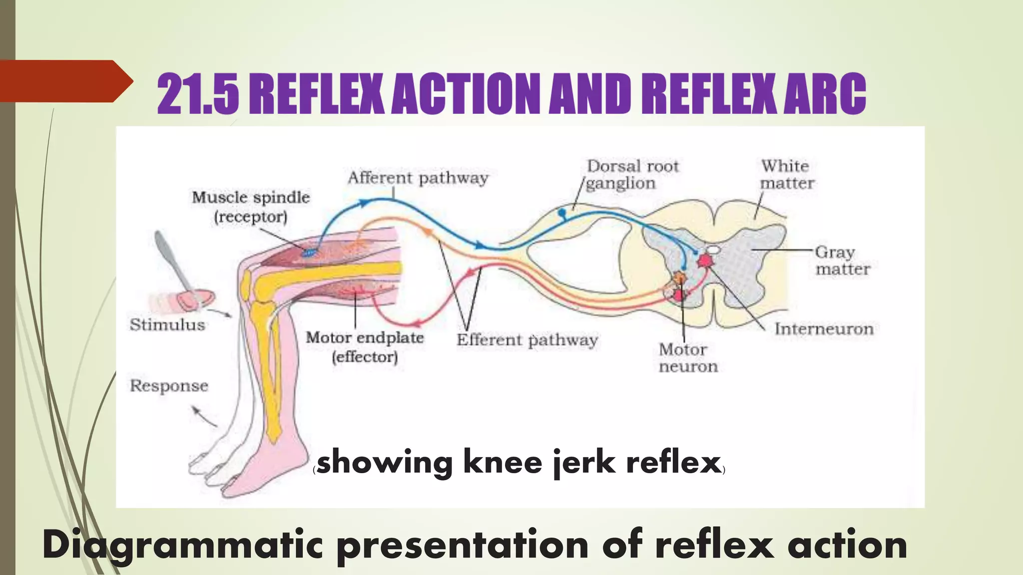
3. Coordination between different body systems like the neural and endocrine systems is explained. Sensory receptors and effector organs are described in the context of reflex arcs.