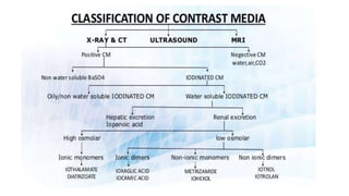
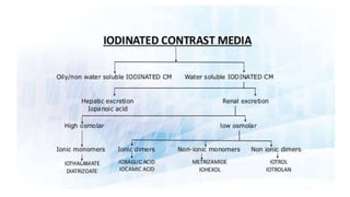
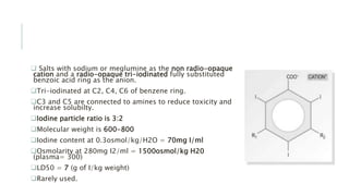
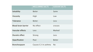
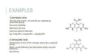
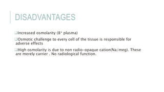
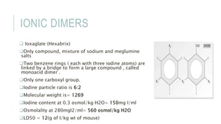
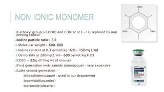
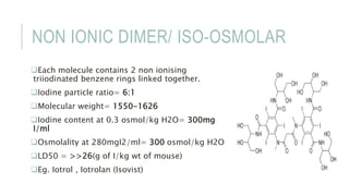
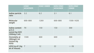
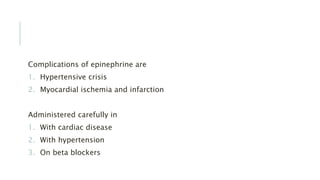
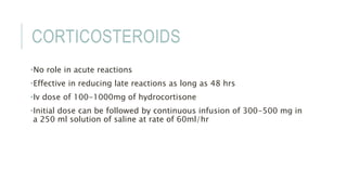
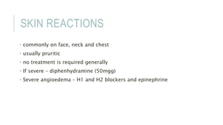
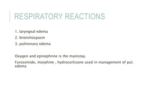
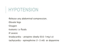
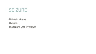
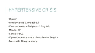
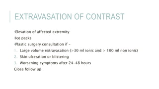
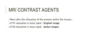
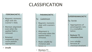
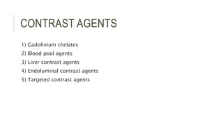
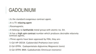
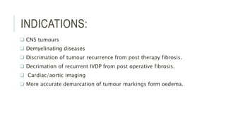
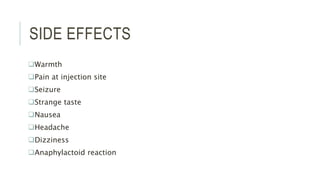
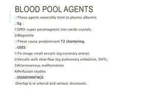
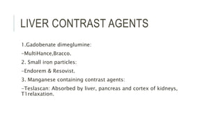
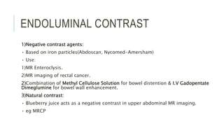
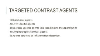
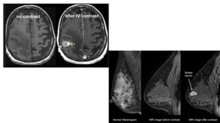
INTRAVENOUS CONTRAST
AGENTS
DR. ARUSHI GUPTA
DNB RADIO-DIAGNOSIS
CONTENT
 Introduction
 Iodinated contrast agents
- classification
- application
- adverse reactions
 USG contrast agents
 MRI contrast agents
INTRODUCTION
Difference in optical density between different parts of a image on a
radiograph.
Contrast media is a substance which when introduced into the body will
increase the radiographic contrast in an are where it was less before.
>90% are excreted through passive glomerular filtration.
<1% by liver and intestine.
 half life 30-60 mins
IODINE
Atomic weight 53
Atomic number 127
Total iodine content in the body is 50mg
 Iodine is preferred because :
•High contrast density
•Allow firm binding to highly variable benzene ring
•Low toxicity
Not suitable for MRI.
USEFUL FACTS
1) Radioopacity depends on
Iodine concentration of the solution, so dependent on the number of
iodine atoms in each molecule of the contrast media.
2) Iodine-particle ratio
Number of iodine atoms in per molecule to the number of osmotically
active particles in per molecule of solute in solution.
3) High radiopacity and low osmolarity are desirable requirements.
IOPANIC ACID
Is an iodine-containing radiocontrast medium.
 potent inhibitor of thyroid hormone release from thyroid gland, as well as of
peripheral conversion of thyroxine (T4) to triiodothyronine (T3)
Hepatic excretion
 Use:
1) Cholecystography
2) Hyperthyroidism: adjunctive therapy with thioamides (propylthiouracil,
carbimazole).
COVENTIONAL/HIGH
OSMOLAR/IONIC MONOMER
The basic molecule of all water-soluble iodine-
containing contrast media is the benzene ring.
 Benzene itself is not water soluble; to make it
soluble, carboxyl acid (COOH) is added.
Three of the hydrogens in this molecule are
replaced by iodine, rendering it radio-opaque, but
it still remains quite toxic.
 The remaining two hydrogens (R1 and R2) are
replaced by a short chain of hydrocarbons, making
the compound less toxic and more acceptable to
the body.
They are usually prepared as sodium or
meglumine salts as these help to provide solubility.
 Salts with sodium or meglumine as the non radio-opaque
cation and a radio-opaque tri-iodinated fully substituted
benzoic acid ring as the anion.
Tri-iodinated at C2, C4, C6 of benzene ring.
C3 and C5 are connected to amines to reduce toxicity and
increase solubilty.
Iodine particle ratio is 3:2
Molecular weight is 600-800
Iodine content at 0.3osmol/kg/H2O = 70mg I/ml
Osmolarity at 280mg I2/ml = 1500osmol/kg H20
(plasma= 300)
LD50 = 7 (g of I/kg weight)
Rarely used.
MEGLUMINE SALTS SODIUM SALTS
Solubility Better Same
Viscosity High Low
Tolerance Better Less
Blood brain barrier No effect crosses
Vascular effects Less Marked
Diuretic effect Strong Less
Opacification Poor Better
Bronchospasm Causes C/I in asthma No
EXAMPLES
1) DIATRIZOIC ACID
The two side chains , R3 and R5 are replaced by
acetamido group.
Increases solubilty
Decreases toxicity
Improves patient tolerance
Eg. Urograffin, trazograffin , angiograffin
2) IOTHALAMIC ACID
Substitution of one of the nitrogen atoms by a carboxyl
group.
Better neural tolerance but decreased cardio-vascular
tolerance.
Eg. Conray
DISADVANTAGES
Increased osmolarity (8* plasma)
Osmotic challenge to every cell of the tissue is responsible for
adverse effects
High osmolarity is due to non radio-opaque cation(Na/meg). These
are merely carrier . No radiological function.
IONIC DIMERS
 Ioxaglate (Hexabrix)
Only compound, mixture of sodium and meglumine
salts
Two benzene rings ( each with three iodine atoms) are
linked by a bridge to form a large compound , called
monoacid dimer .
Only one carboxyl group.
Iodine particle ratio is 6:2
Molecular weight is= 1269
Iodine content at 0.3 osmol/kg H2O= 150mg I/ml
Osmolality at 280mgI2/ml= 560 osmol/kg H2O
LD50 = 12(g of I/kg wt of mouse)
NON IONIC MONOMER
Carboxyl group (-COOH) and CONH2 at C-1 is replaced by non
ionising radical
Iodine particle ratio= 3:1
 Molecular weight= 600-800
 Iodine content at 0.3 osmol/kg H2O= 150mg I/ml
 Osmolality at 280mgI2/ml= 600 osmol/kg H2O
LD50 = 22(g of I/kg wt of mouse)
First generation metrizamide (amnipaque) – very expensive
Later second generation –
Iohexol(omnipaque) : used in our department
Iopamidol(iopamiro)
Iopromide(ultravist)
NON IONIC DIMER/ ISO-OSMOLAR
Each molecule contains 2 non ionising
triiodinated benzene rings linked together.
Iodine particle ratio= 6:1
Molecular weight= 1550-1626
Iodine content at 0.3 osmol/kg H2O= 300mg
I/ml
Osmolality at 280mgI2/ml= 300 osmol/kg H2O
LD50 = >>26(g of I/kg wt of mouse)
Eg. Iotrol , Iotrolan (Isovist)
IONIC
MONOMER
IONIC DIMER NON IONIC
MONOMER
NON IONIC
DIMER
Iodine particle
ratio
3:2 6:2 3:1 6:1
Molecular
weight
600-800 1269 600-800 1500-1626
Iodine content
at 0.3
osmol/kg H2O
(300mg I/ml)
70 150 150 300
Osmolality at
280mgI2/ml
(osmol/kg
H2O)
1500 600 600 300
LD50 (g of I/kg
wt of mouse)
7 12 22 >>36
ADDITIVES USED
1) Stabilizer: Ca or Na EDTA
2) Buffers: Stabilizes pH during storage, Na acid phosphates
3) Preservatives: Generally not disclosed by the manufacturers.
IDEAL IV CONTRAST AGENT
• Water soluble
• Heat/chemical/storage stability
• Non-antigenic
• Available at the right viscosity and density
• Low viscosity, making them easy to administer
• Persistent enough in the area of interest to allow its visualisation
• Selective excretion by the patient when the examination is complete
• Same osmolarity as plasma or lower
• Non-toxic, both locally and systemically
• Low cost
IMPORTANT FACTS
Contrast media used for myelography- non-ionic CM.
CM used for cerebral angiography- only meglumine salt.
Least osmolar- Ioxaglate (Hexabrix).
Most hyperosmolar- Iohexol.
Max nausea & vomiting- Ioxaglate (Hexabrix).
Bronchospasm- Meglumine salts.
Viscosity:
• Increase with concentration
• Higher for dimers(big size)
• High viscosity interferes with mixing of contrast media with plasma & body
fluids.
• Least viscosity- Omnipaque240
Meticulous heparinization is required during angiography as incidence of
thromboembolic phenomenon is high when CM is mixed with blood.
TOXICITY
Reactions unrelated to contrast media
Hyperosmolarity
Chemotoxic
Immunological
REACTIONS UNRELATED TO
CONTRAST MEDIA
1) Pyrogenic (unsterile injection)
stop injection , reassure patient , cover with blanket. No need for
medication.
2) vasovagal – in anxious patients
3) hypertensive attacks in patients with pheochromocytoma
4) excessive dehydration, hypoglycemia.
HYPER OSMOLARITY
More with conventional contrast media
1) erythrocyte damage
2) capillary endothelium damage
3) vasodilatation
4) hypervolemia
5) cardiovascular effects
6) vascular pain
7) disturbance of BBB
8) thrombosis and thrombophlebitis
CHEMOTOXIC
Are due to cations especially Na.
•Neurons
•Myocardial cells
•Capillary endothelium
•RBC
•Kidney
1. decreased renal perfusion
2. glomerular injury
3. tubular injury
4. contrast media precipitation that block tubules.
IMMUNOLOGICAL TOXICITY
Mechanism :
•Deactivation of ACE : ACE deactivates bradykinin
•Damage to endothelium which initiates activation system
•Activation of complements, kinins, coagulation system
•Inhibition of cholinesterase with consequent vagal over stimulation
•Release of histamine, bradykinin , serotonin
1. Anaphylactic reaction
2. Analphylactoid reaction
HIGH RISK GROUP
Prior reaction to contrast media
History of allergy
Cardiac disease
Asthma
Diabetes
Old age
Neonates
Sickle cell anemia , pheochromocytoma
TREATMENT
Two basic rules :
1. make sure drugs for allergy are available before injecting the
contrast
2. never leave the patient unattended until the examination is
complete
General principles:
1. oxygen
2. epinephrine
3. corticosteroids.
OXYGEN
1. oxygen and equipment for assisting ventilation should be readily
available.
2. high dose oxygen at 10-12L/min via face mask
3. should be used in any patient with respiratory distress
EPINEPHRINE
Single most important drug
•Powerful sympathomimetic agent (activates alpha and beta receptors)
•Produces peripheral vasoconstriction, increased cardiac contractibility
and cardiac rate and smooth muscle bronchodilatation.
•2 dilutions:
1. 1 in 1000 – 1mg epinephrine in 1ml of fluid. For s.c or i.m use
2. 1 in 10000 – 1 mg epinephrine in 10 ml of fluid. For i.v use
Complications of epinephrine are
1. Hypertensive crisis
2. Myocardial ischemia and infarction
Administered carefully in
1. With cardiac disease
2. With hypertension
3. On beta blockers
CORTICOSTEROIDS
•No role in acute reactions
•Effective in reducing late reactions as long as 48 hrs
•Iv dose of 100-1000mg of hydrocortisone
•Initial dose can be followed by continuous infusion of 300-500 mg in
a 250 ml solution of saline at rate of 60ml/hr
SKIN REACTIONS
• commonly on face, neck and chest
• usually pruritic
• no treatment is required generally
• If severe – diphenhydramine (50mgg)
• Severe angioedema – H1 and H2 blockers and epinephrine
RESPIRATORY REACTIONS
1. laryngeal edema
2. bronchospasm
3. pulmonary edema
Oxygen and epinephrine is the mainstay.
Furosemide, morphine , hydrocortisone used in management of pul.
edema
HYPOTENSION
•Release any abdominal compression.
•Elevate legs
•Oxygen
•Isotonic i.v fluids
•If severe
bradycardia – atropine slowly (0.6-1mg I.v)
tachycardia – epinephrine (1-3 ml) or dopamine
SEIZURE
•Maintain airway
•Oxygen
•Diazepam 5mg i.v slowly
HYPERTENSIVE CRISIS
•Oxygen
•Nitroglycerine 0.4mg tab s.l
•If no response- nifedipine – 10mg tab
•Monitor BP
•Consider ECG
•If pheochromocytome – phentolamine 5mg i.v
•Frusemide 40mg i.v slowly
EXTRAVASATION OF CONTRAST
•Elevation of affected extremity
•Ice packs
•Plastic surgery consultation if –
1. Large volume extravasation (>30 ml ionic and > 100 ml non ionic)
2. Skin ulceration or blistering
3. Worsening symptoms after 24-48 hours
Close follow up
USG CONTRAST AGENTS
 Increases the echogenicity of the blood - increases tissue contrast
 Microscopic gas filled bubbles – reflect sound waves. Size <8um
(smaller than rbc).
 Consists of a gas core surrounded by a lipid shell
 Avoid gases with tendency to coalesce and form emboli.
 Main mechanism : interaction between microbubbles and the US
beam
Backscattering due to different impedances of liquids and gas
Bubble resonance
Bubble rupture which is dependent on the acoustic power of the
transmitted USG
GENERATIONS OF ECHO
ENHANCERS
First : unstabilised bubbles in indocyanine green
(cant survive pulmonary passage , therefore used
only for cardiac and large veins study)
Second : longer lasting bubbles coated with shells of
protein or lipids.
Third : encapsulated emulsions or bubbles , offer
high reflectivity.
NEWER APPLICATIONS
 ECHOVIST : evaluation of fallopian tube patency
 LEVOVIST : reflux sonography to detect or exclude VUR
 KnorX : oral agent – uniform echogencity of stomach and
pancreas
IDEAL QUALITIES
High echogenicity
Low attenuation
Low blood solubility
Low diffusivity
Pass through pulmonary bed
No biological effect
Smaller size
Prolonged persistence
APPLICATIONS
1) Evaluating normal, increased or decreased vascularity.
2) Detecting vascular stenosis & occlusions.
3) Improving neoplasm detection.
4) Analysing & characterizing tumour neovascularity.
5) Differentiating normal variants such as renal column of bertin from
neoplasm.
6) Echocardiography – cardiac cavities, valves, coronary artery &
myocardial viability
MRI CONTRAST AGENTS
Must alter the relaxation of the protons within the tissues.
If T1 relaxation is more rapid – brighter image
If T2 relaxation is more rapid – darker images
FERROMAGNETIC
• Magnetic moments
align with the
scanner’s field.
• Maintain alignment
even when the
applied field is
removed.
• Retained
magnetism may
cause particle
aggregation
• Unsafe
PARAMAGNETIC
Eg Gadolinium
• Magnetic moments
align with scanners
field.
• Alignment is
overcome when the
applied field is
removed.
• Made soluble by
chelation and
hence i.v
• Shortens T1
SUPERPARAMAGNETIC
Eg. Ferrite
• Aggregations of
paramagnetic ions
in a crystalline
lattice.
• Reduces T2
relaxation time
• Less soluble
because of
chemical structure
CLASSIFICATION
CONTRAST AGENTS
1) Gadolinium chelates
2) Blood pool agents
3) Liver contrast agents
4) Endoluminal contrast agents
5) Targeted contrast agents
GADOLINIUM
Is the standard exogenous contrast agent.
It is T1 relaxing agent
Paramagnetic.
It belongs to lanthanide metal group with atomic no. 64.
It has a high spin contrast number which produces desirable relaxivity
contrast agents.
Three agents have been approved by FDA, they are-
1) Gd-HP-DO3A: Gadoteridol/ProHance (non ionic)
2) Gd-DTPA : Gadopentetate diglumine/Magnevist (ionic)
3) Gd-DTPA-BMA: Gadodiamide/Omniscan (nonionic)
These function as extracellular contrast agents.
They are rapidly excreted by glomerular filteration
half lives: 1 – 2hrs.
As these compounds are excreted by renal excretion, caution shoud be
taken in renal impaired patients.
S/E: Nausea(3 –5%)
Dose: 0.1 to 0.3mmol/kg body weight
Disadvantages:
1) Enhancement is non specific neither organ specific or pathology specific.
2) Short window for imaging of blood vessels as it is diluted in blood stream
and excreted rapidly.
INDICATIONS:
 CNS tumours
 Demyelinating diseases
 Discrimation of tumour recurrence from post therapy fibrosis.
 Decrimation of recurrent IVDP from post operative fibrosis.
 Cardiac/aortic imaging
 More accurate demarcation of tumour markings form oedema.
SIDE EFFECTS
Warmth
Pain at injection site
Seizure
Strange taste
Nausea
Headache
Dizziness
Anaphylactoid reaction
BLOOD POOL AGENTS
These agents reversibly bind to plasma albumin
Eg :
1)SPIO-super paramagnetic iron oxide crystals
2)Magnetite
These cause predominant T2 shortening.
USES:
1)To image small vessels (eg:coronary artery).
2)Vessels with slow flow (eg pulmonary embolism, DVT),
3)Arteriovenous malformation
4)Perfusion studies
DISADVANTAGE:
Overlap b/w arterial and venous structures.
LIVER CONTRAST AGENTS
1.Gadobenate dimeglumine:
-MultiHance,Bracco.
2. Small iron particles:
-Endorem & Resovist.
3. Manganese containing contrast agents:
-Teslascan: Absorbed by liver, pancreas and cortex of kidneys,
T1relaxation.
ENDOLUMINAL CONTRAST
1)Negative contrast agents:
• Based on iron particles(Abdoscan, Nycomed-Amersham)
• Use:
1)MR Enteroclysis.
2)MR imaging of rectal cancer.
2)Combination of Methyl Cellulose Solution for bowel distention & I.V Gadopentate
Dimeglumine for bowel wall enhancement.
3)Natural contrast:
• Blueberry juice acts as a negative contrast in upper abdominal MR imaging.
• eg MRCP
TARGETED CONTRAST AGENTS
1) Blood pool agents
2) Liver specific agents
3) Necrosis specific agents (bis-gadolinium-mesoporphyrin)
4) Lymphographic contrast agents
5) Agents targeted at inflammation detection.
THANK YOU !

Intravenous contrast agents

  • 1.
    INTRAVENOUS CONTRAST AGENTS DR. ARUSHIGUPTA DNB RADIO-DIAGNOSIS
  • 2.
    CONTENT  Introduction  Iodinatedcontrast agents - classification - application - adverse reactions  USG contrast agents  MRI contrast agents
  • 3.
    INTRODUCTION Difference in opticaldensity between different parts of a image on a radiograph. Contrast media is a substance which when introduced into the body will increase the radiographic contrast in an are where it was less before. >90% are excreted through passive glomerular filtration. <1% by liver and intestine.  half life 30-60 mins
  • 6.
    IODINE Atomic weight 53 Atomicnumber 127 Total iodine content in the body is 50mg  Iodine is preferred because : •High contrast density •Allow firm binding to highly variable benzene ring •Low toxicity Not suitable for MRI.
  • 7.
    USEFUL FACTS 1) Radioopacitydepends on Iodine concentration of the solution, so dependent on the number of iodine atoms in each molecule of the contrast media. 2) Iodine-particle ratio Number of iodine atoms in per molecule to the number of osmotically active particles in per molecule of solute in solution. 3) High radiopacity and low osmolarity are desirable requirements.
  • 8.
    IOPANIC ACID Is aniodine-containing radiocontrast medium.  potent inhibitor of thyroid hormone release from thyroid gland, as well as of peripheral conversion of thyroxine (T4) to triiodothyronine (T3) Hepatic excretion  Use: 1) Cholecystography 2) Hyperthyroidism: adjunctive therapy with thioamides (propylthiouracil, carbimazole).
  • 9.
    COVENTIONAL/HIGH OSMOLAR/IONIC MONOMER The basicmolecule of all water-soluble iodine- containing contrast media is the benzene ring.  Benzene itself is not water soluble; to make it soluble, carboxyl acid (COOH) is added. Three of the hydrogens in this molecule are replaced by iodine, rendering it radio-opaque, but it still remains quite toxic.  The remaining two hydrogens (R1 and R2) are replaced by a short chain of hydrocarbons, making the compound less toxic and more acceptable to the body. They are usually prepared as sodium or meglumine salts as these help to provide solubility.
  • 10.
     Salts withsodium or meglumine as the non radio-opaque cation and a radio-opaque tri-iodinated fully substituted benzoic acid ring as the anion. Tri-iodinated at C2, C4, C6 of benzene ring. C3 and C5 are connected to amines to reduce toxicity and increase solubilty. Iodine particle ratio is 3:2 Molecular weight is 600-800 Iodine content at 0.3osmol/kg/H2O = 70mg I/ml Osmolarity at 280mg I2/ml = 1500osmol/kg H20 (plasma= 300) LD50 = 7 (g of I/kg weight) Rarely used.
  • 11.
    MEGLUMINE SALTS SODIUMSALTS Solubility Better Same Viscosity High Low Tolerance Better Less Blood brain barrier No effect crosses Vascular effects Less Marked Diuretic effect Strong Less Opacification Poor Better Bronchospasm Causes C/I in asthma No
  • 12.
    EXAMPLES 1) DIATRIZOIC ACID Thetwo side chains , R3 and R5 are replaced by acetamido group. Increases solubilty Decreases toxicity Improves patient tolerance Eg. Urograffin, trazograffin , angiograffin 2) IOTHALAMIC ACID Substitution of one of the nitrogen atoms by a carboxyl group. Better neural tolerance but decreased cardio-vascular tolerance. Eg. Conray
  • 13.
    DISADVANTAGES Increased osmolarity (8*plasma) Osmotic challenge to every cell of the tissue is responsible for adverse effects High osmolarity is due to non radio-opaque cation(Na/meg). These are merely carrier . No radiological function.
  • 14.
    IONIC DIMERS  Ioxaglate(Hexabrix) Only compound, mixture of sodium and meglumine salts Two benzene rings ( each with three iodine atoms) are linked by a bridge to form a large compound , called monoacid dimer . Only one carboxyl group. Iodine particle ratio is 6:2 Molecular weight is= 1269 Iodine content at 0.3 osmol/kg H2O= 150mg I/ml Osmolality at 280mgI2/ml= 560 osmol/kg H2O LD50 = 12(g of I/kg wt of mouse)
  • 15.
    NON IONIC MONOMER Carboxylgroup (-COOH) and CONH2 at C-1 is replaced by non ionising radical Iodine particle ratio= 3:1  Molecular weight= 600-800  Iodine content at 0.3 osmol/kg H2O= 150mg I/ml  Osmolality at 280mgI2/ml= 600 osmol/kg H2O LD50 = 22(g of I/kg wt of mouse) First generation metrizamide (amnipaque) – very expensive Later second generation – Iohexol(omnipaque) : used in our department Iopamidol(iopamiro) Iopromide(ultravist)
  • 16.
    NON IONIC DIMER/ISO-OSMOLAR Each molecule contains 2 non ionising triiodinated benzene rings linked together. Iodine particle ratio= 6:1 Molecular weight= 1550-1626 Iodine content at 0.3 osmol/kg H2O= 300mg I/ml Osmolality at 280mgI2/ml= 300 osmol/kg H2O LD50 = >>26(g of I/kg wt of mouse) Eg. Iotrol , Iotrolan (Isovist)
  • 17.
    IONIC MONOMER IONIC DIMER NONIONIC MONOMER NON IONIC DIMER Iodine particle ratio 3:2 6:2 3:1 6:1 Molecular weight 600-800 1269 600-800 1500-1626 Iodine content at 0.3 osmol/kg H2O (300mg I/ml) 70 150 150 300 Osmolality at 280mgI2/ml (osmol/kg H2O) 1500 600 600 300 LD50 (g of I/kg wt of mouse) 7 12 22 >>36
  • 18.
    ADDITIVES USED 1) Stabilizer:Ca or Na EDTA 2) Buffers: Stabilizes pH during storage, Na acid phosphates 3) Preservatives: Generally not disclosed by the manufacturers.
  • 19.
    IDEAL IV CONTRASTAGENT • Water soluble • Heat/chemical/storage stability • Non-antigenic • Available at the right viscosity and density • Low viscosity, making them easy to administer • Persistent enough in the area of interest to allow its visualisation • Selective excretion by the patient when the examination is complete • Same osmolarity as plasma or lower • Non-toxic, both locally and systemically • Low cost
  • 20.
    IMPORTANT FACTS Contrast mediaused for myelography- non-ionic CM. CM used for cerebral angiography- only meglumine salt. Least osmolar- Ioxaglate (Hexabrix). Most hyperosmolar- Iohexol. Max nausea & vomiting- Ioxaglate (Hexabrix). Bronchospasm- Meglumine salts. Viscosity: • Increase with concentration • Higher for dimers(big size) • High viscosity interferes with mixing of contrast media with plasma & body fluids. • Least viscosity- Omnipaque240 Meticulous heparinization is required during angiography as incidence of thromboembolic phenomenon is high when CM is mixed with blood.
  • 21.
    TOXICITY Reactions unrelated tocontrast media Hyperosmolarity Chemotoxic Immunological
  • 22.
    REACTIONS UNRELATED TO CONTRASTMEDIA 1) Pyrogenic (unsterile injection) stop injection , reassure patient , cover with blanket. No need for medication. 2) vasovagal – in anxious patients 3) hypertensive attacks in patients with pheochromocytoma 4) excessive dehydration, hypoglycemia.
  • 23.
    HYPER OSMOLARITY More withconventional contrast media 1) erythrocyte damage 2) capillary endothelium damage 3) vasodilatation 4) hypervolemia 5) cardiovascular effects 6) vascular pain 7) disturbance of BBB 8) thrombosis and thrombophlebitis
  • 24.
    CHEMOTOXIC Are due tocations especially Na. •Neurons •Myocardial cells •Capillary endothelium •RBC •Kidney 1. decreased renal perfusion 2. glomerular injury 3. tubular injury 4. contrast media precipitation that block tubules.
  • 25.
    IMMUNOLOGICAL TOXICITY Mechanism : •Deactivationof ACE : ACE deactivates bradykinin •Damage to endothelium which initiates activation system •Activation of complements, kinins, coagulation system •Inhibition of cholinesterase with consequent vagal over stimulation •Release of histamine, bradykinin , serotonin 1. Anaphylactic reaction 2. Analphylactoid reaction
  • 26.
    HIGH RISK GROUP Priorreaction to contrast media History of allergy Cardiac disease Asthma Diabetes Old age Neonates Sickle cell anemia , pheochromocytoma
  • 27.
    TREATMENT Two basic rules: 1. make sure drugs for allergy are available before injecting the contrast 2. never leave the patient unattended until the examination is complete General principles: 1. oxygen 2. epinephrine 3. corticosteroids.
  • 28.
    OXYGEN 1. oxygen andequipment for assisting ventilation should be readily available. 2. high dose oxygen at 10-12L/min via face mask 3. should be used in any patient with respiratory distress
  • 29.
    EPINEPHRINE Single most importantdrug •Powerful sympathomimetic agent (activates alpha and beta receptors) •Produces peripheral vasoconstriction, increased cardiac contractibility and cardiac rate and smooth muscle bronchodilatation. •2 dilutions: 1. 1 in 1000 – 1mg epinephrine in 1ml of fluid. For s.c or i.m use 2. 1 in 10000 – 1 mg epinephrine in 10 ml of fluid. For i.v use
  • 30.
    Complications of epinephrineare 1. Hypertensive crisis 2. Myocardial ischemia and infarction Administered carefully in 1. With cardiac disease 2. With hypertension 3. On beta blockers
  • 31.
    CORTICOSTEROIDS •No role inacute reactions •Effective in reducing late reactions as long as 48 hrs •Iv dose of 100-1000mg of hydrocortisone •Initial dose can be followed by continuous infusion of 300-500 mg in a 250 ml solution of saline at rate of 60ml/hr
  • 32.
    SKIN REACTIONS • commonlyon face, neck and chest • usually pruritic • no treatment is required generally • If severe – diphenhydramine (50mgg) • Severe angioedema – H1 and H2 blockers and epinephrine
  • 33.
    RESPIRATORY REACTIONS 1. laryngealedema 2. bronchospasm 3. pulmonary edema Oxygen and epinephrine is the mainstay. Furosemide, morphine , hydrocortisone used in management of pul. edema
  • 34.
    HYPOTENSION •Release any abdominalcompression. •Elevate legs •Oxygen •Isotonic i.v fluids •If severe bradycardia – atropine slowly (0.6-1mg I.v) tachycardia – epinephrine (1-3 ml) or dopamine
  • 35.
  • 36.
    HYPERTENSIVE CRISIS •Oxygen •Nitroglycerine 0.4mgtab s.l •If no response- nifedipine – 10mg tab •Monitor BP •Consider ECG •If pheochromocytome – phentolamine 5mg i.v •Frusemide 40mg i.v slowly
  • 37.
    EXTRAVASATION OF CONTRAST •Elevationof affected extremity •Ice packs •Plastic surgery consultation if – 1. Large volume extravasation (>30 ml ionic and > 100 ml non ionic) 2. Skin ulceration or blistering 3. Worsening symptoms after 24-48 hours Close follow up
  • 38.
    USG CONTRAST AGENTS Increases the echogenicity of the blood - increases tissue contrast  Microscopic gas filled bubbles – reflect sound waves. Size <8um (smaller than rbc).  Consists of a gas core surrounded by a lipid shell  Avoid gases with tendency to coalesce and form emboli.  Main mechanism : interaction between microbubbles and the US beam Backscattering due to different impedances of liquids and gas Bubble resonance Bubble rupture which is dependent on the acoustic power of the transmitted USG
  • 39.
    GENERATIONS OF ECHO ENHANCERS First: unstabilised bubbles in indocyanine green (cant survive pulmonary passage , therefore used only for cardiac and large veins study) Second : longer lasting bubbles coated with shells of protein or lipids. Third : encapsulated emulsions or bubbles , offer high reflectivity.
  • 41.
    NEWER APPLICATIONS  ECHOVIST: evaluation of fallopian tube patency  LEVOVIST : reflux sonography to detect or exclude VUR  KnorX : oral agent – uniform echogencity of stomach and pancreas
  • 42.
    IDEAL QUALITIES High echogenicity Lowattenuation Low blood solubility Low diffusivity Pass through pulmonary bed No biological effect Smaller size Prolonged persistence
  • 43.
    APPLICATIONS 1) Evaluating normal,increased or decreased vascularity. 2) Detecting vascular stenosis & occlusions. 3) Improving neoplasm detection. 4) Analysing & characterizing tumour neovascularity. 5) Differentiating normal variants such as renal column of bertin from neoplasm. 6) Echocardiography – cardiac cavities, valves, coronary artery & myocardial viability
  • 46.
    MRI CONTRAST AGENTS Mustalter the relaxation of the protons within the tissues. If T1 relaxation is more rapid – brighter image If T2 relaxation is more rapid – darker images
  • 47.
    FERROMAGNETIC • Magnetic moments alignwith the scanner’s field. • Maintain alignment even when the applied field is removed. • Retained magnetism may cause particle aggregation • Unsafe PARAMAGNETIC Eg Gadolinium • Magnetic moments align with scanners field. • Alignment is overcome when the applied field is removed. • Made soluble by chelation and hence i.v • Shortens T1 SUPERPARAMAGNETIC Eg. Ferrite • Aggregations of paramagnetic ions in a crystalline lattice. • Reduces T2 relaxation time • Less soluble because of chemical structure CLASSIFICATION
  • 48.
    CONTRAST AGENTS 1) Gadoliniumchelates 2) Blood pool agents 3) Liver contrast agents 4) Endoluminal contrast agents 5) Targeted contrast agents
  • 49.
    GADOLINIUM Is the standardexogenous contrast agent. It is T1 relaxing agent Paramagnetic. It belongs to lanthanide metal group with atomic no. 64. It has a high spin contrast number which produces desirable relaxivity contrast agents. Three agents have been approved by FDA, they are- 1) Gd-HP-DO3A: Gadoteridol/ProHance (non ionic) 2) Gd-DTPA : Gadopentetate diglumine/Magnevist (ionic) 3) Gd-DTPA-BMA: Gadodiamide/Omniscan (nonionic)
  • 50.
    These function asextracellular contrast agents. They are rapidly excreted by glomerular filteration half lives: 1 – 2hrs. As these compounds are excreted by renal excretion, caution shoud be taken in renal impaired patients. S/E: Nausea(3 –5%) Dose: 0.1 to 0.3mmol/kg body weight Disadvantages: 1) Enhancement is non specific neither organ specific or pathology specific. 2) Short window for imaging of blood vessels as it is diluted in blood stream and excreted rapidly.
  • 51.
    INDICATIONS:  CNS tumours Demyelinating diseases  Discrimation of tumour recurrence from post therapy fibrosis.  Decrimation of recurrent IVDP from post operative fibrosis.  Cardiac/aortic imaging  More accurate demarcation of tumour markings form oedema.
  • 52.
    SIDE EFFECTS Warmth Pain atinjection site Seizure Strange taste Nausea Headache Dizziness Anaphylactoid reaction
  • 53.
    BLOOD POOL AGENTS Theseagents reversibly bind to plasma albumin Eg : 1)SPIO-super paramagnetic iron oxide crystals 2)Magnetite These cause predominant T2 shortening. USES: 1)To image small vessels (eg:coronary artery). 2)Vessels with slow flow (eg pulmonary embolism, DVT), 3)Arteriovenous malformation 4)Perfusion studies DISADVANTAGE: Overlap b/w arterial and venous structures.
  • 54.
    LIVER CONTRAST AGENTS 1.Gadobenatedimeglumine: -MultiHance,Bracco. 2. Small iron particles: -Endorem & Resovist. 3. Manganese containing contrast agents: -Teslascan: Absorbed by liver, pancreas and cortex of kidneys, T1relaxation.
  • 55.
    ENDOLUMINAL CONTRAST 1)Negative contrastagents: • Based on iron particles(Abdoscan, Nycomed-Amersham) • Use: 1)MR Enteroclysis. 2)MR imaging of rectal cancer. 2)Combination of Methyl Cellulose Solution for bowel distention & I.V Gadopentate Dimeglumine for bowel wall enhancement. 3)Natural contrast: • Blueberry juice acts as a negative contrast in upper abdominal MR imaging. • eg MRCP
  • 56.
    TARGETED CONTRAST AGENTS 1)Blood pool agents 2) Liver specific agents 3) Necrosis specific agents (bis-gadolinium-mesoporphyrin) 4) Lymphographic contrast agents 5) Agents targeted at inflammation detection.
  • 58.