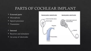
COCHLEAR IMPLANT
DR. TANIYA SHEORAN
Junior resident
MGM Medical college, Indore
Surgically placed electrical device that receive sound and
transmit the resulting electrical signals to electrodes
implanted in the cochlea.
These signals stimulate cochlea , allowing patient to
hear.
Also known as bionic ear.
PARTS OF COCHLEAR IMPLANT
 External parts
 Microphone
 Speech processor
 Transmitter
 Internal
 Receiver and stimulator
 An array of electrodes
 EXTERNAL PART : converts acoustic signal to electric signal which is
transmitted to internal coil using radiofrequency
 INTERNAL RECIEVER AND STIMULATOR : contains internal magnet,
telemetry coil and hermetically sealed electronics system.
 Latest generation of CI are compatible with 1.5 Tesla MRI without magnet
removal.
 INTRACOCHLEAR ELECTRODE : group of individual wires ending at a
contact point along silicone casing.
 Smaller electrodes – reduce insertion trauma
 Inserted by the round window or through small cochleostomies with
diameter of 0.5-0.8mm.
CANDIDACY FOR CI
 THREE GROUPS OF CANDIDATES:
1. Post lingually deafened children and adults
2. Prelingually deafened children
3. Early pre and peri lingually deafened adults and adolescents
 Severe to profound bilateral sensorineural hearing loss with little or insufficient benefit from
conventional hearing aids.
 In adults with post lingual hearing loss:
 Speech audiometry tested with hearing aids in a free field setting at 65–75 dB sound pressure
level with a score of ≤ 50%.
 In congenitally deaf children or (young) children with prelingual hearing loss:
 Auditory brainstem response audiometry (ABR) with no responses or responses at 80 dB or
more.
 Free field test with hearing aids after trial of at least 3 months, with responses only above 60
dB or no responses at all (up to 100–120 dB stimulation).
 In early deafened adults and adolescents:
 Speech audiometry tested with hearing aids in a free field setting at 65–75 dB sound pressure
level with a score of ≤ 50% + additional evaluation of speech intelligibility and lip-reading
skills.
 CHILDREN
 12 months to 7 years of age in pre lingually deaf
 B/L severe to profound SNHL with PTA 90dB or greater in better ear
 No appreciable benefit with hearing aids
 No medical or anatomic contraindication
 Motivated parents
 In case of HL due to meningitis, implantation should be considered before 12 months of
age.
 ADULTS
 18 years or more
 B/L severe to profound SNHL
 Post lingual deaf
 No appreciable benefit from hearing aid
 Sentence recognition test <50%
 No medical or anatomical contraindication
 Severe to profound deafness is defined as the ability to hear only sounds
louder than 90dBHL at 2kHz and 4kHz without hearing aids.
 Hearing aids should be used for at least 3 months unless inappropriate or
contraindicated.
 Adequate benefit with hearing aids is defined as:
 ❍ For adults, a score of 50% or greater on Bamford– Kowal–Bench sentence
testing at a sound level of 70dBSPL.
 ❍ For children, speech, language and listening skills appropriate to age,
developmental stage and cognitive ability.
 Simultaneous bilateral CI is recommended for children and for adults who
are blind or who have other disabilities that increase their reliance on
auditory stimuli as a primary sensory mechanism for spatial awareness.
CONTRAINDICATIONS
 Absolute contraindications
 • Cochlear nerve aplasia, confirmed by appropriate testing
 • Cochlear aplasia, Michel deformity
 • Deafness of central origin
 Relative contraindications
 • Cochlear nerve hypoplasia
 • Bilateral cochlear ossification, confirmed on MRI and not accessible via drill-out
procedure
 • Psychiatric pathology
MODES OF STIMULATION OF AUDITORY SYSTEM IN CI
 THREE MODES
1. Electrical stimulation – when there is no residual hearing left in both ears
2. Electro-acoustic stimulation – HYBRID – lower frequencies stimulated
acoustically via hearing and higher frequencies via CI
3. Bimodal stimulation – one ear uses CI, one ear uses high gain hearing aid
CODING STRATEGY
 Methods by which pitch, loudness and timing of sound are translated into
series of electrical impulses
 Two methods – SIMULTANEOUS and NON SIMULTAANEOUS
SIMULTANEOUS NON SIMULTANEOUS
Activation of more than one
electrode at the same time
Stimulate each electrode serially, no
electrode bypassed
Available with advanced bionics Available with all three devices
Problems of signal interference Faster sequential stimulation -
better speech recognition
Which ear to implant?
 Better hearing ear
 Recent deaf ear
 Radiographically favourable anatomy (well pneumatized mastoid, normal
facial nerve anatomy, inner ear and patent cochlea)
 Least obstructed labyrinth
PRE OP ASSESSMENT
 MEDICAL EVALUATION
 History – genetic hearing loss/auditory neuropathy
 Audiological – determine type and severity of HL/duration of
HL/tympanometry/acoustic reflexes
 ELECTROPHYSIOLOGICAL TEST
 ABR – verify audiometric result/identify patient with auditory dys-
synchrony/ rule out possibility of functional deafness
 Speech perception test
 Adults : monosyllabic test and hearing in noise test
 Children : Early speech perception and Meaword intelligibility by picture
identification
 PSYCHOLOGICAL EVALUATION
 No un realistic expectations
 RADIOLOGICAL
 HRCT – allows precise demonstration of fine middle ear structures, with
thin slices of less than 1 mm.
 preferred direction of axial scanning or reconstruction should be at the
plane of the lateral semicircular canal.
 inner ear morphology/patency of cochlea/position of facial nerve/size of
facial recess/height of jugular bulb
 MRI – labyrinthine ossificans/ cochlear nerve/ CNS abnormalities
OUTCOMES
 CHILDREN
 Earlier age – better language
development
 Implanted after 3 yrs – struggle to
catch up
 Neural plasticity
 ADULTS
 Duration of severe or profound deafness –
most important factor in predicting speech
perception outcomes.
 Deterioration in spiral ganglion cell
population and central auditory pathways
with long period of auditory deprivation.
 Duration of deafness – deciding which ear
to implant (better ear implanted, putting
residual hearing at risk to ensure optimal
outcome)
 Patients with binaural hearing, bilateral cochlear implant or bimodal
hearing have better outcomes.
 Advantages : sound localization, spatial acuity, improved speech
understanding.
 Music perception : generally poor
SURGERY
COCHLEAR IMPLANTS
MED-EL Cochlear implant
 PRE OPERATIVE MEDICATIONS
 In adults 24 hours, starting 45 minute prior to incision:
 • Clindamycin 600 mg 3 times a day for 1 day
 • Ceftazidime 2 g 3 times a day for 1 day
 In children 24 hours, starting 45 minute prior to incision:
 • Clindamycin 40 mg/kg/day in 3–4 doses (max. 1.8 g)
 • Ceftazidime 100–150 mg/kg/day in 3–4 doses (max. 6 g)
 PRE OPERATIVE IMMUNISATION
 Children planned for implant should be up to date for age appropriate
vaccination >=2 weeks before surgery
 Children <2 years – PCV 13 ( pneumococcal conjugate vaccine)
 Children >2 years – PPV 23 (pneumococcal polysaccharide vaccine)
 All children should receive three doses of PCV before 1 year of age.
 For age 5-60 years – PPV 23, single dose is indicated
 Perioperative Concerns
 Start of the antibiotic infusion must be at least 30 minutes before incision.
 Hair shaving to be done just before incision.
 adequate cleaning and skin sterilization.
 changing of surgical gloves before insertion.
 use of clean tools for implant handling.
 reduction of the chances of hematoma development.
 reduction of operation room door movements.
 Facial Nerve Monitoring
 The surgeon should be prepared to skeletonize the facial nerve canal, and
the use of the facial nerve monitor and stimulator can be very helpful in
these cases.
 During surgery, the use of facial nerve monitoring and stimulation precludes
the use of muscle relaxant.
 Coagulation
 Bipolar coagulation should be used around delicate middle ear and mastoid
structures such as the facial nerve, the sigmoid sinus, and the middle or
posterior fossa dura since the equipment allows precise coagulation with
minimal risk of spreading current and heat to surrounding structures.
 Once the implant is in place all coagulation should be avoided and
preferably the coagulation device should be switched off to prevent artifacts
in the neural response measurements.
 Electrophysiologic testing
1. Impendence measurement
 confirms the integrity of the electrodes on the electrode array in the cochlea
 provides information on individual electrode integrity, such as short or open
circuits.
 High impedance can be due to a bubble of air in the cochlea, improper
placement of the electrode (not in a “fluid-rich” environment), or improper
placement of the ground electrode.
2. Electrically Evoked Stapedial Reflex Threshold (ESRT) Measurement
 can be done for the different electrodes
3. Neural Response/Electrically Evoked Compound Action Potential (ECAP)
Measurement
 confirms the reaction of the auditory nerve.
 It is rapid, it is not degraded by motion artifacts, and it is unaffected by type
or depth of anesthesia, making the neural measurements feasible in both the
operating room and in the outpatient clinic.
4. Spread of excitation (SOE)
 provide information regarding the selectivity of neural excitation fields
around each electrode; when these overlap it may suggest presence of a tip
fold over.
 Corticosteroids
 can be considered to avoid an immune reaction within the cochlea and
consequent negative intracochlear effects.
 Systemic, local (middle ear/round window), and even intracochlear
administration has been investigated.
 improves the chances of residual hearing preservation.
 Four types of skin incisions:
1. Normal retro auricular incision
2. Wide C shaped retro auricular
insicion
3. Lazy S incision
4. Minimal access incision
 Musculoperiosteal Layer
 adequately cover the cochlear implant and electrode arrays postoperatively
 Care must be taken not to create too large a pocket underneath the temporal
muscle, or between temporal muscle, the periosteal flaps, and the skin: this
can lead to a void filled with hematoma and to risk of infection.
 Flap thickness should not be more than 6mm.
 Mastoidectomy and Posterior Tympanotomy
 The mastoidectomy can be limited when no pathology is present.
 An internal rim of cortical bone can be left along the cavity to hold the
electrode array.
 Widening the posterior tympanotomy improves the visibility of the cochlea and
round window niche and improves the working space, and it also increases the
illumination by the microscope.
 Exposure of the round window :
 The overhang and inferior bony projection need to be removed to visualize
the complete round window.
 In particular, the superior part needs to be removed to enable one to reach
the scala tympani at the right axial line for insertion.
 This would be from a posterior-superior toward anterior-inferior position
 Drill-out of the Receiver Well
 For some implants a receiver-stimulator well has to be drilled out; templates
of the implants are used, provided by the supplier.
 For others only a pocket needs to be created or the implant can be fixated
with screws
 Fixation of the Implant
 Two small bony canals, superior and inferior of the receiver well, are
created for fixation of the implant using a nonresorbable tie-down suture.
 In children the skull can be too thin for these bony canals, but tight suturing
of the temporal muscle over the implant is necessary.
 Care must be taken not to place the fixating suture over the fragile
electrodes, which could result in damage to the electrode
 Cochleostomy
 usually performed in the widest and easiest accessible scala, the scala
tympani.
 The scala tympani approach can be performed through the round window,
with or without widening of the entrance, or through a separate bony
cochleostomy created inferiorly to the round window.
 The round window approach gives safe and easy access to the scala
tympani, with a minimum amount of drilling on the cochlea, good chances
of saving the residual hearing, and the smallest likelihood of vestibular
complaints postoperatively.
 Some surgeons prefer a separate cochleostomy, or in some cases a separate
cochleostomy is necessary when the anatomical conditions make round
window insertion impossible.
 This cochleostomy is created inferiorly from the round window, giving
access also to the scala tympani
 Insertion of the Cochlear Electrode
 important consideration in cochlear implantation is the prevention of blood
and bone dust from entering the cochlea after cochleostomy.
 Hyaluronic acid solution can be used for this purpose. (lubricant)
 With a precurved implant an “advanced-off stylet” technique should be
used: after insertion of the first part of the electrode array (usually there is a
mark on the array), the last part is gently pushed off the stylet, while the
stylet is kept in a stable position.
 Monopolar cautery should be removed.
 Electrophysiologic Testing
 Before the cochlear electrode is fixated, preoperative electrophysiologic
testing has to be performed.
 Impedance, stapedius reflex, and neural response measurements give
information on the correct functioning and position of the electrode.
 Fixation of the Implant
 Performed with small rectangular pieces of fascia harvested from the
temporal muscle.
 Fixation in the posterior tympanotomy can also be achieved using a small
piece of muscle.
 Fibrin glue can be used additionally in both locations.
 Closure
 Subsequently, the musculoperiosteal flaps and skin are closed in 2 or 3
layers.
 In young children the skin can be sutured using an intracutaneous,
absorbable suture.
 A tight circular bandage remains for 24 to 48 hours.
 Especially in children care needs to be taken for proper fixation of the head
bandage.
 In the postoperative period additional protection of the implant and wound
can be useful, for example, by the wearing of a hat, cap, or loose-fitting head
bandage especially in children who are unstable and tumble often
COMPLICATIONS
THANK YOU!

COCHLEAR IMPLANT - BASIC GUIDE FOR JUNIOR RESIDENTS

  • 1.
    COCHLEAR IMPLANT DR. TANIYASHEORAN Junior resident MGM Medical college, Indore
  • 3.
    Surgically placed electricaldevice that receive sound and transmit the resulting electrical signals to electrodes implanted in the cochlea. These signals stimulate cochlea , allowing patient to hear. Also known as bionic ear.
  • 5.
    PARTS OF COCHLEARIMPLANT  External parts  Microphone  Speech processor  Transmitter  Internal  Receiver and stimulator  An array of electrodes
  • 7.
     EXTERNAL PART: converts acoustic signal to electric signal which is transmitted to internal coil using radiofrequency  INTERNAL RECIEVER AND STIMULATOR : contains internal magnet, telemetry coil and hermetically sealed electronics system.  Latest generation of CI are compatible with 1.5 Tesla MRI without magnet removal.  INTRACOCHLEAR ELECTRODE : group of individual wires ending at a contact point along silicone casing.  Smaller electrodes – reduce insertion trauma  Inserted by the round window or through small cochleostomies with diameter of 0.5-0.8mm.
  • 8.
  • 9.
     THREE GROUPSOF CANDIDATES: 1. Post lingually deafened children and adults 2. Prelingually deafened children 3. Early pre and peri lingually deafened adults and adolescents
  • 10.
     Severe toprofound bilateral sensorineural hearing loss with little or insufficient benefit from conventional hearing aids.  In adults with post lingual hearing loss:  Speech audiometry tested with hearing aids in a free field setting at 65–75 dB sound pressure level with a score of ≤ 50%.  In congenitally deaf children or (young) children with prelingual hearing loss:  Auditory brainstem response audiometry (ABR) with no responses or responses at 80 dB or more.  Free field test with hearing aids after trial of at least 3 months, with responses only above 60 dB or no responses at all (up to 100–120 dB stimulation).  In early deafened adults and adolescents:  Speech audiometry tested with hearing aids in a free field setting at 65–75 dB sound pressure level with a score of ≤ 50% + additional evaluation of speech intelligibility and lip-reading skills.
  • 11.
     CHILDREN  12months to 7 years of age in pre lingually deaf  B/L severe to profound SNHL with PTA 90dB or greater in better ear  No appreciable benefit with hearing aids  No medical or anatomic contraindication  Motivated parents  In case of HL due to meningitis, implantation should be considered before 12 months of age.
  • 12.
     ADULTS  18years or more  B/L severe to profound SNHL  Post lingual deaf  No appreciable benefit from hearing aid  Sentence recognition test <50%  No medical or anatomical contraindication
  • 13.
     Severe toprofound deafness is defined as the ability to hear only sounds louder than 90dBHL at 2kHz and 4kHz without hearing aids.  Hearing aids should be used for at least 3 months unless inappropriate or contraindicated.  Adequate benefit with hearing aids is defined as:  ❍ For adults, a score of 50% or greater on Bamford– Kowal–Bench sentence testing at a sound level of 70dBSPL.  ❍ For children, speech, language and listening skills appropriate to age, developmental stage and cognitive ability.  Simultaneous bilateral CI is recommended for children and for adults who are blind or who have other disabilities that increase their reliance on auditory stimuli as a primary sensory mechanism for spatial awareness.
  • 14.
    CONTRAINDICATIONS  Absolute contraindications • Cochlear nerve aplasia, confirmed by appropriate testing  • Cochlear aplasia, Michel deformity  • Deafness of central origin  Relative contraindications  • Cochlear nerve hypoplasia  • Bilateral cochlear ossification, confirmed on MRI and not accessible via drill-out procedure  • Psychiatric pathology
  • 15.
    MODES OF STIMULATIONOF AUDITORY SYSTEM IN CI  THREE MODES 1. Electrical stimulation – when there is no residual hearing left in both ears 2. Electro-acoustic stimulation – HYBRID – lower frequencies stimulated acoustically via hearing and higher frequencies via CI 3. Bimodal stimulation – one ear uses CI, one ear uses high gain hearing aid
  • 16.
    CODING STRATEGY  Methodsby which pitch, loudness and timing of sound are translated into series of electrical impulses  Two methods – SIMULTANEOUS and NON SIMULTAANEOUS SIMULTANEOUS NON SIMULTANEOUS Activation of more than one electrode at the same time Stimulate each electrode serially, no electrode bypassed Available with advanced bionics Available with all three devices Problems of signal interference Faster sequential stimulation - better speech recognition
  • 17.
    Which ear toimplant?  Better hearing ear  Recent deaf ear  Radiographically favourable anatomy (well pneumatized mastoid, normal facial nerve anatomy, inner ear and patent cochlea)  Least obstructed labyrinth
  • 19.
    PRE OP ASSESSMENT MEDICAL EVALUATION  History – genetic hearing loss/auditory neuropathy  Audiological – determine type and severity of HL/duration of HL/tympanometry/acoustic reflexes  ELECTROPHYSIOLOGICAL TEST  ABR – verify audiometric result/identify patient with auditory dys- synchrony/ rule out possibility of functional deafness
  • 20.
     Speech perceptiontest  Adults : monosyllabic test and hearing in noise test  Children : Early speech perception and Meaword intelligibility by picture identification  PSYCHOLOGICAL EVALUATION  No un realistic expectations
  • 21.
     RADIOLOGICAL  HRCT– allows precise demonstration of fine middle ear structures, with thin slices of less than 1 mm.  preferred direction of axial scanning or reconstruction should be at the plane of the lateral semicircular canal.  inner ear morphology/patency of cochlea/position of facial nerve/size of facial recess/height of jugular bulb  MRI – labyrinthine ossificans/ cochlear nerve/ CNS abnormalities
  • 23.
    OUTCOMES  CHILDREN  Earlierage – better language development  Implanted after 3 yrs – struggle to catch up  Neural plasticity  ADULTS  Duration of severe or profound deafness – most important factor in predicting speech perception outcomes.  Deterioration in spiral ganglion cell population and central auditory pathways with long period of auditory deprivation.  Duration of deafness – deciding which ear to implant (better ear implanted, putting residual hearing at risk to ensure optimal outcome)
  • 24.
     Patients withbinaural hearing, bilateral cochlear implant or bimodal hearing have better outcomes.  Advantages : sound localization, spatial acuity, improved speech understanding.  Music perception : generally poor
  • 25.
  • 27.
  • 32.
     PRE OPERATIVEMEDICATIONS  In adults 24 hours, starting 45 minute prior to incision:  • Clindamycin 600 mg 3 times a day for 1 day  • Ceftazidime 2 g 3 times a day for 1 day  In children 24 hours, starting 45 minute prior to incision:  • Clindamycin 40 mg/kg/day in 3–4 doses (max. 1.8 g)  • Ceftazidime 100–150 mg/kg/day in 3–4 doses (max. 6 g)
  • 33.
     PRE OPERATIVEIMMUNISATION  Children planned for implant should be up to date for age appropriate vaccination >=2 weeks before surgery  Children <2 years – PCV 13 ( pneumococcal conjugate vaccine)  Children >2 years – PPV 23 (pneumococcal polysaccharide vaccine)  All children should receive three doses of PCV before 1 year of age.  For age 5-60 years – PPV 23, single dose is indicated
  • 34.
     Perioperative Concerns Start of the antibiotic infusion must be at least 30 minutes before incision.  Hair shaving to be done just before incision.  adequate cleaning and skin sterilization.  changing of surgical gloves before insertion.  use of clean tools for implant handling.  reduction of the chances of hematoma development.  reduction of operation room door movements.
  • 35.
     Facial NerveMonitoring  The surgeon should be prepared to skeletonize the facial nerve canal, and the use of the facial nerve monitor and stimulator can be very helpful in these cases.  During surgery, the use of facial nerve monitoring and stimulation precludes the use of muscle relaxant.
  • 36.
     Coagulation  Bipolarcoagulation should be used around delicate middle ear and mastoid structures such as the facial nerve, the sigmoid sinus, and the middle or posterior fossa dura since the equipment allows precise coagulation with minimal risk of spreading current and heat to surrounding structures.  Once the implant is in place all coagulation should be avoided and preferably the coagulation device should be switched off to prevent artifacts in the neural response measurements.
  • 37.
     Electrophysiologic testing 1.Impendence measurement  confirms the integrity of the electrodes on the electrode array in the cochlea  provides information on individual electrode integrity, such as short or open circuits.  High impedance can be due to a bubble of air in the cochlea, improper placement of the electrode (not in a “fluid-rich” environment), or improper placement of the ground electrode. 2. Electrically Evoked Stapedial Reflex Threshold (ESRT) Measurement  can be done for the different electrodes
  • 39.
    3. Neural Response/ElectricallyEvoked Compound Action Potential (ECAP) Measurement  confirms the reaction of the auditory nerve.  It is rapid, it is not degraded by motion artifacts, and it is unaffected by type or depth of anesthesia, making the neural measurements feasible in both the operating room and in the outpatient clinic. 4. Spread of excitation (SOE)  provide information regarding the selectivity of neural excitation fields around each electrode; when these overlap it may suggest presence of a tip fold over.
  • 40.
     Corticosteroids  canbe considered to avoid an immune reaction within the cochlea and consequent negative intracochlear effects.  Systemic, local (middle ear/round window), and even intracochlear administration has been investigated.  improves the chances of residual hearing preservation.
  • 42.
     Four typesof skin incisions: 1. Normal retro auricular incision 2. Wide C shaped retro auricular insicion 3. Lazy S incision 4. Minimal access incision
  • 43.
     Musculoperiosteal Layer adequately cover the cochlear implant and electrode arrays postoperatively  Care must be taken not to create too large a pocket underneath the temporal muscle, or between temporal muscle, the periosteal flaps, and the skin: this can lead to a void filled with hematoma and to risk of infection.  Flap thickness should not be more than 6mm.
  • 45.
     Mastoidectomy andPosterior Tympanotomy  The mastoidectomy can be limited when no pathology is present.  An internal rim of cortical bone can be left along the cavity to hold the electrode array.  Widening the posterior tympanotomy improves the visibility of the cochlea and round window niche and improves the working space, and it also increases the illumination by the microscope.  Exposure of the round window :  The overhang and inferior bony projection need to be removed to visualize the complete round window.  In particular, the superior part needs to be removed to enable one to reach the scala tympani at the right axial line for insertion.  This would be from a posterior-superior toward anterior-inferior position
  • 46.
     Drill-out ofthe Receiver Well  For some implants a receiver-stimulator well has to be drilled out; templates of the implants are used, provided by the supplier.  For others only a pocket needs to be created or the implant can be fixated with screws
  • 47.
     Fixation ofthe Implant  Two small bony canals, superior and inferior of the receiver well, are created for fixation of the implant using a nonresorbable tie-down suture.  In children the skull can be too thin for these bony canals, but tight suturing of the temporal muscle over the implant is necessary.  Care must be taken not to place the fixating suture over the fragile electrodes, which could result in damage to the electrode
  • 48.
     Cochleostomy  usuallyperformed in the widest and easiest accessible scala, the scala tympani.  The scala tympani approach can be performed through the round window, with or without widening of the entrance, or through a separate bony cochleostomy created inferiorly to the round window.  The round window approach gives safe and easy access to the scala tympani, with a minimum amount of drilling on the cochlea, good chances of saving the residual hearing, and the smallest likelihood of vestibular complaints postoperatively.  Some surgeons prefer a separate cochleostomy, or in some cases a separate cochleostomy is necessary when the anatomical conditions make round window insertion impossible.  This cochleostomy is created inferiorly from the round window, giving access also to the scala tympani
  • 49.
     Insertion ofthe Cochlear Electrode  important consideration in cochlear implantation is the prevention of blood and bone dust from entering the cochlea after cochleostomy.  Hyaluronic acid solution can be used for this purpose. (lubricant)  With a precurved implant an “advanced-off stylet” technique should be used: after insertion of the first part of the electrode array (usually there is a mark on the array), the last part is gently pushed off the stylet, while the stylet is kept in a stable position.  Monopolar cautery should be removed.
  • 50.
     Electrophysiologic Testing Before the cochlear electrode is fixated, preoperative electrophysiologic testing has to be performed.  Impedance, stapedius reflex, and neural response measurements give information on the correct functioning and position of the electrode.
  • 51.
     Fixation ofthe Implant  Performed with small rectangular pieces of fascia harvested from the temporal muscle.  Fixation in the posterior tympanotomy can also be achieved using a small piece of muscle.  Fibrin glue can be used additionally in both locations.
  • 52.
     Closure  Subsequently,the musculoperiosteal flaps and skin are closed in 2 or 3 layers.  In young children the skin can be sutured using an intracutaneous, absorbable suture.  A tight circular bandage remains for 24 to 48 hours.  Especially in children care needs to be taken for proper fixation of the head bandage.  In the postoperative period additional protection of the implant and wound can be useful, for example, by the wearing of a hat, cap, or loose-fitting head bandage especially in children who are unstable and tumble often
  • 54.
  • 55.