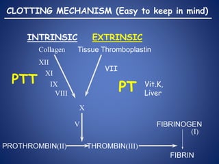
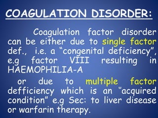
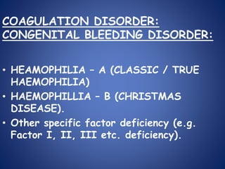
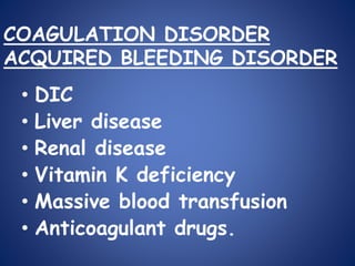
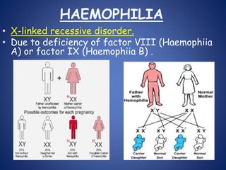
WELCOME
TO ALL
Coagulopathy in
children
Dr. Mohammed Saiful Islam
Resident ( Phase:A),
Department of Paediatric Surgery, BSMMU.
Bangladesh.
20/09/2016, Venue: Paediatric surgery class room. 2
Objectives
• To define & understand the extent of
coagulopathy.
• A review of coagulation pathways.
• Discussions on Platelet disorders.
• Key points discussions associated with
coagulation factor disorders.
• Perioperative strategies to meet the
challenges of coagulopathy in case of
children.
Definition:
Group of medical
conditions resulting from deficiency
of clotting factors, inhibition of
coagulation process or excessive
activity of the fibrinolytic system.
Reff: de Gruchy 5th
Bleeding
disorders
Coagulopathy
Extent of Coagulopaty:
• VESSEL WALL ABNORMALITIES:
• PLATELETS DISORDER:
• COAGULATION DISORDER:
INTRINSIC EXTRINSIC
PROTHROMBIN(II) THROMBIN(III)
FIBRINOGEN
FIBRIN
(I)
V
X
Tissue ThromboplastinCollagen
XII
XI
IX
VIII
VII
PT
PTT Vit.K,
Liver
CLOTTING MECHANISM (Easy to keep in mind)
PLATELETS DISORDERS:
• QUANTITATIVE PLATELETS
DISORDERs.
• QUALITATIVE PLATELET
DYSFUNCTION.
QUANTITATIVE PLATELETS DISORDER
(Thrombocytopenia)
Mechanisms:
1. Failure of megakaryocytic maturation.
2. Excessive platelets consumption after
their release into circulation i.e ITP,
DIC etc.
3. Platelets sequestration in enlarged
spleen i.e HYPERSPLEENISM.
QUANTITATIVE PLATELETS DISORDER
(Continued…)
S/S:
• Petechial cutaneous bleeding,
• Intracranial bleeding,
• Oozing from mucus membrane & skin
surface.
Lab: Decreased platelets count
and prolong bleeding time.
(Thrombocytopenia) Causes:
Marrow Disorder
• Aplastic anemia.
• Haematological
malignancy.
• Myelodysplastic
disorder.
• B12 defficiency.
Non Marrow Disorder
• Immune disorders.
• ITP, Drug induced.
• Sec: ALL, SLE.
• Post transfusion.
• DIC.
• Human immunodeficiency
syndrome.
• Hyperspleenism.
• Haemangiomas.
• Sepsis.
• Viral infection.
Thrombocytopenia (Continued…)
• Management:
– Rx Underlying cause.
– Platelet transfusion.
IDIOPATHIC THROMBOCYTOPENIC
PURPURA(ITP)
• An autoimmune antibody IgG is
formed against unknown antigen of
platelets membrane/surface.
• Antipletelet antibody binds to
complement, but platelets are not
destroyed by direct lysis.
• Destruction takes place in spleen,
where spleenic macrophages
destroyes antibody coated platelets.
IDIOPATHIC THROMBOCYTOPENIC PURPURA. (Continued…)
Clinical Features:
Often precipitated by viral infection and
usually self limiting.
 Asymptomatic, not febrile.
 Present with mucosal/skin bleeding,
mennorrhagia, purpura, petechiae.
 Females are more affected (2:1
male/female ratio).
IDIOPATHIC THROMBOCYTOPENIC PURPURA. (Continued…)
Δ LAB:
Platelets below 10,000 /ml.
Bone marrow will appear normal.
Rx
PREDNISONONE: 1-2 mg/kg/day.
Immunoglobulin: 1g/kg/day 2-3 days.
DANAZOLE: 600mg/day response rate is 50%
IMMUNOSUPPERESSIVE DRUGS: i-e vincristine,
vinblastine, azathioprine, cyclosporin,
cyclophosphomide.
SPLEENECTOMY.
Prognosis:
Good, if disease is initially controlled with
prednisolone,
Spleenectomy is definite Rx.
QUALITATIVE PLATELET DISORDER
CONGENITAL:
• Glanzmann’s
thrombasthenia.
• Bernard souliar
syndrome.
• Von Willibrand’s
disease.
ACQUIRED
• Myeloproliferative
disorder.
• Uremia.
• Drugs i-e NSAIDS
Aspirin.
• Autoantibody.
• Fibrin degradation
products.
QUALITATIVE PLATELET DISORDER
GLANZMANN’s THROMBASTHENIA:
Autosomal recessive disorder.
Lack of receptors (glycoprotein Ib & IIIa)
for fibrinogen on platelets.
Platelets fails to aggregate in respons to
ADP, collagen, thrombin.
Clinical Features: Mucosal bleeding
LAB:
Platelets no’s and morphology are normal
B.T is prolonged
Rx:
Platelet transfusion
QUALITATIVE PLATELET DISORDER
BERNARD SOULIER SYNDROME:
Autosomal recessive intrinsic platelets disorder.
Due to lack of glycoprotein (Gp1b), receptor for
vonWillibrand’s factor.
Clinical Features:
Presents with mucosal bleeding and post operative
oozes.
LAB:
Thrombocytopenia may be present, and Platelets
are abnormally large in size.
BT is prolonged
Von Willibrand’s factor Normal .
Rx:
Platelet transfusion
QUALITATIVE PLATELET DISORDER
VON-WILLIBRAND’S DISEASE:
• Autosomal dominant (gene for vWF
is located on chromosome 12.)
• vWF is synthesized by endothelial
cells and megakaryocytes.
• It acts as carrier protein for
factor VIII by non-covalent bond.
• A defect therefore leads to
decreased plasma factor VIII level.
VON-WILLIBRAND’S DISEASE (Continued….)
• It also forms bridges between
platelets and sub endothelium.
• There fore defect of vWF leads
to prolonged bleeding.
VON-WILLIBRAND’S DISEASE (Continued….)
Figure: Structure and function of factor VIII-von Willebrand factor (vWF) complex.
VON-WILLIBRAND’S DISEASE (Continued….)
Clinical Features:
• Mucosal bleeding (mild-massive)
LAB:
• Reduced level of vWF.
• Secondary reduction in factor VIII.
• Prolonged bleeding time (B.T).
VON-WILLIBRAND’S DISEASE (Continued….)
Rx:
• MILD HAEMORRHAGES:
Desmopressin 0.3 μg/kg, after
which vWF levels usually raise 3 units in
30-90 minutes.
• MASSIVE HAEMORRHAGES:
Factor VIII transfusion.
COAGULATION DISORDER:
Coagulation factor disorder
can be either due to single factor
def., i.e. a “congenital deficiency”,
e.g factor VIII resulting in
HAEMOPHILIA-A
or due to multiple factor
defficiency which is an ‘’acquired
condition” e.g Sec: to liver disease
or warfarin therapy.
• HEAMOPHILIA – A (CLASSIC / TRUE
HAEMOPHILIA)
• HAEMOPHILLIA – B (CHRISTMAS
DISEASE).
• Other specific factor deficiency (e.g.
Factor І, ІІ, ІІІ etc. deficiency).
COAGULATION DISORDER:
CONGENITAL BLEEDING DISORDER:
COAGULATION DISORDER
ACQUIRED BLEEDING DISORDER
• DIC
• Liver disease
• Renal disease
• Vitamin K deficiency
• Massive blood transfusion
• Anticoagulant drugs.
HAEMOPHILIA
• X-linked recessive disorder.
• Due to deficiency of factor VIII (Haemophiia
A) or factor IX (Haemophiia B) .
HAEAMOPHILIA (Continued….)
C/F:
• Bleeding occurs as bruising at the age of 6
months.
• Trauma results in excessively bleeding.
• Recurrent bleeding /hemorrhage in knee, elbow,
ankle, and hip. (Hemarthrosis).
• Mucus membrane /internal bleeding of mouth,
lips, gums, brain and kidney also occur.
• Muscle haematoma esp. calf and Psoas muscle .
Rx
• Cryoprecipitate or Factor VIII infusion ( in
Haemophiia A) & Factor IX infusion (in
Haemophilia B)
HAEAMOPHILIA (Continued….)
LONG TERM COMPLICATIONs
COMPLICATION due to repeated
hemorrhage:
– Arthropathy of large joints eg knee, elbow
– Muscle atrophy due to haematoma
– Mononeuropathy due to pressure of
haematoma.
COMPLICATION due to therapy
– Antifactor VIII antibody develops.
– Virus transmission Hepatitis B,C + HIV etc.
HAEAMOPHILIA (Continued….)
Figure: Haemarthrosis & Arthropathy.
SURGERY IN A CHILD WITH
HAEMOPHILLIA:
• In minor Surgery:
Factor VIII should be at least 50% of
the normal predicted value.
• In Major Surgery:
 Factor VIII should be 100%.
 Half life of Factor VIII is 8 to 12
hours.
 Factor VIII should be substituted just
before surgery & 12 hours after the
initial dose ( Target is to pass the
time of reactionary haemorrhage).
Calculation of Factor VІІІ
% Desired Χ Wt in Kg
• Amount of Factor VІІІ =
K
Here, value of K is 1.5 for Haemophilia A
& 0.75 for Haemophilia B.
DISSAMINATED INTRAVASCULAR
COAGULATION
Disseminated Intravascular
Coagulation (DIC) is a thrombo-
haemorrhagic disorder appearing as a
secondary complication of several diseases,
where there is consumption of platelets
and clotting factors in the circulation as
well as secondary activation of plasminogen
fibrinolytic system.
DIC:
CAUSES
A Infection:
1. Gram negative
septicaemia
2. Meningococcaemia
3. Pneumococcaemia
4.Malaria
B. Massive injury:
1. Massive trauma
2. Extensive burn
(severe burn)
3. Extensive surgery
(brain, lung, prostate, etc.)
C. Carcinoma:
1. Pancreas
2. Lungs
3. Stomach
4. Prostate, etc.
D. Others:
1. Transfusion
reaction
2. Fat embolism
3. Snake bite
4. Shock, etc.
Fig: Pathophysiology of disseminated intravascular coagulation.
DIC (Continued….)
CLINICAL FEATURES:
• Bleeding & thrombosis, bleeding is more
than thrombosis.
• Features of complications.
LAB:
• Thrombocytopenia
• Prolong PT
• APTT may be normal/increased
• Low fibrinogen
• Increased level FDP/D-dimmer
Treatment of DIC
Rx. Underlying cause.
General Measures: Correction & prevention of :
• Dehydration
• Renal failure
• Acidosis and
• Shock
Replacement:
• Platelets transfusion if platelets counts below
10,000/ml
• Cryoprecipitate to maintain plasma fibrinogen
level above 150 mg/dl
• FFP
• Heparin, if there is DVT, Pulmonary thrombosis.
WARCRAFT FOR SURGERY IN
A CHILD HAVING
ANTICOAGULANT THERAPY
• If patient on oral anticoaguant:
Dose adjustment for INR within 2.5 to 3.5.
During surgery INR should be 1.5 or less.
• If patient on Parenteral anticoaguant:
Per operative APTT should be within 1.5 to
2.5 times of normal range.
WARCRAFT FOR SURGERY (Continued….)
• During emergency surgery:
 Inj. Vitamin K should be
administered.
FFP & Factor concentrate of ІІ,
VІІ, ІX & X should be kept in hand.
Per operative & Post operative
monitoring of PT, INR.
WARCRAFT FOR SURGERY (Continued….)
• During elective surgery:
• Minor surgery:
» Oral anticoagulant should be stopped 2
to 3 days prior to surgery.
• Major surgery:
» Oral anticoagulant should be stopped 4
to 5 days prior to surgery.
» Then low molecular weight heparin
should be started.
» Heparin should be stopped 4 hours prior
to surgery & restarted 12 hours after
surgery.
THANK YOU

Coagulopathy in children

  • 1.
  • 2.
    Coagulopathy in children Dr. MohammedSaiful Islam Resident ( Phase:A), Department of Paediatric Surgery, BSMMU. Bangladesh. 20/09/2016, Venue: Paediatric surgery class room. 2
  • 3.
    Objectives • To define& understand the extent of coagulopathy. • A review of coagulation pathways. • Discussions on Platelet disorders. • Key points discussions associated with coagulation factor disorders. • Perioperative strategies to meet the challenges of coagulopathy in case of children.
  • 4.
    Definition: Group of medical conditionsresulting from deficiency of clotting factors, inhibition of coagulation process or excessive activity of the fibrinolytic system. Reff: de Gruchy 5th
  • 5.
  • 6.
    Extent of Coagulopaty: •VESSEL WALL ABNORMALITIES: • PLATELETS DISORDER: • COAGULATION DISORDER:
  • 7.
    INTRINSIC EXTRINSIC PROTHROMBIN(II) THROMBIN(III) FIBRINOGEN FIBRIN (I) V X TissueThromboplastinCollagen XII XI IX VIII VII PT PTT Vit.K, Liver CLOTTING MECHANISM (Easy to keep in mind)
  • 8.
    PLATELETS DISORDERS: • QUANTITATIVEPLATELETS DISORDERs. • QUALITATIVE PLATELET DYSFUNCTION.
  • 9.
    QUANTITATIVE PLATELETS DISORDER (Thrombocytopenia) Mechanisms: 1.Failure of megakaryocytic maturation. 2. Excessive platelets consumption after their release into circulation i.e ITP, DIC etc. 3. Platelets sequestration in enlarged spleen i.e HYPERSPLEENISM.
  • 10.
    QUANTITATIVE PLATELETS DISORDER (Continued…) S/S: •Petechial cutaneous bleeding, • Intracranial bleeding, • Oozing from mucus membrane & skin surface. Lab: Decreased platelets count and prolong bleeding time.
  • 11.
    (Thrombocytopenia) Causes: Marrow Disorder •Aplastic anemia. • Haematological malignancy. • Myelodysplastic disorder. • B12 defficiency. Non Marrow Disorder • Immune disorders. • ITP, Drug induced. • Sec: ALL, SLE. • Post transfusion. • DIC. • Human immunodeficiency syndrome. • Hyperspleenism. • Haemangiomas. • Sepsis. • Viral infection.
  • 12.
    Thrombocytopenia (Continued…) • Management: –Rx Underlying cause. – Platelet transfusion.
  • 13.
    IDIOPATHIC THROMBOCYTOPENIC PURPURA(ITP) • Anautoimmune antibody IgG is formed against unknown antigen of platelets membrane/surface. • Antipletelet antibody binds to complement, but platelets are not destroyed by direct lysis. • Destruction takes place in spleen, where spleenic macrophages destroyes antibody coated platelets.
  • 14.
    IDIOPATHIC THROMBOCYTOPENIC PURPURA.(Continued…) Clinical Features: Often precipitated by viral infection and usually self limiting.  Asymptomatic, not febrile.  Present with mucosal/skin bleeding, mennorrhagia, purpura, petechiae.  Females are more affected (2:1 male/female ratio).
  • 15.
    IDIOPATHIC THROMBOCYTOPENIC PURPURA.(Continued…) Δ LAB: Platelets below 10,000 /ml. Bone marrow will appear normal. Rx PREDNISONONE: 1-2 mg/kg/day. Immunoglobulin: 1g/kg/day 2-3 days. DANAZOLE: 600mg/day response rate is 50% IMMUNOSUPPERESSIVE DRUGS: i-e vincristine, vinblastine, azathioprine, cyclosporin, cyclophosphomide. SPLEENECTOMY. Prognosis: Good, if disease is initially controlled with prednisolone, Spleenectomy is definite Rx.
  • 16.
    QUALITATIVE PLATELET DISORDER CONGENITAL: •Glanzmann’s thrombasthenia. • Bernard souliar syndrome. • Von Willibrand’s disease. ACQUIRED • Myeloproliferative disorder. • Uremia. • Drugs i-e NSAIDS Aspirin. • Autoantibody. • Fibrin degradation products.
  • 17.
    QUALITATIVE PLATELET DISORDER GLANZMANN’sTHROMBASTHENIA: Autosomal recessive disorder. Lack of receptors (glycoprotein Ib & IIIa) for fibrinogen on platelets. Platelets fails to aggregate in respons to ADP, collagen, thrombin. Clinical Features: Mucosal bleeding LAB: Platelets no’s and morphology are normal B.T is prolonged Rx: Platelet transfusion
  • 18.
    QUALITATIVE PLATELET DISORDER BERNARDSOULIER SYNDROME: Autosomal recessive intrinsic platelets disorder. Due to lack of glycoprotein (Gp1b), receptor for vonWillibrand’s factor. Clinical Features: Presents with mucosal bleeding and post operative oozes. LAB: Thrombocytopenia may be present, and Platelets are abnormally large in size. BT is prolonged Von Willibrand’s factor Normal . Rx: Platelet transfusion
  • 19.
    QUALITATIVE PLATELET DISORDER VON-WILLIBRAND’SDISEASE: • Autosomal dominant (gene for vWF is located on chromosome 12.) • vWF is synthesized by endothelial cells and megakaryocytes. • It acts as carrier protein for factor VIII by non-covalent bond. • A defect therefore leads to decreased plasma factor VIII level.
  • 20.
    VON-WILLIBRAND’S DISEASE (Continued….) •It also forms bridges between platelets and sub endothelium. • There fore defect of vWF leads to prolonged bleeding.
  • 21.
    VON-WILLIBRAND’S DISEASE (Continued….) Figure:Structure and function of factor VIII-von Willebrand factor (vWF) complex.
  • 22.
    VON-WILLIBRAND’S DISEASE (Continued….) ClinicalFeatures: • Mucosal bleeding (mild-massive) LAB: • Reduced level of vWF. • Secondary reduction in factor VIII. • Prolonged bleeding time (B.T).
  • 23.
    VON-WILLIBRAND’S DISEASE (Continued….) Rx: •MILD HAEMORRHAGES: Desmopressin 0.3 μg/kg, after which vWF levels usually raise 3 units in 30-90 minutes. • MASSIVE HAEMORRHAGES: Factor VIII transfusion.
  • 24.
    COAGULATION DISORDER: Coagulation factordisorder can be either due to single factor def., i.e. a “congenital deficiency”, e.g factor VIII resulting in HAEMOPHILIA-A or due to multiple factor defficiency which is an ‘’acquired condition” e.g Sec: to liver disease or warfarin therapy.
  • 25.
    • HEAMOPHILIA –A (CLASSIC / TRUE HAEMOPHILIA) • HAEMOPHILLIA – B (CHRISTMAS DISEASE). • Other specific factor deficiency (e.g. Factor І, ІІ, ІІІ etc. deficiency). COAGULATION DISORDER: CONGENITAL BLEEDING DISORDER:
  • 26.
    COAGULATION DISORDER ACQUIRED BLEEDINGDISORDER • DIC • Liver disease • Renal disease • Vitamin K deficiency • Massive blood transfusion • Anticoagulant drugs.
  • 27.
    HAEMOPHILIA • X-linked recessivedisorder. • Due to deficiency of factor VIII (Haemophiia A) or factor IX (Haemophiia B) .
  • 28.
    HAEAMOPHILIA (Continued….) C/F: • Bleedingoccurs as bruising at the age of 6 months. • Trauma results in excessively bleeding. • Recurrent bleeding /hemorrhage in knee, elbow, ankle, and hip. (Hemarthrosis). • Mucus membrane /internal bleeding of mouth, lips, gums, brain and kidney also occur. • Muscle haematoma esp. calf and Psoas muscle . Rx • Cryoprecipitate or Factor VIII infusion ( in Haemophiia A) & Factor IX infusion (in Haemophilia B)
  • 29.
    HAEAMOPHILIA (Continued….) LONG TERMCOMPLICATIONs COMPLICATION due to repeated hemorrhage: – Arthropathy of large joints eg knee, elbow – Muscle atrophy due to haematoma – Mononeuropathy due to pressure of haematoma. COMPLICATION due to therapy – Antifactor VIII antibody develops. – Virus transmission Hepatitis B,C + HIV etc.
  • 30.
  • 31.
    SURGERY IN ACHILD WITH HAEMOPHILLIA: • In minor Surgery: Factor VIII should be at least 50% of the normal predicted value. • In Major Surgery:  Factor VIII should be 100%.  Half life of Factor VIII is 8 to 12 hours.  Factor VIII should be substituted just before surgery & 12 hours after the initial dose ( Target is to pass the time of reactionary haemorrhage).
  • 32.
    Calculation of FactorVІІІ % Desired Χ Wt in Kg • Amount of Factor VІІІ = K Here, value of K is 1.5 for Haemophilia A & 0.75 for Haemophilia B.
  • 33.
    DISSAMINATED INTRAVASCULAR COAGULATION Disseminated Intravascular Coagulation(DIC) is a thrombo- haemorrhagic disorder appearing as a secondary complication of several diseases, where there is consumption of platelets and clotting factors in the circulation as well as secondary activation of plasminogen fibrinolytic system.
  • 34.
    DIC: CAUSES A Infection: 1. Gramnegative septicaemia 2. Meningococcaemia 3. Pneumococcaemia 4.Malaria B. Massive injury: 1. Massive trauma 2. Extensive burn (severe burn) 3. Extensive surgery (brain, lung, prostate, etc.) C. Carcinoma: 1. Pancreas 2. Lungs 3. Stomach 4. Prostate, etc. D. Others: 1. Transfusion reaction 2. Fat embolism 3. Snake bite 4. Shock, etc.
  • 35.
    Fig: Pathophysiology ofdisseminated intravascular coagulation.
  • 36.
    DIC (Continued….) CLINICAL FEATURES: •Bleeding & thrombosis, bleeding is more than thrombosis. • Features of complications. LAB: • Thrombocytopenia • Prolong PT • APTT may be normal/increased • Low fibrinogen • Increased level FDP/D-dimmer
  • 37.
    Treatment of DIC Rx.Underlying cause. General Measures: Correction & prevention of : • Dehydration • Renal failure • Acidosis and • Shock Replacement: • Platelets transfusion if platelets counts below 10,000/ml • Cryoprecipitate to maintain plasma fibrinogen level above 150 mg/dl • FFP • Heparin, if there is DVT, Pulmonary thrombosis.
  • 38.
    WARCRAFT FOR SURGERYIN A CHILD HAVING ANTICOAGULANT THERAPY • If patient on oral anticoaguant: Dose adjustment for INR within 2.5 to 3.5. During surgery INR should be 1.5 or less. • If patient on Parenteral anticoaguant: Per operative APTT should be within 1.5 to 2.5 times of normal range.
  • 39.
    WARCRAFT FOR SURGERY(Continued….) • During emergency surgery:  Inj. Vitamin K should be administered. FFP & Factor concentrate of ІІ, VІІ, ІX & X should be kept in hand. Per operative & Post operative monitoring of PT, INR.
  • 40.
    WARCRAFT FOR SURGERY(Continued….) • During elective surgery: • Minor surgery: » Oral anticoagulant should be stopped 2 to 3 days prior to surgery. • Major surgery: » Oral anticoagulant should be stopped 4 to 5 days prior to surgery. » Then low molecular weight heparin should be started. » Heparin should be stopped 4 hours prior to surgery & restarted 12 hours after surgery.
  • 41.