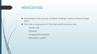
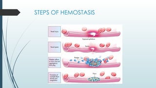
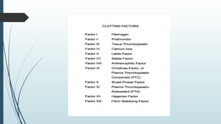
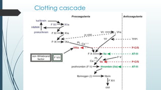
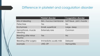
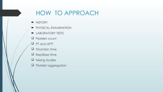
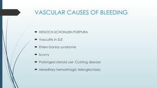
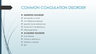
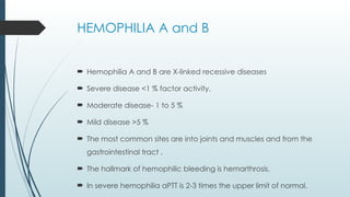
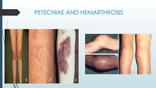
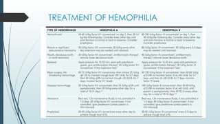
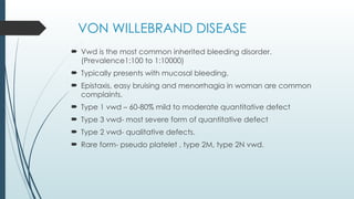
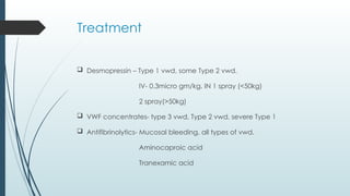
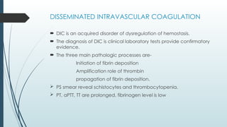
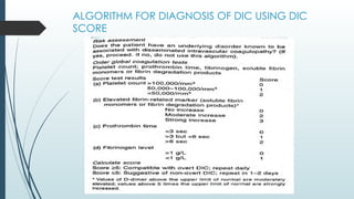
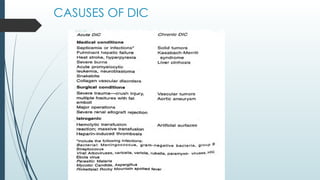
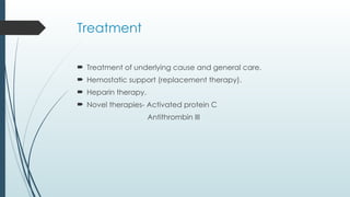
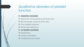
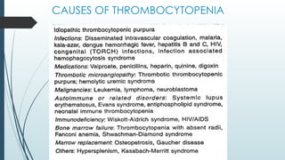
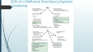
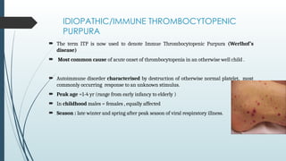
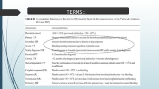
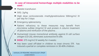
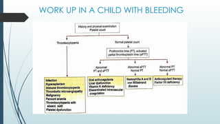
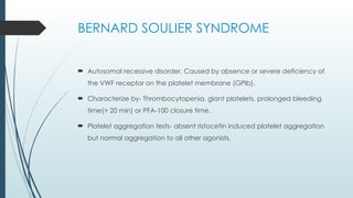
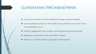
BLEEDING AND PLATELET
DISORDER
PRESENTED BY-
DR. ANURUDDH CHATURVEDI
GUIDED BY-
DR. SHALINI HAJELA
ASSOCIATE PROFESSOR
HEMOSTASIS
 Hemostasis is the process of blood clotting in areas of blood vessel
injury.
 The main components of the hemostatic process are-
Vessel wall
Platelets
Coagulation proteins
Fibrinolytic system
STEPS OF HEMOSTASIS
Clotting cascade
Difference in platelet and coagulation disorder
Platelet disorder Coagulation disorder
Site of bleeding Skin, mucous membrane Soft tissue , joint, muscle
Petechiae Yes No
Ecchymosis Small, superficial Large, deep
Hemarthrosis, muscle
bleeding
Extremely rare Common
Bleeding after minor
trauma
Yes No
Bleeding after surgery Immediate usually mild Delayed
example vWD ,ITP Hemophilia
HOW TO APPROACH
 HISTORY
 PHYSICAL EXAMINATION
 LABORATORY TESTS
 Platelet count
 PT and aPTT
 Thrombin time
 Reptilase time
 Mixing studies
 Platelet aggregation
VASCULAR CAUSES OF BLEEDING
 HENOCH-SCHONLEIN PURPURA
 Vasculitis in SLE
 Ehlers-Danlos syndrome
 Scurvy
 Prolonged steroid use- Cushing disease
 Hereditary hemorrhagic telengiectasia
COMMON COAGULATION DISORDER
 INHERITED DISORDER
 Hemophilia A and B
 Von Willebrand disease
 Specific factor deficiencies
 Factor VII, X, XII deficiency
 Afibrinogenemia
 ACQUIRED DISORDER
 Liver disease
 Vitamin k deficiency
 Warfarin overdose
 DIC
HEMOPHILIA A and B
 Hemophilia A and B are X-linked recessive diseases
 Severe disease <1 % factor activity,
 Moderate disease- 1 to 5 %
 Mild disease >5 %
 The most common sites are into joints and muscles and from the
gastrointestinal tract .
 The hallmark of hemophilic bleeding is hemarthrosis.
 In severe hemophilia aPTT is 2-3 times the upper limit of normal.
PETECHIAE AND HEMARTHROSIS
TREATMENT OF HEMOPHILIA
VON WILLEBRAND DISEASE
 Vwd is the most common inherited bleeding disorder.
(Prevalence1:100 to 1:10000)
 Typically presents with mucosal bleeding.
 Epistaxis, easy bruising and menorrhagia in woman are common
complaints.
 Type 1 vwd – 60-80% mild to moderate quantitative defect
 Type 3 vwd- most severe form of quantitative defect
 Type 2 vwd- qualitative defects.
 Rare form- pseudo platelet , type 2M, type 2N vwd.
Pathophysiology
Treatment
 Desmopressin – Type 1 vwd, some Type 2 vwd.
IV- 0.3micro gm/kg, IN 1 spray (<50kg)
2 spray(>50kg)
 VWF concentrates- type 3 vwd, Type 2 vwd, severe Type 1
 Antifibrinolytics- Mucosal bleeding, all types of vwd.
Aminocaproic acid
Tranexamic acid
DISSEMINATED INTRAVASCULAR COAGULATION
 DIC is an acquired disorder of dysregulation of hemostasis.
 The diagnosis of DIC is clinical laboratory tests provide confirmatory
evidence.
 The three main pathologic processes are-
Initiation of fibrin deposition
Amplification role of thrombin
propagation of fibrin deposition.
 PS smear reveal schistocytes and thrombocytopenia.
 PT, aPTT, TT are prolonged, fibrinogen level is low
ALGORITHM FOR DIAGNOSIS OF DIC USING DIC
SCORE
CASUSES OF DIC
Treatment
 Treatment of underlying cause and general care.
 Hemostatic support (replacement therapy).
 Heparin therapy.
 Novel therapies- Activated protein C
Antithrombin III
PLATELET DISORDERS
Qualitative disorders of platelet
function
 INHERITED DISORDER
 Glanzmann thrombasthenia (GP IIb-IIIa def.)
 Bernard-Soulier syndrome (GP Ib def.)
 Gray platelet syndrome
 Dense body deficiency
 ACQUIRED DISORDER
 Medications
 Chronic renal failure
 Cardiopulmonary bypass
CAUSES OF THROMBOCYTOPENIA
D/D of childhood thrombocytopenic
syndrome
IDIOPATHIC/IMMUNE THROMBOCYTOPENIC
PURPURA
 The term ITP is now used to denote Immue Thrombocytopenic Purpura (Werlhof’s
disease)
 Most common cause of acute onset of thrombocytopenia in an otherwise well child .
 Autoimmune disorder characterised by destruction of otherwise normal platelet, most
commonly occurring response to an unknown stimulus.
 Peak age =1-4 yr (range from early infancy to elderly )
 In childhood males = females , equally affected
 Season : late winter and spring after peak season of viral respiratory illness.
TERMS:
 CBC including blood smear and platelet count.
Bone marrow aspiration: Indications:
a)Abnormal WBC count or differential or unexplained anemia
b) Findings on history and physical examination suggestive of bone
marrow failure syndrome or malignancy.
Not indicated in a clinically and hematologically typical case of ITP
Other Investigations: (To rule out other causes, if required):
 Antinuclear antibody (ANA) ,and anti-ds-DNA
 Blood type, Coombs test
 PT, APTT, fibrinogen , and split products of fibrinogen(SPF)
 Liver function tests,
 Blood urea nitrogen (BUN) and Creatinine
 Monospot test and/or EBV, HIV ,parvovirus titers
INVESTIGATIONS
MANUAL OF PEDIATRIC HEMATOLOGY
(PHILIP LANZKOWSKY)
 No therapy other than education and counselling of the family and the
patient for patient with minimal, mild and moderate symptoms
 Treat patient , not the platelet count
 Hospitalization not required in an uncomplicated case
 Hospitalization is appropriate for treatment of child severe bleeding
episodes
 Grading the severity of bleeding should be employed to decide
management of particular patient
TREATMENT PRINCIPLES :
NELSON
Treatment (Contd.)
 First line drugs include: Prednisolone, IVIG, Anti-D
 Second line drugs : High dose dexamethasone or
Methylprednisolone, & Immunosuppressive agents
 SPLENECTOMY should only be considered in pts with CHRONIC ITP
with significant bleeds, who fails to respond to usual first & second
line drugs
TPO Agonists:
 Reduced levels of thrombopoietin (TPO) ,the primary stimulator of
platelet synthesis is now known to occur in patient with ITP
 Thrombopoietin agonist ( ELTROMBOPAG & ROMIPLOSTIM) have
shown encouraging results.
NELSON
Prednisone:
Inhibits phagocytosis of
antibody coated
platelet in spleen
Inhibit platelet
antibody production
Improves capillary
resistance and hence
platelet economy
Prolong platelet survival
Reduced in a stepwise fashion at 5 to 7 day intervals ,
irrespective of the platelet count
Stopped at the end of 21-28 days , REGARDLESS OF THE
RESPONSE.
2mg/kg/day (max 60mg/day )
(in divided doses)
STEROID THERAPY :
MANUAL OF PEDIATRIC HEMATOLOGY
(PHILIP LANZKOWSKY)
In case of intracranial hemorrhage multiple modalities to be
used :
 Platelet transfusion
 IVIG 2g/kg
 High dose corticosteroids /methylprednisolone 500mg/m2 IV
per day for 3 days
 Emergency splenectomy
 Patient refractory to these measures may benefit from
vincristine sulfate 2mg/m2 IV and protein A column treatment
of plasma and reinfusion of the plasma .
 Humanized mouse monoclonal antibody against B cell surface
antigen (CD-20), eliminates autoreactive B cells
 DOSE: 375 mg/m2 IV weekly for 4 weeks
 Has been used off-label in children to treat chronic ITP, has
induced a partial or complete remission in 30-40% children.
MANUAL OF PEDIATRIC HEMATOLOGY (PHILIP
LANZKOWSKY)
WORK UP IN A CHILD WITH BLEEDING
BERNARD SOULIER SYNDROME
 Autosomal recessive disorder, Caused by absence or severe deficiency of
the VWF receptor on the platelet membrane (GPIb).
 Characterize by- Thrombocytopenia, giant platelets, prolonged bleeding
time(> 20 min) or PFA-100 closure time.
 Platelet aggregation tests- absent ristocetin induced platelet aggregation
but normal aggregation to all other agonists.
GLANZMANN THROMBASTHENIA
 Caused by deficiency of the platelet fibrinogen receptor αIIb-β3.
 Severe platelet dysfunction that yields severe platelet dysfunction and a
normal platelet count.
 Platelet aggregation test- positive with all agonists except ristocetin.
 Diagnosis confirmed by flow cytometric analysis.
 Reports of curative therapy using stem cell transplant.
LIVER DISORDER
 All the clotting factor except factor VIII are produced by liver.
 Only 15% have significant bleeding because of concomitant reuction in
anticoagulation proteins.
 Mainly due to decreased synthesis of all proteins.
 Treatment Vit k – infants 1mg/24hr, children 2-5mg/24hr,
adolescents- 5-10mg.
FFP and cryoprecipitate
Desmopressin (0.3microgm/kg IV)
VIT K DEFICIENCY DISORDER
 Plays vital role in the production of vit k dependent coagulation
factor.
 Vit k dependent factors are- factors II, VII, IX, X, protein C and S.
 Leads to increase PT and aPTT.
 Precise diagnosis- Assay of proteins in vit K absence (PIVKA).
 Prevalence of late vit k deficiency bleeding in breastfed infants
without prophylaxis is 20 cases per 100,000.
REFERENCES
 Nelson textbook of pediatrics 21st
edition.
 OP Ghai textbook of pediatrics 9th
edition.
 Emergency in children Meherbhan singh.
 Lanzowsky’s manual of pediatrics hematology and oncology.
THANK YOU

bleeding disorders.ppt presentation in pediatrics

  • 1.
    BLEEDING AND PLATELET DISORDER PRESENTEDBY- DR. ANURUDDH CHATURVEDI GUIDED BY- DR. SHALINI HAJELA ASSOCIATE PROFESSOR
  • 2.
    HEMOSTASIS  Hemostasis isthe process of blood clotting in areas of blood vessel injury.  The main components of the hemostatic process are- Vessel wall Platelets Coagulation proteins Fibrinolytic system
  • 3.
  • 5.
  • 6.
    Difference in plateletand coagulation disorder Platelet disorder Coagulation disorder Site of bleeding Skin, mucous membrane Soft tissue , joint, muscle Petechiae Yes No Ecchymosis Small, superficial Large, deep Hemarthrosis, muscle bleeding Extremely rare Common Bleeding after minor trauma Yes No Bleeding after surgery Immediate usually mild Delayed example vWD ,ITP Hemophilia
  • 7.
    HOW TO APPROACH HISTORY  PHYSICAL EXAMINATION  LABORATORY TESTS  Platelet count  PT and aPTT  Thrombin time  Reptilase time  Mixing studies  Platelet aggregation
  • 8.
    VASCULAR CAUSES OFBLEEDING  HENOCH-SCHONLEIN PURPURA  Vasculitis in SLE  Ehlers-Danlos syndrome  Scurvy  Prolonged steroid use- Cushing disease  Hereditary hemorrhagic telengiectasia
  • 9.
    COMMON COAGULATION DISORDER INHERITED DISORDER  Hemophilia A and B  Von Willebrand disease  Specific factor deficiencies  Factor VII, X, XII deficiency  Afibrinogenemia  ACQUIRED DISORDER  Liver disease  Vitamin k deficiency  Warfarin overdose  DIC
  • 10.
    HEMOPHILIA A andB  Hemophilia A and B are X-linked recessive diseases  Severe disease <1 % factor activity,  Moderate disease- 1 to 5 %  Mild disease >5 %  The most common sites are into joints and muscles and from the gastrointestinal tract .  The hallmark of hemophilic bleeding is hemarthrosis.  In severe hemophilia aPTT is 2-3 times the upper limit of normal.
  • 11.
  • 12.
  • 13.
    VON WILLEBRAND DISEASE Vwd is the most common inherited bleeding disorder. (Prevalence1:100 to 1:10000)  Typically presents with mucosal bleeding.  Epistaxis, easy bruising and menorrhagia in woman are common complaints.  Type 1 vwd – 60-80% mild to moderate quantitative defect  Type 3 vwd- most severe form of quantitative defect  Type 2 vwd- qualitative defects.  Rare form- pseudo platelet , type 2M, type 2N vwd.
  • 14.
  • 15.
    Treatment  Desmopressin –Type 1 vwd, some Type 2 vwd. IV- 0.3micro gm/kg, IN 1 spray (<50kg) 2 spray(>50kg)  VWF concentrates- type 3 vwd, Type 2 vwd, severe Type 1  Antifibrinolytics- Mucosal bleeding, all types of vwd. Aminocaproic acid Tranexamic acid
  • 16.
    DISSEMINATED INTRAVASCULAR COAGULATION DIC is an acquired disorder of dysregulation of hemostasis.  The diagnosis of DIC is clinical laboratory tests provide confirmatory evidence.  The three main pathologic processes are- Initiation of fibrin deposition Amplification role of thrombin propagation of fibrin deposition.  PS smear reveal schistocytes and thrombocytopenia.  PT, aPTT, TT are prolonged, fibrinogen level is low
  • 17.
    ALGORITHM FOR DIAGNOSISOF DIC USING DIC SCORE
  • 18.
  • 19.
    Treatment  Treatment ofunderlying cause and general care.  Hemostatic support (replacement therapy).  Heparin therapy.  Novel therapies- Activated protein C Antithrombin III
  • 20.
  • 21.
    Qualitative disorders ofplatelet function  INHERITED DISORDER  Glanzmann thrombasthenia (GP IIb-IIIa def.)  Bernard-Soulier syndrome (GP Ib def.)  Gray platelet syndrome  Dense body deficiency  ACQUIRED DISORDER  Medications  Chronic renal failure  Cardiopulmonary bypass
  • 22.
  • 23.
    D/D of childhoodthrombocytopenic syndrome
  • 24.
    IDIOPATHIC/IMMUNE THROMBOCYTOPENIC PURPURA  Theterm ITP is now used to denote Immue Thrombocytopenic Purpura (Werlhof’s disease)  Most common cause of acute onset of thrombocytopenia in an otherwise well child .  Autoimmune disorder characterised by destruction of otherwise normal platelet, most commonly occurring response to an unknown stimulus.  Peak age =1-4 yr (range from early infancy to elderly )  In childhood males = females , equally affected  Season : late winter and spring after peak season of viral respiratory illness.
  • 25.
  • 26.
     CBC includingblood smear and platelet count. Bone marrow aspiration: Indications: a)Abnormal WBC count or differential or unexplained anemia b) Findings on history and physical examination suggestive of bone marrow failure syndrome or malignancy. Not indicated in a clinically and hematologically typical case of ITP Other Investigations: (To rule out other causes, if required):  Antinuclear antibody (ANA) ,and anti-ds-DNA  Blood type, Coombs test  PT, APTT, fibrinogen , and split products of fibrinogen(SPF)  Liver function tests,  Blood urea nitrogen (BUN) and Creatinine  Monospot test and/or EBV, HIV ,parvovirus titers INVESTIGATIONS MANUAL OF PEDIATRIC HEMATOLOGY (PHILIP LANZKOWSKY)
  • 27.
     No therapyother than education and counselling of the family and the patient for patient with minimal, mild and moderate symptoms  Treat patient , not the platelet count  Hospitalization not required in an uncomplicated case  Hospitalization is appropriate for treatment of child severe bleeding episodes  Grading the severity of bleeding should be employed to decide management of particular patient TREATMENT PRINCIPLES : NELSON
  • 28.
    Treatment (Contd.)  Firstline drugs include: Prednisolone, IVIG, Anti-D  Second line drugs : High dose dexamethasone or Methylprednisolone, & Immunosuppressive agents  SPLENECTOMY should only be considered in pts with CHRONIC ITP with significant bleeds, who fails to respond to usual first & second line drugs TPO Agonists:  Reduced levels of thrombopoietin (TPO) ,the primary stimulator of platelet synthesis is now known to occur in patient with ITP  Thrombopoietin agonist ( ELTROMBOPAG & ROMIPLOSTIM) have shown encouraging results. NELSON
  • 29.
    Prednisone: Inhibits phagocytosis of antibodycoated platelet in spleen Inhibit platelet antibody production Improves capillary resistance and hence platelet economy Prolong platelet survival Reduced in a stepwise fashion at 5 to 7 day intervals , irrespective of the platelet count Stopped at the end of 21-28 days , REGARDLESS OF THE RESPONSE. 2mg/kg/day (max 60mg/day ) (in divided doses) STEROID THERAPY : MANUAL OF PEDIATRIC HEMATOLOGY (PHILIP LANZKOWSKY)
  • 30.
    In case ofintracranial hemorrhage multiple modalities to be used :  Platelet transfusion  IVIG 2g/kg  High dose corticosteroids /methylprednisolone 500mg/m2 IV per day for 3 days  Emergency splenectomy  Patient refractory to these measures may benefit from vincristine sulfate 2mg/m2 IV and protein A column treatment of plasma and reinfusion of the plasma .  Humanized mouse monoclonal antibody against B cell surface antigen (CD-20), eliminates autoreactive B cells  DOSE: 375 mg/m2 IV weekly for 4 weeks  Has been used off-label in children to treat chronic ITP, has induced a partial or complete remission in 30-40% children. MANUAL OF PEDIATRIC HEMATOLOGY (PHILIP LANZKOWSKY)
  • 31.
    WORK UP INA CHILD WITH BLEEDING
  • 32.
    BERNARD SOULIER SYNDROME Autosomal recessive disorder, Caused by absence or severe deficiency of the VWF receptor on the platelet membrane (GPIb).  Characterize by- Thrombocytopenia, giant platelets, prolonged bleeding time(> 20 min) or PFA-100 closure time.  Platelet aggregation tests- absent ristocetin induced platelet aggregation but normal aggregation to all other agonists.
  • 33.
    GLANZMANN THROMBASTHENIA  Causedby deficiency of the platelet fibrinogen receptor αIIb-β3.  Severe platelet dysfunction that yields severe platelet dysfunction and a normal platelet count.  Platelet aggregation test- positive with all agonists except ristocetin.  Diagnosis confirmed by flow cytometric analysis.  Reports of curative therapy using stem cell transplant.
  • 34.
    LIVER DISORDER  Allthe clotting factor except factor VIII are produced by liver.  Only 15% have significant bleeding because of concomitant reuction in anticoagulation proteins.  Mainly due to decreased synthesis of all proteins.  Treatment Vit k – infants 1mg/24hr, children 2-5mg/24hr, adolescents- 5-10mg. FFP and cryoprecipitate Desmopressin (0.3microgm/kg IV)
  • 35.
    VIT K DEFICIENCYDISORDER  Plays vital role in the production of vit k dependent coagulation factor.  Vit k dependent factors are- factors II, VII, IX, X, protein C and S.  Leads to increase PT and aPTT.  Precise diagnosis- Assay of proteins in vit K absence (PIVKA).  Prevalence of late vit k deficiency bleeding in breastfed infants without prophylaxis is 20 cases per 100,000.
  • 38.
    REFERENCES  Nelson textbookof pediatrics 21st edition.  OP Ghai textbook of pediatrics 9th edition.  Emergency in children Meherbhan singh.  Lanzowsky’s manual of pediatrics hematology and oncology.
  • 39.