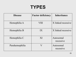
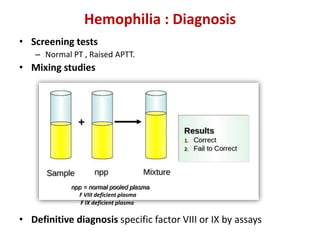
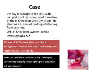
Case
6yr boy is brought to the OPD with
complaints of recurrent painful swelling
of the Lt Knee joint since 2yr of age. He
also has a history of prolonged bleeding
from cut sites.
One maternal uncle of the child died due
to prolonged bleeding following a minor
surgery.
O/E, No petechiae/purpura.
Lt Knee joint swollen, tender
Hemophilia and
Coagulation
Disorders
Dr Nishant Verma
Hemostatic Mechanism
 Platelet adhesion
 Platelet aggregation
 Clot formation
 Clot stabilization
 Limitation of clotting to the site of
injury by regulatory anticoagulants,
and
 Re-establishment of vascular patency
through fibrinolysis and vascular
healing
von Willebrand‘s
Factor (vWF)
binds to
subendothelial
collagen.
Conformational changes occur in
vWF allow it to bind to the GP
Ib/IX receptor on platelets,
causing adhesion
BERNARD- SOULIER SYNDROME
The GP IIb/IIIa
receptor complex
changes conformation
allowing binding of
fibrinogen.
Fibrinogen acts as
a glue binding
platelets together.
GLANZMANN’s Thrombasthenia
Clotting Factors
I Fibrinogen
II Prothrombin
V Labile factor, proaccelerin
VII Stable factor or proconvertin
VIII Antihemophilic factor (AHF)
IX Christmas factor
X Stuart-Power factor
XI Plasma thromboplastin antecedent
XII Hageman factor
XIII Fibrin Stabilizing Factor
Waterfall cascade:
Prothrombin
Fibrinogen
Factor X
Common
Pathway
Fibrin
Thrombin
Factor Xa
HEMOPHILIA
Overview of fibrinolytic mechanism
von Willebrand Factor
Classification of bleeding disorders
• Primary Hemostatic defect
– Platelet disorder
• Congenital
• Acquired
– Von Willebrand Disease
• Coagulation Disorders (Clotting factor deficiency)
– Acquired
– Inherited
• Vascular
Clinical characteristic Primary Hemostatic Defect Coagulation Disorder
Site of bleeding Skin, mucous membrane Soft tissues, muscles, joints
Bleeding after minor cuts Yes No
Petechiae Yes No
Ecchymosis Small, superficial Larger, deeper
Hemarthrosis Rare Common
Bleeding after trauma/surgery Immediate Delayed
Example Platelet defect, vWD Hemophilia
Classification of bleeding disorders
Source: Nathan and Oski’s Hematology of Infancy and Childhood. 7th Edition, Pg 1450
Clinical Approach to bleeding disorders
History
Physical Examination
Laboratory Evaluation
Clinical Approach to bleeding disorders
History
Nature of bleeding-
- Immediate vs delayed
- Superficial vs deep
- Surgical / dental history
Family H/O bleeding-
- Others involved ?
- Males only? (x-linked)
- Consecutive generations
Medication history-
-NSAID, Heparin (patients with central lines)
Others- Liver / renal disease
Clinical Approach to bleeding disorders
Physical Examination
Bruises-
- Number
- Location
- Site
Petechiae
Joint bleeding
Other Physical findings-
- Jaundice
- Skeletal deformity
- Hepatosplenomegaly
Clinical Approach to bleeding disorders
Screening Laboratory Evaluation
• Platelet count / morphology
• Coagulation profile
– Prothrombin time (PT)
– Activated partial thromboplastin time (APTT)
– Bleeding time (BT)
BT / CT
• Bleeding time
– 3-9 min
• Clotting time
– 3-6 min
Coagulation profile
• Sample collection
– Citrated tube
– Gently mixed
– Immediate transport
Coagulation profile
• PT
– Method
– Normal – 10-11s
– INR = (Patient PT/Control PT)ISI
– Isolated  PT
• APTT
– Method
– Normal – 26-35s
– Isolated  APTT
–  PT +  APTT
• TT
TT
Advanced tests
• Factor assays
• Testing for vWD
• Platelet function analyzer (PFA 100)
Classification of bleeding disorders
• Primary Hemostatic defect
– Platelet disorder
• Congenital
• Acquired
– Von Willebrand Disease
• Coagulation Disorders (Clotting factor deficiency)
– Acquired
– Inherited
• Vascular
Coagulation Disorders: Acquired
• Vitamin K deficiency
• Liver disease
• Accelerated destruction of coagulation factors
• Inhibitors of coagulation
• Miscellaneous
Vitamin K Dependent Proteins
Dietary
Vitamin K
Vitamin K
Reductase
Vitamin K deficiency
Liver Disease or Vit. K deficiency
 Hemorrhagic disease of the newborn
 Biliary obstruction (neonatal cholestatic disorders)
 Malabsorption of vitamin K (celiac disease, ulcerative colitis)
 Drugs
 Vit.K antagonists – warfarin, phenytoin
 Broad-spectrum antibiotics – alter gut flora
 Manifestations
 Diagnosis: PT, APTT
 Management
 Vitamin K oral / sc / iv
 Repeat PT after 6hr
 Prevention
 Prophylactic Vit K to at risk population
Vitamin K deficiency
Coagulation Disorders: Inherited
Hemophilia A
Hemophilia B
Factor XIII Deficiency
Prothrombin Deficiency
Factor V Deficiency
Factor VII Deficiency
Factor X Deficiency
Factor XI Deficiency
Factor XII Deficiency
Prekillikrein Deficiency
High Molecular Weight Kininogen Deficiency
a2-antiplasmin Deficiency
Plasminogen Activator Inhibitor Deficiency
Incidence 1 in 5000 to 10,000
Rare
27
Often called ‘the disease of kings’
because it was carried by many members
of Europe’s royal family. Queen Victoria
of England was a carrier of Hemophilia
HEMOPHILIA
• X-linked recessive
• Hemophilia A (FVIII def): 80-85%
Hemophilia B (FIX def)
• Mutations of the clotting factor gene
• Family H/O bleeding common,
- generally affects males on the maternal side
- 1/3 no family history – due to new mutations
TYPES
Disease Factor deficiency Inheritance
Hemophilia A VIII X linked recessive
Hemophilia B IX X linked recessive
Hemophilia C XI Autosomal
recessive
Parahemophilia V Autosomal
recessive
29
30
Distribution Clotting factor
activity
Severe hemophilia 50% <1%
Moderate hemophilia 10% 1-5%
Mild hemophilia 30-40% 5-40%
Severity of Hemophilia is defined by measured
level of clotting factor activity
HEMOPHILIA
32
• Bleeding can happen
anywhere in the body.
• Following an
injury / surgery or
rarely spontaneous.
CLINICAL MANIFESTATIONS
33
CLINICAL MANIFESTATIONS
Musculoskeletal bleeding
– Deep bleeding into joints and muscles
– Begin when child reaches toddler age.
– In toddlers ankle the most common
site.
– Later knees and elbow become common
sites.
34
• Target joint
– Repeated bleeds
Hemophilic arthropathy
35
Other manifestations
• Intracranial haemorrhage
• Hematuria
• Traumatic bleeding
• Venipuncture
Hemophilia : Diagnosis
• Screening tests
– Normal PT , Raised APTT.
• Mixing studies
• Definitive diagnosis specific factor VIII or IX by assays
F VIII deficient plasma
F IX deficient plasma
37
Carrier state and Genetic testing
Three approaches:
1. Patient and family history
2. Coagulation-based assays: unreliable
3. DNA testing: GOLD standard
Prenatal diagnosis
Case
6yr boy is brought to the OPD with
complaints of recurrent painful swelling
of the Lt Knee joint since 2yr of age. He
also has a history of prolonged bleeding
from cut sites.
O/E, Lt Knee joint swollen, tender
Investigations ???
PT- Normal, APTT – 90sec (Control – 25sec)
Mixing study: Corrected with factor IX deficient plasma
Factor VIII assay: < 1% activity
Went to a dentist for tooth extraction. Developed
uncontrolled bleeding following the procedure. How
will you manage ?
Hemophilia: Management
Issues to be considered
• Lifestyle modifications
• Available therapeutic options
• Inhibitors complicating Hemophilia
• Prophylactic factor therapy
• Transfusion transmitted infections
Hemophilia: Management
Lifestyle modifications: Goal - Prevention of bleeding.
- Avoid drugs that affect platelet function -NSAIDs
- paracetamol - safe for analgesia.
- Regular exercise to promote strong muscles, protect joints, and
improve fitness.
- Avoid contact sports ; swimming and cycling encouraged.
- Recognize early signs of bleeding - a tingling sensation or “aura”.
- trained to seek treatment at this stage.
- Carry identification indicating the diagnosis, severity, and contact
information .
Hemophilia: Management
Available pharmacological agents
• Factor concentrates
• Cryoprecipitate
• Fresh Frozen Plasma and Cryo-Poor Plasma
• Adjuvant Pharmacological Options
– Desmopressin (DDAVP)
– Tranexamic acid
– Epsilon aminocaproic acid (EACA)
Hemophilia: Management
Factor VIII Factor IX
•Half-life – approx. 8–12 hours. • About 18-24 hours.
•Each FVIII unit/ per kg i.v. will raise
plasma FVIII level approximately 2%.
• Each FIX unit per kg i.v. will raise
plasma FIX level approx. 0.7 to 1.0%.
•Dose of factor VIII= desired % rise x
body wt (kg) x 0.5
• Dose of factor IX= desired % rise x
body wt (kg) x 1.4
Factor concentrates
Type of Hemorrhage Desired factor level Duration (days)
(longer if indicated)
Hemophilia A Hemophilia B
Joint 10%–20% 10%–20% 1–2
Muscle (except iliopsoas) 10%–20% 10%–20% 2–3
Iliopsoas
• initial
•maintenance
20%–40%
10%–20%
15%–30%
10%–20%
1-2
3-5
CNS/head
•initial
•maintenance
50%–80%
30%–50%
20%–40%
50%–80%
30%–50%
20%–40%
1-3
4-7
8-14
Throat and neck
• initial
•maintenance
30%–50%
10%–20%
30%–50%
10%–20%
1-3
4-7
Gastrointestinal
• initial
• maintenance
30%–50%
10%–20%
30%–50%
10%–20%
1–3
4–7
Renal 20%–40% 15%–30% 3–5
Deep laceration 20%–40% 15%–30% 5-7
Surgery (major)
• Pre-op
• Post-op
60%–80%
30%–40%
20%–30%
10%–20%
50%–70%
30%–40%
20%–30%
10%–20%
1–3
4–6
7–14
WHF
Recommendations
for target factor
levels
Hemophilia: Management
Cryoprecipitate
- Prepared by slow thawing of FFP at 4°C for 10–24 hours.
- Contains – FVIII, vWF, fibrinogen, & FXIII (not FIX or XI).
- supernatant - cryo-poor plasma and contains other coagulation
factors VII, IX, X, and XI.
- FVIII /bag of cryoppt is 60-100 units (avg-80 units) in a 30-40 ml
vol.
-does not contain factor IX, so no use in Haemophilia B
Concerns :
- factor content of individual packs variable.
- not subjected to viral inactivation procedures
Hemophilia: Management
Fresh Frozen Plasma
• FFP can be used to treat both hemophilia A &B
• 1 U FFP contains about 160-250ml plasma with activity of ~80%.
• Rate and total dose limited by the risk of acute or chronic circulatory
overload.
• How to use
– Thaw.
– Transfuse over how many minutes.
– Reusing after thawing
• Disadvantages:
– No viral inactivation
– F level >20-25% difficult to achieve
Hemophilia: Management
Desmopressin
• Only effective in mild hemophilia A - single i.v.
infusion of 0.3 mg/kg expected to boost FVIII
level 3-6 fold
• Ineffective in severe hemophilia A
• No value in hemophilia B - does not affect FIX
levels
• Nasal spray available - useful for home
treatment of minor bleeding problems.
Hemophilia: Management
Tranexamic acid / EACA
• Antifibrinolytic agent, competitively inhibits activation of
plasminogen to plasmin.
• Valuable in controlling bleeding from mucosal surfaces (e.g.,
oral bleeding, epistaxis, menorrhagia)
- dental surgery
- eruption of teeth
• Tranexa dose for children - 25 mg/kg up to three times daily
- 500 mg tablet can be crushed, dissolved in water for
topical use on bleeding mucosal lesions.
48
Management of Hemophilic arthropathy
• Analgesics, ice packs ( 5 minutes on, 10
minutes off, for as long as the joint feels hot),
avoidance of weight bearing and immobilisation.
• Factor replacement- most important
• Physiotherapy
Hemophilia: Management
Inhibitors:
• Suspected - when no / inadequate response to factor
replacement.
• Detected by:
– Measuring factor levels after factor replacement
– Mixing studies
• Treatment:
– low-responders - specific factor at a much higher dose
– High responders - alternative agents like bypassing agents : as
recombinant factor VIIa and prothrombin complex
concentrates.
Hemophilia: Management
Prophylactic Therapy
• Administration of clotting factors at regular intervals to prevent
bleeding
- Patients with clotting factor level > 1% seldom have
spontaneous bleeding
• 25-40 IU/kg of clotting factor concentrates
- 3 times/week for hemophilia A
- twice a week for hemophilia B
• Expensive but preservation of joint function & improved QOL
• Administered by subcutaneous access port of central line
51
Comprehensive care
• Comprehensive team including hemophilia
specialist, nurse coordinator, social worker,
psychologist, physiotherapist, orthopaedic
surgeon, primary care physician, financial
counsellor and sometimes infectious disease
specialist
• Provided primarily through comprehensive
hemophilia treatment centres
Case
6yr boy is brought to the OPD with
complaints of recurrent painful swelling
of the Lt Knee joint since 2yr of age. He
also has a history of prolonged bleeding
from cut sites.
O/E, Lt Knee joint swollen, tender
Investigations ???
PT- Normal, APTT – 90sec (Control – 25sec)
Mixing study: Corrected with factor IX deficient plasma
Factor VIII assay: < 1% activity
Went to a dentist for tooth extraction. Developed
uncontrolled bleeding following the procedure. How
will you manage ?
Classification of bleeding disorders
• Primary Hemostatic defect
– Platelet disorder
• Qualitative
• Quantitative
– Von Willebrand Disease
• Coagulation Disorders (Clotting factor deficiency)
– Acquired
– Inherited
• Vascular
Classification of bleeding disorders
• Primary Hemostatic defect
– Platelet disorder
• Qualitative
• Quantitative BERNARD- SOULIER
SYNDROME
GLANZMANN’s Thrombasthenia
Diagnosis
•BT
•Platelet counts
•Failure to agglutinate by Ristocetin
•PFA
•Flowcytometry
•Genetic testing
Classification of bleeding disorders
• Primary Hemostatic defect
– Platelet disorder
• Qualitative
• Quantitative (Thrombocytopenia)
Impaired production
•Aplastic anemia
•Leukemia
•MDS
•B12/Folate deficiency
•Hereditary (TAR)
Increased destruction
•ITP
•SLE
•Thrombotic
microangiopathy (HUS)
Sequestration
•Hyperspenism
SPURIOUS THROMBOCYTOPENIA
Case
2 yr girl is brought to the ER with complaints of red
colored spots over entire body for last 3 days.
H/O, URI 2wk back.
O/E, Afebrile. No Pallor
Spleen : just palpable
DDx ?
Investigations ?
Platelet count: 12000/mm3
Pathogenesis of ITP
Definitions
• Newly diagnosed ITP: diagnosis to 3 months
• Persistent ITP: 3 to 12 months from diagnosis
• Chronic ITP: lasting for more than 12 months
Immune Thrombocytopenia (ITP)
Platelet count less than 100 × 109/L in absence of other causes or
disorders that may be associated with thrombocytopenia
Source: The American Society of Hematology 2011 evidence-based practice guideline for immune thrombocytopenia
Investigations
• Complete blood count
• Peripheral smear
• Bone marrow aspiration ???
Treatment of Newly diagnosed ITP
General Tt
– Education
– Activity limitation
– No NSAIDs
– Careful follow up
Observation only Vs Pharmacological Tt
Case
2 yr girl is brought to the ER with
complaints of red colored spots over
entire body for last 3 days.
H/O, URI 2wk back.
O/E, Afebrile. No Pallor
Spleen : just palpable
Observation only
Daily follow up advised.
Comes next day with Epistaxis and gum bleeding.
What next???
Treatment of Newly diagnosed ITP
General Tt
– Education
– Activity limitation
– No NSAIDs
– Careful follow up
Observation only
1st line Pharmacological Tt
• Corticosteroids
• IVIG
• Anti D
Vs
1st line Medical Therapies in ITP
IVIG
Y YAnti D
RBC
Anti D
1st line Medical Therapies in ITP
Corticosteroids
2nd line Treatment options
• Rituximab
• High dose Dexamethasone
• Other immunosuppressants
• Splenectomy
Classification of bleeding disorders
• Primary Hemostatic defect
– Platelet disorder
• Qualitative
• Quantitative
– von Willebrand Disease
• Coagulation Disorders (Clotting factor deficiency)
– Acquired
– Inherited
• Vascular
von Willebrand Disease
• Pathophysiology
• Types
• Manifestations
• Treatment
DIC
• Causes
• Manifestations
• Diagnosis
• Treatment
Activation of
coagulation
cascade
Thank You

Bleeding_and_Coagulation_disorders_2015_2_lectures.pptx

  • 1.
    Case 6yr boy isbrought to the OPD with complaints of recurrent painful swelling of the Lt Knee joint since 2yr of age. He also has a history of prolonged bleeding from cut sites. One maternal uncle of the child died due to prolonged bleeding following a minor surgery. O/E, No petechiae/purpura. Lt Knee joint swollen, tender
  • 2.
  • 3.
    Hemostatic Mechanism  Plateletadhesion  Platelet aggregation  Clot formation  Clot stabilization  Limitation of clotting to the site of injury by regulatory anticoagulants, and  Re-establishment of vascular patency through fibrinolysis and vascular healing
  • 4.
    von Willebrand‘s Factor (vWF) bindsto subendothelial collagen. Conformational changes occur in vWF allow it to bind to the GP Ib/IX receptor on platelets, causing adhesion BERNARD- SOULIER SYNDROME
  • 5.
    The GP IIb/IIIa receptorcomplex changes conformation allowing binding of fibrinogen. Fibrinogen acts as a glue binding platelets together. GLANZMANN’s Thrombasthenia
  • 6.
    Clotting Factors I Fibrinogen IIProthrombin V Labile factor, proaccelerin VII Stable factor or proconvertin VIII Antihemophilic factor (AHF) IX Christmas factor X Stuart-Power factor XI Plasma thromboplastin antecedent XII Hageman factor XIII Fibrin Stabilizing Factor
  • 7.
  • 8.
  • 9.
  • 10.
  • 11.
    Classification of bleedingdisorders • Primary Hemostatic defect – Platelet disorder • Congenital • Acquired – Von Willebrand Disease • Coagulation Disorders (Clotting factor deficiency) – Acquired – Inherited • Vascular
  • 12.
    Clinical characteristic PrimaryHemostatic Defect Coagulation Disorder Site of bleeding Skin, mucous membrane Soft tissues, muscles, joints Bleeding after minor cuts Yes No Petechiae Yes No Ecchymosis Small, superficial Larger, deeper Hemarthrosis Rare Common Bleeding after trauma/surgery Immediate Delayed Example Platelet defect, vWD Hemophilia Classification of bleeding disorders Source: Nathan and Oski’s Hematology of Infancy and Childhood. 7th Edition, Pg 1450
  • 13.
    Clinical Approach tobleeding disorders History Physical Examination Laboratory Evaluation
  • 14.
    Clinical Approach tobleeding disorders History Nature of bleeding- - Immediate vs delayed - Superficial vs deep - Surgical / dental history Family H/O bleeding- - Others involved ? - Males only? (x-linked) - Consecutive generations Medication history- -NSAID, Heparin (patients with central lines) Others- Liver / renal disease
  • 15.
    Clinical Approach tobleeding disorders Physical Examination Bruises- - Number - Location - Site Petechiae Joint bleeding Other Physical findings- - Jaundice - Skeletal deformity - Hepatosplenomegaly
  • 16.
    Clinical Approach tobleeding disorders Screening Laboratory Evaluation • Platelet count / morphology • Coagulation profile – Prothrombin time (PT) – Activated partial thromboplastin time (APTT) – Bleeding time (BT)
  • 17.
    BT / CT •Bleeding time – 3-9 min • Clotting time – 3-6 min
  • 18.
    Coagulation profile • Samplecollection – Citrated tube – Gently mixed – Immediate transport
  • 19.
    Coagulation profile • PT –Method – Normal – 10-11s – INR = (Patient PT/Control PT)ISI – Isolated  PT • APTT – Method – Normal – 26-35s – Isolated  APTT –  PT +  APTT • TT TT
  • 20.
    Advanced tests • Factorassays • Testing for vWD • Platelet function analyzer (PFA 100)
  • 21.
    Classification of bleedingdisorders • Primary Hemostatic defect – Platelet disorder • Congenital • Acquired – Von Willebrand Disease • Coagulation Disorders (Clotting factor deficiency) – Acquired – Inherited • Vascular
  • 22.
    Coagulation Disorders: Acquired •Vitamin K deficiency • Liver disease • Accelerated destruction of coagulation factors • Inhibitors of coagulation • Miscellaneous
  • 23.
    Vitamin K DependentProteins Dietary Vitamin K Vitamin K Reductase
  • 24.
    Vitamin K deficiency LiverDisease or Vit. K deficiency  Hemorrhagic disease of the newborn  Biliary obstruction (neonatal cholestatic disorders)  Malabsorption of vitamin K (celiac disease, ulcerative colitis)  Drugs  Vit.K antagonists – warfarin, phenytoin  Broad-spectrum antibiotics – alter gut flora
  • 25.
     Manifestations  Diagnosis:PT, APTT  Management  Vitamin K oral / sc / iv  Repeat PT after 6hr  Prevention  Prophylactic Vit K to at risk population Vitamin K deficiency
  • 26.
    Coagulation Disorders: Inherited HemophiliaA Hemophilia B Factor XIII Deficiency Prothrombin Deficiency Factor V Deficiency Factor VII Deficiency Factor X Deficiency Factor XI Deficiency Factor XII Deficiency Prekillikrein Deficiency High Molecular Weight Kininogen Deficiency a2-antiplasmin Deficiency Plasminogen Activator Inhibitor Deficiency Incidence 1 in 5000 to 10,000 Rare
  • 27.
    27 Often called ‘thedisease of kings’ because it was carried by many members of Europe’s royal family. Queen Victoria of England was a carrier of Hemophilia HEMOPHILIA
  • 28.
    • X-linked recessive •Hemophilia A (FVIII def): 80-85% Hemophilia B (FIX def) • Mutations of the clotting factor gene • Family H/O bleeding common, - generally affects males on the maternal side - 1/3 no family history – due to new mutations
  • 29.
    TYPES Disease Factor deficiencyInheritance Hemophilia A VIII X linked recessive Hemophilia B IX X linked recessive Hemophilia C XI Autosomal recessive Parahemophilia V Autosomal recessive 29
  • 30.
    30 Distribution Clotting factor activity Severehemophilia 50% <1% Moderate hemophilia 10% 1-5% Mild hemophilia 30-40% 5-40% Severity of Hemophilia is defined by measured level of clotting factor activity
  • 31.
  • 32.
    32 • Bleeding canhappen anywhere in the body. • Following an injury / surgery or rarely spontaneous. CLINICAL MANIFESTATIONS
  • 33.
    33 CLINICAL MANIFESTATIONS Musculoskeletal bleeding –Deep bleeding into joints and muscles – Begin when child reaches toddler age. – In toddlers ankle the most common site. – Later knees and elbow become common sites.
  • 34.
    34 • Target joint –Repeated bleeds Hemophilic arthropathy
  • 35.
    35 Other manifestations • Intracranialhaemorrhage • Hematuria • Traumatic bleeding • Venipuncture
  • 36.
    Hemophilia : Diagnosis •Screening tests – Normal PT , Raised APTT. • Mixing studies • Definitive diagnosis specific factor VIII or IX by assays F VIII deficient plasma F IX deficient plasma
  • 37.
    37 Carrier state andGenetic testing Three approaches: 1. Patient and family history 2. Coagulation-based assays: unreliable 3. DNA testing: GOLD standard Prenatal diagnosis
  • 38.
    Case 6yr boy isbrought to the OPD with complaints of recurrent painful swelling of the Lt Knee joint since 2yr of age. He also has a history of prolonged bleeding from cut sites. O/E, Lt Knee joint swollen, tender Investigations ??? PT- Normal, APTT – 90sec (Control – 25sec) Mixing study: Corrected with factor IX deficient plasma Factor VIII assay: < 1% activity Went to a dentist for tooth extraction. Developed uncontrolled bleeding following the procedure. How will you manage ?
  • 39.
    Hemophilia: Management Issues tobe considered • Lifestyle modifications • Available therapeutic options • Inhibitors complicating Hemophilia • Prophylactic factor therapy • Transfusion transmitted infections
  • 40.
    Hemophilia: Management Lifestyle modifications:Goal - Prevention of bleeding. - Avoid drugs that affect platelet function -NSAIDs - paracetamol - safe for analgesia. - Regular exercise to promote strong muscles, protect joints, and improve fitness. - Avoid contact sports ; swimming and cycling encouraged. - Recognize early signs of bleeding - a tingling sensation or “aura”. - trained to seek treatment at this stage. - Carry identification indicating the diagnosis, severity, and contact information .
  • 41.
    Hemophilia: Management Available pharmacologicalagents • Factor concentrates • Cryoprecipitate • Fresh Frozen Plasma and Cryo-Poor Plasma • Adjuvant Pharmacological Options – Desmopressin (DDAVP) – Tranexamic acid – Epsilon aminocaproic acid (EACA)
  • 42.
    Hemophilia: Management Factor VIIIFactor IX •Half-life – approx. 8–12 hours. • About 18-24 hours. •Each FVIII unit/ per kg i.v. will raise plasma FVIII level approximately 2%. • Each FIX unit per kg i.v. will raise plasma FIX level approx. 0.7 to 1.0%. •Dose of factor VIII= desired % rise x body wt (kg) x 0.5 • Dose of factor IX= desired % rise x body wt (kg) x 1.4 Factor concentrates
  • 43.
    Type of HemorrhageDesired factor level Duration (days) (longer if indicated) Hemophilia A Hemophilia B Joint 10%–20% 10%–20% 1–2 Muscle (except iliopsoas) 10%–20% 10%–20% 2–3 Iliopsoas • initial •maintenance 20%–40% 10%–20% 15%–30% 10%–20% 1-2 3-5 CNS/head •initial •maintenance 50%–80% 30%–50% 20%–40% 50%–80% 30%–50% 20%–40% 1-3 4-7 8-14 Throat and neck • initial •maintenance 30%–50% 10%–20% 30%–50% 10%–20% 1-3 4-7 Gastrointestinal • initial • maintenance 30%–50% 10%–20% 30%–50% 10%–20% 1–3 4–7 Renal 20%–40% 15%–30% 3–5 Deep laceration 20%–40% 15%–30% 5-7 Surgery (major) • Pre-op • Post-op 60%–80% 30%–40% 20%–30% 10%–20% 50%–70% 30%–40% 20%–30% 10%–20% 1–3 4–6 7–14 WHF Recommendations for target factor levels
  • 44.
    Hemophilia: Management Cryoprecipitate - Preparedby slow thawing of FFP at 4°C for 10–24 hours. - Contains – FVIII, vWF, fibrinogen, & FXIII (not FIX or XI). - supernatant - cryo-poor plasma and contains other coagulation factors VII, IX, X, and XI. - FVIII /bag of cryoppt is 60-100 units (avg-80 units) in a 30-40 ml vol. -does not contain factor IX, so no use in Haemophilia B Concerns : - factor content of individual packs variable. - not subjected to viral inactivation procedures
  • 45.
    Hemophilia: Management Fresh FrozenPlasma • FFP can be used to treat both hemophilia A &B • 1 U FFP contains about 160-250ml plasma with activity of ~80%. • Rate and total dose limited by the risk of acute or chronic circulatory overload. • How to use – Thaw. – Transfuse over how many minutes. – Reusing after thawing • Disadvantages: – No viral inactivation – F level >20-25% difficult to achieve
  • 46.
    Hemophilia: Management Desmopressin • Onlyeffective in mild hemophilia A - single i.v. infusion of 0.3 mg/kg expected to boost FVIII level 3-6 fold • Ineffective in severe hemophilia A • No value in hemophilia B - does not affect FIX levels • Nasal spray available - useful for home treatment of minor bleeding problems.
  • 47.
    Hemophilia: Management Tranexamic acid/ EACA • Antifibrinolytic agent, competitively inhibits activation of plasminogen to plasmin. • Valuable in controlling bleeding from mucosal surfaces (e.g., oral bleeding, epistaxis, menorrhagia) - dental surgery - eruption of teeth • Tranexa dose for children - 25 mg/kg up to three times daily - 500 mg tablet can be crushed, dissolved in water for topical use on bleeding mucosal lesions.
  • 48.
    48 Management of Hemophilicarthropathy • Analgesics, ice packs ( 5 minutes on, 10 minutes off, for as long as the joint feels hot), avoidance of weight bearing and immobilisation. • Factor replacement- most important • Physiotherapy
  • 49.
    Hemophilia: Management Inhibitors: • Suspected- when no / inadequate response to factor replacement. • Detected by: – Measuring factor levels after factor replacement – Mixing studies • Treatment: – low-responders - specific factor at a much higher dose – High responders - alternative agents like bypassing agents : as recombinant factor VIIa and prothrombin complex concentrates.
  • 50.
    Hemophilia: Management Prophylactic Therapy •Administration of clotting factors at regular intervals to prevent bleeding - Patients with clotting factor level > 1% seldom have spontaneous bleeding • 25-40 IU/kg of clotting factor concentrates - 3 times/week for hemophilia A - twice a week for hemophilia B • Expensive but preservation of joint function & improved QOL • Administered by subcutaneous access port of central line
  • 51.
    51 Comprehensive care • Comprehensiveteam including hemophilia specialist, nurse coordinator, social worker, psychologist, physiotherapist, orthopaedic surgeon, primary care physician, financial counsellor and sometimes infectious disease specialist • Provided primarily through comprehensive hemophilia treatment centres
  • 52.
    Case 6yr boy isbrought to the OPD with complaints of recurrent painful swelling of the Lt Knee joint since 2yr of age. He also has a history of prolonged bleeding from cut sites. O/E, Lt Knee joint swollen, tender Investigations ??? PT- Normal, APTT – 90sec (Control – 25sec) Mixing study: Corrected with factor IX deficient plasma Factor VIII assay: < 1% activity Went to a dentist for tooth extraction. Developed uncontrolled bleeding following the procedure. How will you manage ?
  • 53.
    Classification of bleedingdisorders • Primary Hemostatic defect – Platelet disorder • Qualitative • Quantitative – Von Willebrand Disease • Coagulation Disorders (Clotting factor deficiency) – Acquired – Inherited • Vascular
  • 54.
    Classification of bleedingdisorders • Primary Hemostatic defect – Platelet disorder • Qualitative • Quantitative BERNARD- SOULIER SYNDROME GLANZMANN’s Thrombasthenia Diagnosis •BT •Platelet counts •Failure to agglutinate by Ristocetin •PFA •Flowcytometry •Genetic testing
  • 55.
    Classification of bleedingdisorders • Primary Hemostatic defect – Platelet disorder • Qualitative • Quantitative (Thrombocytopenia) Impaired production •Aplastic anemia •Leukemia •MDS •B12/Folate deficiency •Hereditary (TAR) Increased destruction •ITP •SLE •Thrombotic microangiopathy (HUS) Sequestration •Hyperspenism SPURIOUS THROMBOCYTOPENIA
  • 56.
    Case 2 yr girlis brought to the ER with complaints of red colored spots over entire body for last 3 days. H/O, URI 2wk back. O/E, Afebrile. No Pallor Spleen : just palpable DDx ? Investigations ? Platelet count: 12000/mm3
  • 57.
  • 58.
    Definitions • Newly diagnosedITP: diagnosis to 3 months • Persistent ITP: 3 to 12 months from diagnosis • Chronic ITP: lasting for more than 12 months Immune Thrombocytopenia (ITP) Platelet count less than 100 × 109/L in absence of other causes or disorders that may be associated with thrombocytopenia Source: The American Society of Hematology 2011 evidence-based practice guideline for immune thrombocytopenia
  • 59.
    Investigations • Complete bloodcount • Peripheral smear • Bone marrow aspiration ???
  • 60.
    Treatment of Newlydiagnosed ITP General Tt – Education – Activity limitation – No NSAIDs – Careful follow up Observation only Vs Pharmacological Tt
  • 61.
    Case 2 yr girlis brought to the ER with complaints of red colored spots over entire body for last 3 days. H/O, URI 2wk back. O/E, Afebrile. No Pallor Spleen : just palpable Observation only Daily follow up advised. Comes next day with Epistaxis and gum bleeding. What next???
  • 62.
    Treatment of Newlydiagnosed ITP General Tt – Education – Activity limitation – No NSAIDs – Careful follow up Observation only 1st line Pharmacological Tt • Corticosteroids • IVIG • Anti D Vs
  • 63.
    1st line MedicalTherapies in ITP IVIG Y YAnti D RBC Anti D
  • 64.
    1st line MedicalTherapies in ITP Corticosteroids
  • 65.
    2nd line Treatmentoptions • Rituximab • High dose Dexamethasone • Other immunosuppressants • Splenectomy
  • 66.
    Classification of bleedingdisorders • Primary Hemostatic defect – Platelet disorder • Qualitative • Quantitative – von Willebrand Disease • Coagulation Disorders (Clotting factor deficiency) – Acquired – Inherited • Vascular
  • 67.
    von Willebrand Disease •Pathophysiology • Types • Manifestations • Treatment
  • 68.
    DIC • Causes • Manifestations •Diagnosis • Treatment Activation of coagulation cascade
  • 69.

Editor's Notes

  • #5 vWF circulates in plasma, is stored in Weibel-Palade bodies in endothelial cells and is also present in the alpha granules of the platelets. Plasma vWF transports and stabilizes factor VIII and protects it from inactivation by activated protein C.
  • #9 Blood coagulation reaction can be described as ocurring in 3 overlapping phases: Initiation, Propagation and Termination. Initiation: Perforation results in delivery of blood and with it circulating FVIIa and platelets to the extravascular space rich in membrane bound TF. This activates the extrinsic pathway. The TF:VIIA complex activates F IX and X to IXa and Xa. F Xa activates small amount of prothrombin to thrombin, which activates more platelets, and converts F V and VIII to Va and VIIIa. Propagation: The activated F VIIIa combines with F IXa to form the intrinsic factor Xase (FVIIIa-FIXa complex). >90% of factor Xa is produced by this intrinsic factor Xase. FXa combines with FVa to form FVa – FXa prothrombinase complex, which converts most prothrombin to thrombin.
  • #19 1 part citrate:9 part blood. Gently mixed by inversion 3-4 times. Tested within 2hr (room temp), 4hr (preserved in cold)
  • #20 PT: adding thromboplastin to citrated plasma APTT: adding partial thromboplastin (no tissue factor) to citrated plasma, followed by controlled activation of contact factor.
  • #24 T ½ of factor VII – 6hr, IX-1d, X-1.5d, prothrombin- 2.5days
  • #26 Manifestations include mild to moderate bleeding, ecchymoses, oozing from iv sites, and rarely internal bleeding. PT and APTT may both be abnormal. Decreased levels of > 1 VK dependent factors. Do a LFT to detect liver disease. IV administration only in emergent conditions (risk of anaphylactoid rkn); Oral route if absorption is unimpaired.
  • #27 Highlighted Pink: Markedly prolonged APTT, but no bleeding
  • #31 Clotting factor activity is defined as the activity present in 1 ml of fresh plasma from normal donors
  • #32 Blood coagulation reaction can be described as ocurring in 3 overlapping phases: Initiation, Propagation and Termination. Initiation: Perforation results in delivery of blood and with it circulating FVIIa and platelets to the extravascular space rich in membrane bound TF. This activates the extrinsic pathway. The TF:VIIA complex activates F IX and X to IXa and Xa. F Xa activates small amount of prothrombin to thrombin, which activates more platelets, and converts F V and VIII to Va and VIIIa. Propagation: The activated F VIIIa combines with F IXa to form the intrinsic factor Xase (FVIIIa-FIXa complex). >90% of factor Xa is produced by this intrinsic factor Xase. FXa combines with FVa to form FVa – FXa prothrombinase complex, which converts most prothrombin to thrombin.
  • #36 After s.c., i.d., and small i.m. injections apply firm finger pressure for at least 5 min. Large i.m. injections should be avoided.
  • #37 Mixing studies: With control plasma- confirms factor deficiency and not circulating inhibitors as cause of APTT prolongation. With FVIII deficient plasma (from known patients) - suggests FIX deficiency. With FIX deficient plasma (from known patients) - suggests FVIII deficiency
  • #38 A woman is a definite carrier if (i)her father has hemophilia, (ii)she has one son with hemophilia and a 1st degree male relative with hemophilia, (iii)she has two sons with hemophilia. - A possible carrier if (i)she has one or more maternal relatives with hemophilia, (ii)she has one son with hemophilia & no other affected relative.
  • #55 Platelet count is normal in GT but may be reduced in BSS. Acquired qualitative defect: NSAIDs, Uremia. Failure to agglutinate by Ristocetin seen in BSS
  • #56 Spurious thrombocytopenia: If history and examination are normal, a low plt count may just be a lab artifact. Causes include: platelet activation during blood collection, in vitro agglutination by EDTA dependent antibodies etc. On smear examination in such cases, large clumps of agglutinated platelets may be found at the periphery of blood film, or platelets may be adherent to leucocytes and form platelet “satellites”
  • #57 Why not Hemophilia? Aplastic anemia? Leukemia? Peak age of ITP: 2-6yr; Splenomegaly present in 10% children with ITP; Anemia in proportion to the blood loss.
  • #58 Factors that trigger platelet autoantibody formation in ITP is not well understood.
  • #60 BM reveals normal or increased megakaryocytes in bone marrow. Bone marrow examination is unnecessary in patients with the typical features of ITP outlined above, irrespective of the age of the patient. The presence of abnormalities in the history, physical examination, or the complete blood count and peripheral blood smear should be further investigated, e.g. with a bone marrow examination or other appropriate investigations, before the diagnosis of ITP is made.
  • #61 In adults, all cases with plt count <30000 are treated with pharmacological Tt. But in children most cases resolve on its own, so Tt reserved for severe cases only.
  • #62 Why not Hemophilia? Aplastic anemia? Leukemia? Peak age of ITP: 2-6yr; Splenomegaly present in 10% children with ITP; Anemia in proportion to the blood loss.
  • #64 Prerequisites for use of Anti D: Rh +, Not Anemic, Non-splenectomized Quickest response with IVIG : (response starts in 1-3 days and peaks in 2-7 days) Anti D: (response starts in 1-3 days and peaks in 3-7 days) Corticosteroids: (response starts in 4-14 days and peaks in 7-28 days)
  • #65 Glucocorticoids act by several mechanisms, including inhibition of phagocytosis and antibody synthesis, improved platelet production and increased microvascular stability.
  • #68 Manifestations: mucocutaneous bleeding, menorrhagia Type I treated with desmopressin. Type 2: variable Tt. Type 3: FFP or vWF concentrates.
  • #69 Schistocytes in P/S.