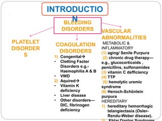
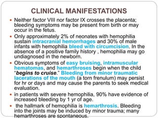
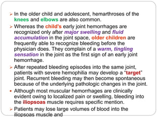
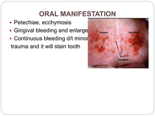
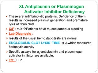
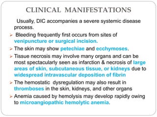
SlideShare a Scribd company logo
TRISHA DAS SARMA
1ST YEAR PGT
DEPARTMENT OF PEDODONTICS AND
PREVENTIVE DENTISTRY
BLEEDING DISORDERS IN
CHILDREN
.
BLEEDING
DISORDERS
PLATELET
DISORDER
S
COAGULATION
DISORDERS
1) Congenital
• Clotting Factor
Disorders e.g.-
Haemophilia A & B
• VWD
2) Aquired
• Vitamin K
deficiency
• Liver disease
• Other disorders—
DIC, fibrinogen
deficiency
VASCULAR
ABNORMALITIES
METABOLIC &
INFLAMMATORY
(1) aging/ Senile Purpura
(2) chronic drug therapy—
e.g., glucocorticoids ,
penicillins, sulfonamides
(3) vitamin C defficiency
(4) TTP
(5) hemolytic uremic
syndrome
(6) Henoch-Schönlein
purpura
HEREDITARY
1) hereditary hemorrhagic
telangiectasia (Osler-
Rendu-Weber disease).
INTRODUCTIO
N
 DIMINISHED PLATELET PRODUCTION
 ACCELELERATED PLATELET DESTRUCTION
PRIMARY
IMMUNE MEDIATED
 Autoimmune thrombocytopenic purpura
 Idiopathic secondary-infections
 pregnancy
 Alloimmune neonatal posttransfusion purpura
 Autoimmune diseases
 MDS
 Lymphoproliferative disorders
 ABNORMAL DISTRIBUTION OR POOLING
 Splenomegaly  sequestration of platelets
 Hypothermia
 Massive transfusion
SECONDARY
•DRUG INDUCED
NONIMMUNE
THROMBOCYTOPENI
A
• Thrombotic
thrombocytopenic
purpura
• DIC
•Kassabach merit
Syndrome
•Neonatal
Thrombocytopenia
•Congenital
Thrombocytopenia
•Thrombocytopenia In
Dengue
CLINICAL DISTINCTION
DISORDERS DUE
TO CLOTTING
FACTORS
DUE TO PLATELETS AND
VESSEL DISORDERS
SITE OF BLEEDING DEEP SKIN,MUCOUS MEMBRANES
ECCHYMOSIS RARE CHARACTERISTIC
PETECHIAE LARGE, DEEP SMALL, SUPERFICIAL
DEEP DISSECTING
HEMATOMAS
CHARACTERISTIC RARE
SUPERFICIAL
ECCHYMOSES
COMMON -LARGE
N SOLITARY
CHARACTERISTIC-SMALL N
MULTIPLE
HEMARTHROSIS CHARACTERISTIC RARE
DELAYED BLEEDING COMMON RARE
SEX MALES MC IN FEMALES
BLEEDING FROM
SUPERFICIAL CUTS
MINIMAL PERSISTENT N PROFUSE
PETECHIAE
1-2 mm red or
purple spot on
skin caused by
minor bleed from
PURPURA
3-10 mm red or
purple spot that
do not blanch
on applying
ECCHYMOSIS
Subcutaneous
bleeding spot
with diameter >
1 cm
 Hemophilia A (factor VIII deficiency) and hemophilia
B (factor IX deficiency) ,also called CHRISTMAS
DISEASE ,are the most common and serious congenital
coagulation factor deficiencies.
 although haemophilia B is passed down from parents to
children, about 1/3 rd of cases are caused by a
spontaneous mutation, a change in gene.
 Hemophilia C is the bleeding disorder associated with
reduced levels of factor XI.
 Reduced levels of the contact factors (factor XII, high
molecular weight kininogen, and prekallikrein) are
associated with ↑↑activated partial thromboplastin time
(APTT; also referred to as PTT), but are not associated
with hemorrhage.
 Other coagulation factor deficiencies that are less
A. Hereditary Clotting Factor Deficiencies
 With mild factor VIII hemophilia, the patient's
endogenously produced factor VIII can be released by
the administration of desmopressin acetate intranasally.
(Stimate) (DDAVP)
 Advantage The risk of exposing the patient with mild
hemophilia to transfusion-transmitted diseases and the
cost of recombinant products warrant the use of
desmopressin, if it is effective.
 The dose is 150 μg (1 puff) for children weighing <50 kg
and 300 μg (2 puffs) for children and young adults
weighing >50 kg.
 Most centers administer a trial of desmopressin to
determine the level of factor VIII achieved after its
infusion.
 Disadvantage
 In patients with moderate or severe factor VIII
deficiency, the stored levels of factor VIII in the body are
inadequate, and desmopressin treatment is ineffective.
1. Hemophilia A or B
PATHOPHYSIOLOGY
 Factors VIII and IX participate in a complex required for
the activation of factor X. Together with phospholipid and
calcium, they form the “tenase,” or factor X-activating,
complex.
 After injury,
the initial hemostatic event is formation of the platelet
plug,
together with the generation of the fibrin clot that
prevents further hemorrhage. In hemophilia A or B, clot
formation is delayed and is not robust.Inadequate thrombin generation leads to failure to form
a tightly cross-linked fibrin clot to support the platelet
plug. Patients with hemophilia slowly form a soft, friable
clot.
When untreated, bleeding occurs in a closed space,
such as a joint, cessation of bleeding may be the
result of tamponade.
The friable clot causes rebleeding during the
physiologic lysis of clots or with minimal new trauma.
CLINICAL MANIFESTATIONS
 Neither factor VIII nor factor IX crosses the placenta;
bleeding symptoms may be present from birth or may
occur in the fetus.
 Only approximately 2% of neonates with hemophilia
sustain intracranial hemorrhages and 30% of male
infants with hemophilia bleed with circumcision. In the
absence of a positive family history , hemophilia may go
undiagnosed in the newborn.
 Obvious symptoms of easy bruising, intramuscular
hematomas, and hemarthroses begin when the child
“begins to cruise.” Bleeding from minor traumatic
lacerations of the mouth (a torn frenulum) may persist
for hr or days and may cause the parents to seek medical
evaluation.
 in patients with severe hemophilia, 90% have evidence of
increased bleeding by 1 yr of age.
 the hallmark of hemophilia is hemarthrosis. Bleeding
into the joints may be induced by minor trauma; many
hemarthroses are spontaneous.
 In the older child and adolescent, hemarthroses of the
knees and elbows are also common.
 Whereas the child's early joint hemorrhages are
recognized only after major swelling and fluid
accumulation in the joint space, older children are
frequently able to recognize bleeding before the
physician does. They complain of a warm, tingling
sensation in the joint as the first sign of an early joint
hemorrhage.
 After repeated bleeding episodes into the same joint,
patients with severe hemophilia may develop a “target”
joint. Recurrent bleeding may then become spontaneous
because of the underlying pathologic changes in the joint.
 Although most muscular hemorrhages are clinically
evident owing to localized pain or swelling, bleeding into
the iliopsoas muscle requires specific mention.
 Patients may lose large volumes of blood into the
iliopsoas muscle and
verge on hypovolemic shock, with only a vague area of
referred pain in the groin.
 The hip is held in a flexed, internally
rotated position due to irritation of the
iliopsoas. The diagnosis is made clinically by the
inability to extend the hip, but must be
confirmed with ultrasonography or CT scan
 Life-threatening bleeding in the patient with hemophilia is
caused by bleeding into vital structures (central nervous
system, upper airway) or by exsanguination (external,
gastrointestinal, or iliopsoas hemorrhage).
ORAL MANIFESTATION
 Petechiae, ecchymosis
 Gingival bleeding and enlargement
 Continuous bleeding d/t minor
trauma and it will stain tooth
LABORATORY FINDINGS AND
DIAGNOSIS
 The laboratory screening test that is affected by a
reduced level of factor VIII or factor IX is PTT. In severe
hemophilia, PTT is usually 2–3 times the upper limit of
normal. (30-50 sec normal)
 Results of the other screening tests of the hemostatic
mechanism (platelet count, bleeding time, prothrombin
time, and thrombin time) are normal.
 In extrinsic pathway the complex of factor VIIa and
tissue factor activates factor X to initiate clotting. In the
laboratory, prothrombin time (PT) measures the
activation of factor X by factor VII and is therefore normal
in patients with factor VIII or factor IX deficiency.
 The specific assay for factors VIII and IX will confirm
the diagnosis of hemophilia
 Unless the patient has an inhibitor to factor VIII or IX, the
mixing of normal plasma with patient plasma results in
correction of PTT. If correction does not occur on mixing,
an inhibitor may be present.
 These antibodies are directed against the active clotting
site and are termed inhibitors. In such patients, the
quantitative Bethesda assay for inhibitors should be
performed to measure the antibody titer.
 Because factor VIII is carried in plasma by von
Willebrand factor, factor VIII : von Willebrand factor is
sometimes used to diagnose carriers of hemophilia.
 Thromboplastin regeneration time differentiate factor
VIII deficiency from factor IX deficiency.
 Successful management depends on the adequate
maintainance of the Antihaemophilic Globulin. Normal
level  50-100. In haemophiliac for good haemostasis,
 severe thrombocytopenia
 severe platelet function disorders, such as Bernard-
Soulier syndrome and Glanzmann thrombasthenia
 type 3 (severe) von Willebrand disease
 and vitamin K deficiency.
GENETICS
 Hemophilia occurs in approximately 1:5,000 males, with
85% having factor VIII deficiency and 10–15% having
factor IX deficiency.
 Hemophilia shows no apparent racial predilection,
appearing in all ethnic groups.
 The genes for factors VIII and IX are carried near the
terminus of the long arm of the X chromosome and
are therefore X-linked traits.
DIFFERENTIAL DIAGNOSIS
 The majority of patients have a reduction in the amount
of clotting factor protein; 5–10% of hemophilia A and
40–50% of hemophilia B has a dysfunctional protein.
 Approximately 45–50% of patients with severe
hemophilia A have the same mutation, in which there is
an internal inversion within the factor VIII gene that
results in no protein being produced. This mutation can
be detected in the blood of patients or carriers and in the
amniotic fluid by molecular techniques.
 In the newborn, factor VIII levels may be artificially
elevated because of the acute phase response elicited by
the birth process. This may cause a mildly affected
patient to have normal or near-normal levels of factor VIII.
 In contrast, factor IX levels are physiologically low in the
newborn. An undetectable level of factor IX is diagnostic
of severe hemophilia B.
 lyonization of the X chromosome female carriers of
hemophilia A or B have sufficient reduction of factor VIII
or factor IX to produce mild bleeding disorders.
CLASSIFICATION
 By definition, 1 international unit (IU) of each factor is
defined as that amount in 1 mL of normal plasma
referenced against a standard established by the World
Health Organization (WHO); thus, 100 mL of normal
plasma has 100 IU/dL (100% activity) of each factor.
 Severe hemophilia is characterized by having <1%
activity of the specific clotting factor, and bleeding is often
spontaneous.
 Patients with moderate hemophilia have levels of 1–5%
and require mild trauma to induce bleeding.
 Individuals with mild hemophilia have levels of >5%,
may go many years before the condition is diagnosed,
and frequently require significant trauma (dental work,
surgery)to cause bleeding.
The hemostatic level for factor VIII is >30–40%,
and for factor IX, it is >25–30%. The lower limit of levels
for factors VIII and IX in normal individuals is
approximately 50%.
TREATMENT
 mild to moderate bleeding levels of factor VIII or
factor IX must be raised to hemostatic levels in the 35–
50% range.
 For life-threatening or major hemorrhages the dose
should aim to achieve levels of 100% activity.
 Calculation of the dose of recombinant factor VIII (FVIII)
& recombinant factor IX (FIX) is as follows:
dose of factor VIII= % desired X body weight (kg) X
1.4
dose of factor IX= % desired X body weight (kg) X
0.5
 For factor VIII, the correction factor is based on the
volume of distribution of factor VIII.
 For factor IX, the correction factor is based on the
volume of distribution and the observed rise in
plasma level after infusion of recombinant factor IX.
ADAPTED FROM MONTGOMERY RR, GILL JC, SCOTT JP: HEMOPHILIA
AND VON WILLEBRAND DISEASE. IN NATHAN DG, ORKIN SH (EDITORS):
NATHAN AND OSKI'S HEMATOLOGY OF INFANCY AND CHILDHOOD, 5TH
ED. PHILADELPHIA, WB SAUNDERS, 1998
TYPE OF
HEMORRHAGE
HEMOPHILIA A HEMOPHILIA B
Hemarthrosi
s
•40 IU/kg factor VIII
concentrate on day 1
• 20 IU/kg on days 2,3,5
until joint function is normal
or back to baseline.
•Consider additional
treatment every other day
for 7–10 days.
• Consider prophylaxis.
•60–80 IU/kg on day 1
•40 IU/kg on days 2,4.
• Consider additional
treatment every other
day for 7–10 days
• Consider prophylaxis
Muscle or
significant
Subcutaneo
•20 IU/kg factor VIII
concentrate
•may need every-other-day
•40 IU/kg factor IX
concentrate
•may need treatment
TYPE OF
HEMORRHAGE
HEMOPHILIA A HEMOPHILIA B
Mouth,
deciduous
tooth, or tooth
extraction
•20 IU/kg factor VIII
concentrate
•antifibrinolytic therapy
• remove loose deciduous
tooth.
•40 IU/kg factor IX
concentrate
•antifibrinolytic
therapy
•remove loose
deciduous tooth.
Epistaxis •Apply pressure for 15–20
min
• pack with petrolatum
gauze
•give antifibrinolytic
therapy
•20 IU/kg factor VIII
concentrate if this
treatment fails.
•Apply pressure for
15–20 min
•pack with petrolatum
gauze; antifibrinolytic
therapy
•30 IU/kg factor IX
concentrate if this
treatment fails.
Major surgery, •50–75 IU/kg factor VIII •120 IU/kg factor IX
TYPE OF
HEMORRHAG
E
HEMOPHILIA A HEMOPHILIA B
•infusion of 2–4 IU/kg/hr to
maintain factor VIII > 100
IU/dl for 24 hr
•then give 2–3 IU/kg/hr
continuously for 5–7 days
to maintain the level at >
50 IU/dl
• an additional 5–7 days at
a level of > 30 IU/dl.
12–24 hr to maintain
factor IX at > 40 IU/dL
for 5–7 days
• > 30 IU/dL for 7 days
Iliopsoas
hemorrhage
•50 IU/kg factor VIII
concentrate
• 25 IU/kg every 12 hr until
asymptomatic
•then 20 IU/kg every other
day for a total of 10–14
•120 IU/kg factor IX
concentrate
• then 50–60 IU/kg
every 12–24 hr to
maintain factor IX at >
40 IU/dL until
TYPE OF
HEMORRHAGE
HEMOPHILIA A HEMOPHILIA B
Hematuria •Bed rest
•1½ × maintenance
fluids
• if not controlled in 1–2
days, 20 IU/kg factor
VIII concentrate
• if not controlled, give
prednisone (unless
HIV-infected).
•Bed rest
•1½ × maintenance
fluids
•if not controlled in
1–2 days, 40 IU/kg
factor IX
concentrate
•if not controlled,
give prednisone
(unless HIV-
infected).
Prophylaxis 20–40 IU/kg factor VIII
concentrate every
other day to achieve a
trough level of ≥ 1%.
30–50 IU/kg factor
IX concentrate
every 2–3 days to
achieve a trough
 In 1985, the genes for both factors VIII and IX were
cloned. Subsequently, recombinant factor VIII and factor
IX concentrates were developed to treat patients with
hemophilia and thereby avoid the infectious risk of
plasma-derived transfusion-transmitted diseases.
 With the availability of
recombinant replacement
products, prophylaxis has
become the standard of care
for most children with severe
hemophilia to prevent
spontaneous bleeding and
early joint deformities.
Fibrinolytic pathway
ACTIVATED THROMBIN (FACTOR IIa)
Fibrinogen soluble fibrin
FACTOR
XIIIa
cross linked fibrin
PLASMINOGEN
plasminogen
activator
PLASMIN
FIBRIN DEGRADATING PRODUCTS (DP) AND
D- DIMERS
PROPHYLAXIS
 Many patients are now given lifelong prophylaxis to
prevent spontaneous joint bleeding.
 Usually, such programs are initiated with first joint
hemorrhage.
 Such programs, although expensive, are highly effective
in preventing or greatly limiting the degree of joint
pathology.
 Treatment is usually provided every 2–3 days to maintain
a measurable plasma level of clotting factor (1–2%) when
assayed just before the next infusion (trough level).
 Because gene therapy may be available within the
lifetime of pediatric patients, keeping joints normal
through prophylaxis is a logical priority.
 If moderate arthropathy develops, prevention of future
bleeding will require higher plasma levels of clotting
factors.
 In the older child who is not given primary prophylaxis,
secondary prophylaxis is frequently initiated if a target
 Although it is easy to advise parents that their child
should avoid trauma, this advice is practically useless.
Toddlers are active, are curious about everything, and
injure themselves easily.
 Effective measures include anticipatory guidance,
including the use of car seats, seatbelts, and bike
helmets, and the importance of avoiding high-risk
behaviors.
 Older boys should be counseled to avoid violent contact
sports, but this is a challenge. Boys with severe
hemophilia often sustain hemorrhages in the absence of
known trauma.
 Early psychosocial intervention helps the family to
achieve a balance between overprotection and
permissiveness.
SUPPORTIVE CARE
CHRONIC COMPLICATIONS
 Long-term complications of hemophilia A and B include
 chronic arthropathy
 the development of an inhibitor to either factor VIII or
factor IX
 and the risk of transfusion-transmitted infectious
diseases.
 untreated hemophilia can cause cyclic recurrent
hemorrhages into specific joints or target joint.
In young children, the joint distends easily and a large
volume of blood may fill the joint until therapy intervenes.
After joint hemorrhage, proteolytic enzymes are released
by white blood cells into the joint space and heme iron
induces macrophage proliferation, leading to inflammation
of the synovium
The synovium thickens and develops frondlike projections
into the joint that are susceptible to being pinched and
may induce further hemorrhage causes severe pain
because the joint may have little space to accommodate
blood
The cartilaginous surface becomes eroded and ultimately
may even expose raw bone, leaving the joint susceptible
 Failure of a bleeding episode to respond to appropriate
replacement therapy is usually the first sign of an
inhibitor.
 Less often, inhibitors are identified during routine follow-
up testing. Inhibitors develop in approximately 25–35% of
patients with hemophilia A; the percentage is lower in
patients with hemophilia B, many of whom make an
inactive dysfunctional protein that renders them less
susceptible to an immune response.
 Highly purified factor IX or recombinant factor IX seems
to increase the frequency of inhibitor development, and
some anti-factor IX inhibitors induce anaphylaxis.
 T/T Many patients who have an inhibitor lose this
inhibitor with continued regular infusions. Others have a
higher titer of antibody with subsequent infusions and
may need to go through desensitization programs, in
which high doses of factor VIII or factor IX are infused in
 Factor IX immune tolerance programs have resulted in
nephrotic syndrome in some patients
 Rituximab has been used in patients with high titer
inhibitors who have failed immune tolerance programs.
 If desensitization fails, bleeding episodes are treated with
either recombinant factor VIIa or activated
prothrombin complex concentrates. The use of these
products bypasses the inhibitor in many instances, but
may increase the risk of thrombosis.
 The child with a bleeding disorder should receive the
appropriate vaccinations against hepatitis B, even though
recombinant products may avoid exposure to transfusion-
transmitted diseases. Patients exposed to plasma-
derived products should be screened periodically for
hepatitis B and C, HIV, and abnormalities in liver
function.
HOW TO MANAGE A
HAEMOPHILIC PATIENT IN
DENTAL CLINIC
?
?
SURGICAL
• For extraction Factor VIII
should be 50-70 %
•Avoid injecting into deep
tissue spaces. e.g.- avoid
block techniques. Use
infiltration anaesthesia
•Atraumatic extraction,
surgical exposure
•Avoid unnecessary trauma
to soft tissue, avoid suturing
PHYSICAL/
MECHANICAL
•Pressure pack
•Gelfoam CHEMICAL
•Avitone
•Surgical oxidised
cellulose
•Tannic acid
•Fibrin glue
MEDICAL
•Tranexamic
acid
• EACA
•DDAVP
•Ethamsylate
POST-OPERATIVE
•Avoid NSAIDS
•Oral hygiene
maintainance
•antibiotics
II. Factor XI Deficiency (Hemophilia C)
 autosomal deficiency associated with mild to moderate
bleeding
 m/c in Ashkenazi Jews. In Israel, 1–3/1,000 are
homozygous for this deficiency. Sephardic Jews are rarely
affected.
 T/t
 replacement of fresh frozen plasma
 Plasma infusions of 1 IU/kg usually increase the plasma
concentration by 2%. Thus, infusion of plasma at 10–15
mL/kg will result in a plasma level of 20–30%. Because
the half-life of factor XI is usually > 48 hr, maintaining
adequate levels of factor XI usually is not difficult.
 Lab diagnosis 
 specific factor XI assays
 In a patient with homozygous deficiency of factor XI, the
 Deficiency of the “contact factors” (factor XII,
prekallikrein, and high molecular weight kininogen)
causes no bleeding symptoms but ↑↑PTT, because
these contact factors function at the step of initiation of
the intrinsic clotting system by the reagent.
 It is important that these individuals be well informed
about the meaning of their clotting factor deficiency
because they do not need treatment, even for major
surgery.
III. Deficiencies of the Contact Factors
(Nonbleeding Disorders)
IV. Factor VII Deficiency
 rare bleeding disorder,usually detected only in the
homozygous state.
 C/F : spontaneous intracranial hemorrhage and
frequent mucocutaneous bleeding.
 Lab diagnosis:
 markedly prolonged PT but normal PTT.
 Factor VII assays show a marked reduction of factor
VII.
 Treatment :
 Because the plasma half-life of factor VII is 2–4
hr, therapy with FFP is difficult and is often
complicated by fluid overload.
 A commercial concentrate of recombinant factor
VIIa this use has not been approved by the FDA.
V. Factor X Deficiency
 rare autosomal disorder , result of either a quantitative
deficiency or a dysfunctional molecule.
 C/F : mucocutaneous and post-traumatic bleeding.
 Lab diagnosis : ↑↑ of both PT and PTT.
 Treatment :
 FFP or prothrombin complex concentrate.
 The half-life of factor X is approximately 30 hr, and its
volume of distribution is similar to that of factor IX. Thus,
1 U/kg will increase the plasma level of factor X by 1%.
 it is rarely a problem in pediatric patients, but systemic
amyloidosis may be associated with factor X deficiency
owing to the adsorption of factor X on the amyloid
protein. Here transfusion therapy often is not successful
because of the rapid clearance of factor X
VI. Prothrombin (Factor II) Deficiency
 caused either by a markedly reduced prothrombin
level (hypoprothrombinemia) or by functionally
abnormal prothrombin (dysprothrombinemia).
 Lab diagnosis
 homozygous patients shows prolonged PT and PTT.
 Factor II, or prothrombin, assays show a markedly
reduced prothrombin level.
 Treatment:
 prothrombin complex concentrates or FFP.
 In prothrombin deficiency, FFP is useful, because the
half-life of prothrombin is 3.5 days. Administration of
1 IU/kg of prothrombin will increase the plasma
activity by 1%.
VII. Factor V Deficiency
 an autosomal recessive disorder
 C/F :
 mild to moderate bleeding termed parahemophilia.
 Hemarthroses occur rarely
 m/c - mucocutaneous bleeding and hematomas
 Severe menorrhagia in women.
 Laboratory evaluation :
 prolonged PTT and PT.
 Specific assays for factor V.
 Treatment :
 FFP is the only currently available therapeutic product
that contains factor V. Factor V is lost rapidly from
stored FFP.
 severe deficiency infusions of FFP at 10 mL/kg every
12 hr.
 In patient with an acquired antibody to factor V do not
bleed because the factor V in platelets prevents
VIII. Combined Deficiency of Factors V
and VIII
 not related to defective genes for this protein but
secondary to the absence of an intracellular transport
protein, ERGIC-53, that is responsible for transporting
factors V and VIII from the endoplasmic reticulum to the
Golgi compartments. ERGIC-53 is encoded on
chromosome 18.
 This explains the paradoxical deficiency of 2 factors, one
encoded on chromosome 1 and the other on the X
chromosome.
IX. Fibrinogen Deficiency
 Congenital afibrinogenemia  rare autosomal recessive
disorder . dysfunctional fibrinogens  dysfibrinogenemia
 C/F : 1)do not bleed as frequently as patients with hemophilia,
rarely have hemarthroses.
2)in the neonatal period  gastrointestinal hemorrhage
or hematomas after vaginal delivery.
 Lab diagnosis : 1)↑↑PT and PTT, thrombin time is
prolonged.
2)an unmeasurable fibrinogen level is
diagnostic. clinical assays for fibrinogen are inhibited by high
doses of heparin. ↑↑reptilase time functional levels of
fibrinogen are low and heparin is not present
 Treatment: 1) Currently, no fibrinogen concentrates are
commercially available. plasma half-life of fibrinogen is 2–4
days treatment with FFP or cryoprecipitate is effective.
X. Factor XIII Deficiency (Fibrin-
Stabilizing Factor or Transglutaminase
Deficiency)
 C/F : symptoms of delayed hemorrhage are secondary to
instability of the fibrin clot. mild bruising, delayed separation
of the umbilical stump beyond 4 wk, poor wound healing,
and recurrent spontaneous abortions in women.
 L/D :1) usual screening tests for hemostasis are normal
2)Screening tests for factor XIII deficiency are based
onthere is increased solubility of the clot because of the
failure of cross linking. The normal clot remains insoluble in
the presence of 5 M urea, whereas in a patient with XIII
deficiency, the clot dissolves.
 Treatment :Because the half-life of factor XIII is 5–7 days
and the hemostatic level is 2–3% activity, infusion of FFP or
cryoprecipitate will correct the deficiency in these patients.
Plasma contains 1 IU/dL, and cryoprecipitate contains 75
IU/bag. In patients with significant bleeding symptoms,
XI. Antiplasmin or Plasminogen
Activator Inhibitor Deficiency
 These are antifibrinolytic proteins. Deficiency of them
results in increased plasmin generation and premature
lysis of fibrin clots.
 C/F : m/c Patients have mucocutaneous bleeding
 Lab Diagnosis :
 results of the usual hemostatic tests are normal
 EUGLOBULIN CLOT LYSIS TIME is ↓which measures
fibrinolytic activity
 Specific assays for α2-antiplasmin and plasminogen
activator inhibitor are available.
 T/t: FFP.
FFP CRYOPRECIPITATE
B. von Willebrand Disease
 The most common hereditary bleeding disorder, and
some reports suggest that it is present in 1–2% of the
general population.
 VWD is inherited autosomally, but most centers report
more affected women than men. Because menorrhagia
is a major symptom, women may be more likely to seek
treatment and thus to be diagnosed.
 VWD is classified on the basis of :
 whether the protein is quantitatively reduced, but not
absent (type 1)
 qualitatively abnormal (type 2)
 absent (type 3)
 Mutations in different loci that code for different functional
domains of the von Willebrand factor (VWF) protein
GENETICS
 Chromosome 12 contains the gene for VWF. In each of
the type 2 variants, specific areas of the molecule are
affected. The phenotype can guide the genetic diagnosis
of the specific mutation.
VON WILLEBRAND DISEASE VARIANTS
 Type 1 VWD
 most common 85% of all cases. Normal function but
↓quantity
 Type 2A VWD
 CAUSE abnormal proteolysis of VWF ↓ in no.
VWF antigen & ↓↓in VWF activity.
 Type 2B VWD
 CAUSE several mutations resulting in “hyperactive”
VWF. The abnormal VWF binds spontaneously to
 clearance of VWF and platelets moderate to severe
thrombocytopenia is common.
 Lab diagnosis: presence of hyperactive 2B VWF binds to
platelets and agglutinates them at low concentrations of
RISTOCETIN, a concentration that would not agglutinate
normal platelets.
 Type 2M VWD
 CAUSE mutations that result in ↓of the platelet-binding
function of VWF. No. of VWF antigen is normal
 factor VIII levels are similar to those of VWF antigen
 Type 2N VWD
 CAUSE ↓ of factor VIII binding by VWF  rapid
clearance of factor VIII that is weakly complexed to VWF
factor VIII level is reduced much more than VWF levels
 also termed autosomal hemophilia.
 Commonly, patients who have symptomatic bleeding are
heterozygotes who have inherited a gene for type 1
VWD from 1 parent and a gene for type 2N VWD from
the other
 Platelet-type (pseudo VWD)
 abnormality of the GPIb receptor on platelets.
Converse abnormality of type 2B where GPIb receptor on
platelets is hyperfunctional and binds plasma VWF
spontaneously.
 This results in thrombocytopenia and a loss of high
molecular weight VWF multimers
 this is a platelet abnormality rather than a plasma
abnormality.
 Type 3 VWD
 homozygous or compound heterozygous inheritance of
VWF deficiency.
 Patients exhibit undetectable plasma levels of VWF and
PATHOPHYSIOLOGY
VWF also serves as the carrier protein for plasma factor
VIII. A severe deficiency of VWF causes a secondary
VWF is a large multimeric glycoprotein that is
synthesized in megakaryocytes and endothelial cells,
stored in platelet α-granules and endothelial cell
Weibel-Palade bodies
During normal hemostasis, VWF adheres to the
subendothelial matrix after vascular damage
the conformation of VWF is changed so that it causes
platelets to adhere to VWF through their GPIb receptor
platelets are then activated, causing the recruitment of
additional platelets at GPIIb-3a receptor with the help
of VWF and exposing phosphatidylserine, which is an
important regulatory step for factor V- and factor VIII–
dependent steps in the clotting cascade
CLINICAL MANIFESTATIONS
 mucocutaneous hemorrhage including excessive
bruising, epistaxis, menorrhagia, and postoperative
hemorrhage after mucosal surgery, such as
tonsillectomy or wisdom tooth extraction. Severity is more
in type 3 VWD
 Because VWF is an acute-phase protein, stress will ↑
its level. Thus, patients may not bleed with procedures
that incur major stress, such as appendectomy and
childbirth(VWF levels may double or triple during
pregnancy), but may bleed excessively at the time of
cosmetic or mucosal surgery.
 gastrointestinal telangiectasia
 Patients with severe type 3 VWD may have joint
hemorrhages or spontaneous central nervous system
hemorrhages.
LABORATORY FINDINGS
 Although ↑↑BT & PTT is seen but they are in normal
range in type 1 VWD.
 quantitative assay for VWF antigen
 test for VWF activity (ristocetin cofactor activity)
 test for plasma factor VIII activity
 determination of VWF structure (VWF multimers)
 platelet count. type 2B & pseudo-VWD may have
lifelong thrombocytopenia.
 Levels of VWF vary with blood type (type O < A < B <
AB)
diagnosis of VWD is dependent on the finding of a
low level of at least 1 of the laboratory measures of
VWF .
D/D abnormalities of platelet number, platelet function,
or the vessel wall. In caring for children, it is important to
remember that the most common cause of such findings
COMPLICATIONS
Complications of bleeding due to VWD are rare. In
adolescent females, blood loss due to menorrhagia can
lead to:
 severe anemia, either acutely, with signs and symptoms
of hypovolemia, or chronically, caused by iron
deficiency
 Individuals with type 3 VWD can manifest joint or
muscle bleeding similar to individuals with hemophilia.
TREATMENT
 motto→↑the plasma level of VWF and factor VIII.
Because the gene for factor VIII is normal in patients with
VWD, elevating the plasma concentration of VWF
permits normal recovery and survival of endogenously
produced factor VIII.
 TYPE 1the synthetic drug DDAVP induces the release of
VWF from endothelial cells. desmopressin (DDAVP)
administration at a dose of 0.3 μg/kg IV will increase the
level of VWF and factor VIII by 3- to 5-fold.
Intranasal DDAVP (Stimate) is particularly helpful for
the outpatient treatment of bleeding episodes. The dose
is 150 μg (1 puff) for children weighing <50 kg and 300
μg (2 puffs) for those weighing >50 kg.
 Type 2 may not respond adequately to DDAVP
because they release an abnormal VWF molecule (most
 replacement therapy  plasma-derived VWF
containing concentrates that also contain factor VIII.
 1 U/kg will increase the plasma level by 1.5%. The
plasma half-life of both factor VIII and VWF is 12 hr, but
the alteration of VWF during fractionation results in half-
lives of 8–10 hr when concentrates are infused.
 Purified or recombinant VWF concentrates
(containing no factor VIII) may become available in the
near future. If only VWF is replaced, endogenous
correction of the factor VIII level takes 12–24 hr.
 Dental extractions and sometimes nosebleeds can be
managed with both DDAVP and an antifibrinolytic agent,
such as ε-aminocaproic acid (Amicar)
 Type 3 VWD
as VWF is both a plasma and a platelet protein. So
need VWF containing concentrate + platelet
concentrates.
LIVER DISEASES
 Because all of the clotting factors are produced exclusively
in the liver except factor VIII, coagulation abnormalities are
very common in patients with severe liver disease d/t
decreased synthesis of coagulation factors
 Patients with severe liver disease characteristically have
normal to increased (not reduced) levels of factor VIII activity
in plasma
 Treatment fresh frozen plasma (FFP) or cryoprecipitate.
 FFP (10–15 mL/kg) contains all clotting factors
 severe hypofibrinogenemia cryoprecipitate at a dose of 1
bag/5 kg body wt.
 Because a reduction in vitamin K–dependent coagulation
factors is common in those with acute or chronic liver
disease, vitamin K therapy can be given as a trial. Vitamin K
can be given orally/SC/ IV (not IM) at a dose of 1 mg/24 hr
for infants, 2–3 mg/24 hr for children, and 5–10 mg/24 hr
for adolescents and adults.
POSTNEONATAL VITAMIN K
DEFICIENCY
 CAUSES
 lack of oral intake of vitamin K
 alterations in the gut flora due to the long-term use of
broad-spectrum antibiotics,
 malabsorption of vitamin K. Intestinal malabsorption of
fats may accompany cystic fibrosis or biliary atresia
and result in a deficiency of fat-soluble dietary vitamin,
with reduced synthesis of vitamin K–dependent clotting
factors (factors II, VII, IX, and X, and protein C and
protein S)
Prophylactic administration of water-soluble
vitamin K orally is indicated in these cases
 warfarin (Coumadin) and related anticoagulants depend
on interference with vitamin K. Rat poison
(superwarfarin) produces a similar deficiency; vitamin K
is a specific antidote.
Disseminated Intravascular
Coagulation
 Thrombotic microangiopathy refers to a
heterogeneous group of conditions, including
disseminated intravascular coagulation (DIC), that result
in consumption of clotting factors(factor V, factor VIII,
prothrombin, fibrinogen, ), platelets, anticoagulant
proteins(protein C, protein S, and antithrombin III) and
procoagulants, resulting in a deficiency of them.
 Any life-threatening pathologic process associated with
hypoxia, acidosis, tissue necrosis, shock, and/or
endothelial damage may trigger DIC
PATHOPHSIOLOGY
CAUSES OF DISSEMINATED INTRAVASCULAR COAGULATION--MODIFIED
FROM MONTGOMERY RR, SCOTT IP: HEMOSTASIS: DISEASES OF THE FLUID
PHASE. IN NATHAN DG, OSKI FA (EDITORS): HEMATOLOGY OF INFANCY AND
CHILDHOOD, 4TH ED., VOL. 2. PHILADELPHIA, WB SAUNDERS, 1993.
INFECTIOUS Meningococcemia (purpura
fulminans)
Other gram-negative bacteria
(Haemophilus, Salmonella,
Escherichia coli)
Gram-positive bacteria (group B
streptococci, staphylococci)
 Rickettsia (Rocky Mountain spotted
fever)
Virus (cytomegalovirus, herpes
simplex, hemorrhagic fevers)
Malaria
 Fungus
TISSUE INJURY Central nervous system trauma
Profound shock or asphyxia
Hypothermia or hyperthermia
Massive burns
MALIGNANCY •Acute promyelocytic leukemia
•Acute monoblastic or myelocytic
leukemia
•Widespread malignancies
(neuroblastoma
VENOM OR TOXIN •Snake bites
•Insect bites
MICROANGIOPATHIC
DISORDERS
• “Severe” thrombotic
thrombocytopenic purpura or
hemolytic-uremic syndrome
•Giant hemangioma (Kasabach-
Merritt syn.)
GASTROINTESTINAL
DISORDERS
•Fulminant hepatitis
• Severe inflammatory bowel
NEWBORN •Maternal toxemia
•Group B streptococcal infections
• Abruptio placentae
• Severe respiratory distress syndrome
•Necrotizing enterocolitis
• Congenital viral disease (cytomegalovirus,
herpes simplex)
•Erythroblastosis fetalis
•Fetal demise of a twin
MISCELLLANEO
US
•Severe acute graft rejection
•Acute hemolytic transfusion reaction
• Severe collagen-vascular disease
• Kawasaki disease
• Heparin-induced thrombosis
•Infusion of “activated” prothrombin
complex concentrates
•Hyperpyrexia/encephalopathy,
•hemorrhagic shock syndrome
CLINICAL MANIFESTATIONS
Usually, DIC accompanies a severe systemic disease
process.
 Bleeding frequently first occurs from sites of
venipuncture or surgical incision.
 The skin may show petechiae and ecchymoses.
 Tissue necrosis may involve many organs and can be
most spectacularly seen as infarction & necrosis of large
areas of skin, subcutaneous tissue, or kidneys due to
widespread intravascular deposition of fibrin
 The hemostatic dysregulation may also result in
thromboses in the skin, kidneys, and other organs
 Anemia caused by hemolysis may develop rapidly owing
to microangiopathic hemolytic anemia.
LABORATORY FINDINGS
 ↑↑ PT,PTT and thrombin times
 Platelet counts may be profoundly depressed.
 The blood smear may contain fragmented, burr, and
helmet-shaped red blood cells (schistocytes).
 As fibrinolytic mechanism is activated, fibrinogen
degradation products (FDPs, D-dimers) appear in the
blood. So D-dimer assay is more specific for diagnosis.
TREATMENT
The first 2 steps in the of DIC are the most critical:
(1) treat the trigger that caused DIC
(2) restore normal homeostasis by correcting the shock,
acidosis, and hypoxia that usually complicate DIC.
Replacement Therapy
•platelet infusions (for
thrombocytopenia)
•cryoprecipitate (for
hypofibrinogenemia)
• fresh frozen plasma (for
replacement of other
coagulation factors and
natural inhibitors).
In DIC associated with sepsis,
a controlled trial of
drotrecogin-α (activated
protein C concentrate [APC])
in adults is given. The role of
these agents in childhood
remains to be defined.
The role of heparin in DIC is
limited to patients who have
vascular thrombosis in
association with DIC.
PLATELET DISORDERS
 Platelets are non-nucleated cellular fragments produced
by megakaryocytes within the bone marrow and other
tissues.
 Platelets circulate with a life span of 10–14 days.
 Thrombopoietin (TPO) is the primary growth factor that
controls platelet production. Levels of TPO are highest in
the thrombocytopenic states associated with decreased
marrow megakaryopoiesis and may be variable in states
of increased platelet production.
 The platelet plays multiple hemostatic roles which
includes primary and secondary haemostasis. The
platelet surface possesses a number of important
receptors for adhesive proteins, including von Willebrand
factor (VWF) and fibrinogen, as well as receptors for
agonists that trigger platelet aggregation, such as
thrombin, collagen, and adenosine diphosphate
 CAUSE
1) Disorders of the bone marrow that inhibit
megakaryopoiesis
2) infiltrative disorders(malignancies, such as acute
lymphocytic leukemia, histiocytosis, lymphomas, and
storage disease)usually affect RBC and WBC
production. (leukopenia, neutropenia, anemia, or
macrocytosis).
 C/F  constitutional aplastic anemia (Fanconi anemia)
radial anomalies, other skeletal anomalies, short stature,
microcephaly, and hyperpigmentation.
 Bone marrow examination should be done in these cases
Thrombocytopenia d/t Acquired
Disorders Causing Decreased
Production
(a) Idiopathic Thrombocytopenic
Purpura
 most common cause of acute onset of
thrombocytopenia in an otherwise well child is
(autoimmune) ITP.ETIOLOGY
Most common :Epstein-Barr virus and HIV.
Epstein-Barr virus–related ITP is usually of short
duration and follows the course of infectious
mononucleosis.
 HIV-associated ITP is usually chronic.
 The reason why some children respond to a
common infection with an autoimmune disease remains
unknown PATHOPHSIOLOGY
50–65% of ITP, 1–4 wk after exposure to a common viral
infection autoantibody directed against the platelet
surface developbinding of the antibody to the platelet
surface  circulating antibody-coated platelets are
recognized by the Fc receptor on the splenic
CLINICAL MANIFESTATIONS
 previously healthy 1–4 yr old child who has sudden onset
of generalized petechiae and purpura. The parents often
state that the child was fine yesterday and now is covered
with bruises and purple dots.
 Often there is bleeding from the gums and mucous
membranes, particularly with profound thrombocytopenia
(platelet count <10 × 109/L).
 There is a history of a preceding viral infection 1–4 wk
before the onset of thrombocytopenia.
 classification system has been proposed from the U.K. to
characterize the severity of bleeding in ITP on the basis of
symptoms and signs, but not platelet count:
 No symptoms
 Mild : bruising and petechiae, occasional minor epistaxis
 Moderate: more severe skin and mucosal lesions,
 Severe: bleeding episodes—menorrhagia, epistaxis,
melena—requiring transfusion or hospitalization,
symptoms interfering seriously with the quality of life
 hepatosplenomegaly or remarkable lymphadenopathy,
suggests other diagnoses (leukemia).
 Fewer than 1% of patients have intracranial
hemorrhage. So the objective of early therapy is to
raise the platelet count to >20 × 109/L and prevent it.
Approximately 20% of patients who present with
acute ITP have persistent thrombocytopenia for > 6 mo
and are said to have chronic ITP.
LABORATORY FINDINGS
 Severe thrombocytopenia (platelet count <20 × 109/L) is
common
 platelet size is normal or increased, reflective of increased
platelet turnover.
 In acute ITP, the hemoglobin value, white blood cell
(WBC) count, and differential count should be normal.
Hemoglobin may be decreased in severe cases
 Bone marrow examination shows normal granulocytic and
erythrocytic series, with characteristically normal or
increased numbers of immature megakaryocytes because
of increased platelet turnover.
 In adolescents with new-onset ITP, an antinuclear
antibody test should be done to evaluate for SLE.
 HIV studies should be done in at-risk populations,
especially sexually active teens.
 Coombs test should be done if there is unexplained
anemia to rule out Evans syndrome (autoimmune
hemolytic anemia and thrombocytopenia) or before
instituting therapy with IV anti-D.
DIFFERENTIAL DIAGNOSIS
 medication that induces drug-dependent antibodies
 splenic sequestration due to previously unappreciated
portal hypertension
 aplastic /Fanconi anemia .
 NONIMMUNE causes hemolytic-uremic syndrome
[HUS], disseminated intravascular coagulation [DIC]
 hypersplenism owing to either liver disease or portal
vein thrombosis.
 Autoimmune thrombocytopenia with insidious onset may
be an initial manifestation of SLE or rarely lymphoma.
 Wiskott-Aldrich syndrome must be considered in
young males found to have low platelet counts,
particularly if there is a history of eczema and recurrent
infection.
TREATMENT
 Antiplatelet antibodies bind to transfused platelets as well
as they do to autologous platelets. Thus, platelet
transfusion in ITP is usually contraindicated unless life-
threatening bleeding is present.
 Initial approaches to the management of ITP include the
following:
1. No therapy other than education and
counseling of the family and patient for patients with
minimal, mild, and moderate symptoms (70-80%).
spontaneous resolution occurs within 6 mo.
2. Intravenous immunoglobulin (IVIg) 0.8–1.0
g/kg/day for 1–2 days induces a rapid rise in platelet
count (usually>20× 109/L) in 95% of patients within 48 hr.
MOA It downregulates Fc-mediated phagocytosis of
antibody-coated platelets.
3. IV anti-D therapy For Rh positive patients, IV anti-D at
a dose of 50–75μg/kg causes a rise in platelet count
to>20× 109/L in 80–90% of patients within 48–72 hr.
MOARBC-antibody complexes bind to macrophage Fc
receptors and interfere with platelet destruction, thereby
causing a rise in platelet count.
D/A a) mild hemolytic anemia in Rh positive individuals
b) ineffective in Rh negative patients.
4. Prednisone 1–4 mg/kg/24 hr appear to induce a more
rapid rise in platelet count than in untreated patients with
ITP. Continued for 2–3 wk or until a rise in platelet count
to>20× 109/L has been achieved, with a rapid taper to
avoid the long-term side effects of it.
 In intracranial hemorrhage  multiple modalities
platelet transfusion+IVIG+high-dose corticosteroids+
prompt surgical consultation, with plans for emergency
splenectomy.
 The spleen is the primary site of both antiplatelet
antibody synthesis and platelet destruction. chronic ITP
and whose symptoms are not easily controlled with
therapy is a candidate for splenectomy.
D/A lifelong risk of overwhelming postsplenectomy
infection
 Before splenectomy, the child should receive
pneumococcal and meningococcal vaccines
 after splenectomy penicillin prophylaxis for a number
of yr.
 AMG 531, a thrombopoiesis-stimulating protein, has
Purpura
 Cause Acquired /Congenital deficiency of a
metalloproteinase that is responsible for cleaving the high
molecular weight multimers of VWF (normal in HUS)
 usually presents in adults and occasionally in adolescents.
 C/F  (a) Initial manifestations nonspecific weakness,
pain, emesis
(b) Microvascular thrombi within the central
nervous system causes neurologic signs aphasia,
blindness, and seizures.
PENTAD of  fever+ microangiopathic hemolytic anemia+
thrombocytopenia+abnormal renal function+ central nervous
system changes that is clinically similar to HUS
 Lab findings  (1) morphologically abnormal RBCs, with
schistocytes, spherocytes, helmet cells + ↑↑reticulocyte
count + thrombocytopenia
( c) Neonatal Thrombocytopenia/ Neonatal
alloimmune thrombocytopenic purpura
(NATP)
 This is the platelet equivalent of Rh disease of the newborn.
Unlike Rh disease, first pregnancies may be severely
affected. Subsequent pregnancies may be even more
severely affected than the first.
 The incidence of NATP is 1/4,000–5,000 live births.
 CAUSE  transplacental transfer of maternal antibodies
directed against fetal platelets that are shared with the
father and recognized as foreign by the maternal immune
system causing severe spontaneous bleeding. The most
common cause is incompatibility for the platelet
alloantigen HPA-1a.
 C/F (A) those of an apparently well child who, within the
1st few days after delivery, has generalized petechiae and
purpura.
 lab diagnosis (a) a normal maternal platelet count,
yet moderate to severe thrombocytopenia in the
newborn
(b) Specific DNA sequence
polymorphisms have been identified that permit
informative prenatal testing to identify at-risk
pregnancies.
(c) Fetal platelet count can be
monitored by percutaneous umbilical blood sampling.
 D/D maternal ITP and more commonly, viral or
bacterial infection. Children born to mothers with ITP
appear to have a lower risk of serious hemorrhage than
infants born with NATP, although severe
thrombocytopenia occurs.
CONDITION
Fetal Alloimmune condition
Congenital infection (e.g., CMV,
toxoplasma, rubella, HIV, syphilis)
Aneuploidy (e.g., trisomy 18,13, or 21, or
triploidy)
Autoimmune condition (e.g., ITP, SLE)
Severe Rh hemolytic disease
Congenital/inherited (e.g., Wiskott-Aldrich
syndrome)
Early-onset
neonatal (<72 hr)
Placental insufficiency (e.g., PET, IUGR,
diabetes)
Perinatal asphyxia
Perinatal infection (e.g., Escherichia coli,
FROM ROBERTS I, MURRAY NA: NEONATAL THROMBOCYTOPENIA:
CAUSES AND MANAGEMENT. ARCH DIS CHILD FETAL NEONATAL ED
2003;88:F359–F364.
Autoimmune condition (e.g., ITP, SLE)
Congenital infection (e.g., CMV, toxoplasma,
rubella, HIV)
Thrombosis (e.g., aortic, renal vein)
Bone marrow replacement (e.g., congenital
leukemia)
Kasabach-Merritt syndrome
Metabolic disease (e.g acidemia)
Congenital/inherited (e.g., TAR, CAMT)
Late-onset neonatal
(>72 hr)
Late-onset sepsis
NEC
Congenital infection (e.g.CMV, toxoplasma,
rubella, HIV)
Autoimmune
Kasabach-Merritt syndrome
 Treatment (1) administration of IVIG & corticosteroids
prenatally to the mother and sometimes corticosteroids to
the infant after delivery. Therapy usually begins in the
2nd trimester and is continued throughout the pregnancy.
(2) Thrombocytopenia in an infant,
whether due to NATP or maternal ITP, usually resolves
within 2–4 mo after delivery. The period of highest risk is
the immediate perinatal period.
(3) Delivery should be performed by
cesarean section. After delivery, if severe
thrombocytopenia persists, transfusion of 1 unit of
washed maternal platelets, that share the maternal
alloantigens will cause a rise in platelet counts to provide
effective hemostasis.
 After there has been 1 affected child, genetic counseling
is critical to inform the parents of the high risk of
thrombocytopenia in subsequent pregnancies.
(d) Congenital Thrombocytopenic
Syndromes
 CONGENITAL AMEGAKARYOCYTIC
THROMBOCYTOPENIA
 Cause mutation in the stem cell TPO receptor that is
essential for the development of all hematopoietic cell
lines.
 C/Fa rare defect in hematopoiesis that usually manifests
within the first few days to wk of life, when the child
presents with petechiae and purpura caused by profound
thrombocytopenia. Findings on physical examination are
normal.
 Lab Examination absence of megakaryocytes in
bone marrow. These patients often progress to marrow
failure (aplasia) over time.
 T/T Bone marrow transplantation
 THROMBOCYTOPENIA ABSENT RADIUS (TAR)
Anomalies mild changes to marked limb shortening,
skeletal abnormalities of the ulna, radius, and lower
extremities. Thumbs are present.
 Intolerance to cow's milk formula (present in 50%) may
complicate management by triggering gastrointestinal
bleeding, ↑↑ thrombocytopenia, eosinophilia, and a
leukemoid reaction.
 frequently remits over the first few yr of life.
 WISKOTT-ALDRICH SYNDROME (WAS)
 X-linked disorder
 Defect in the WAS protein which regulates the
cytoskeletal architecture of both platelets and T
lymphocytes
 C/F thrombocytopenia, with tiny platelets, eczema, and
recurrent infection due to immune deficiency
 Lab Examination bone marrow shows normal number
T/T (1)Successful bone marrow transplantation
(2)Splenectomy often corrects the
thrombocytopenia, suggesting that that the platelets formed
in WAS have accelerated destruction.
Complication Approximately 5% of patients develop
lymphoreticular malignancies.
(e) Kasabach-Merritt Syndrome
 giant hemangioma + localized intravascular coagulation
causing thrombocytopenia and hypofibrinogenemia
 the site of the hemangioma is obvious, but retroperitoneal
and intra-abdominal hemangiomas may require body
imaging for detection.
 Inside the hemangiomaplatelet trapping and activation
of coagulation+ fibrinogen consumption and generation
of fibrin DPs
 The peripheral blood smear shows microangiopathic
changes.
 T/T(1) surgical excision (if possible), laser
photocoagulation, high-dose corticosteroids, local
radiation therapy, and ANTIANGIOGENIC AGENTS,
such as interferon-α2. Over time, most patients who
present in infancy have regression of the hemangioma.
(g) Congenital Abnormalities of Platelet
Function
 BERNARD-SOULIER SYNDROME
 an autosomal recessive disorder.
 Cause
absence or severe deficiency of the VWF receptor (GPIb
complex) on the platelet membrane.
 Lab diagnosis
(1) thrombocytopenia, with giant platelets and markedly
↑↑bleeding time (>20 min).
(2) Platelet aggregation tests show absent ristocetin-
induced platelet aggregation, but normal aggregation
to all other agonists. Ristocetin induces the binding of
VWF to platelets and agglutinates platelets.
 GLANZMANN THROMBASTHENIA autosomal
recessive
 Cause
deficiency of the platelet fibrinogen receptor GPIIb-IIIa,
an integrin complex on the platelet surface that
undergoes conformational changes when platelets are
activated. Fibrinogen binds to this complex when the
platelet is activated and causes platelets to aggregate.
 Lab diagnosis  (1)↑ BT and a normal platelet count.
(2) Platelets have normal size and
morphologic features on the peripheral blood smear.
(3) Aggregation studies show
abnormal or absent aggregation with all agonists used
except ristocetin.
LAB TESTS
 Bleeding time measures the interaction of platelets with the
blood vessel wall and thus is affected by both platelet count
and platelet function.
 platelet function analyzer (PFA-100) measures platelet
adhesion and aggregation in whole blood at high shear when
the blood is exposed to either collagen-epinephrine or
collagen-ADP. More sensitive than BT.
PFA-100 value is
↑↑in VWD as well as in congenital and acquired platelet
function defects
 Both are variably insensitive to mild platelet function
abnormalities.
 Platelet Aggregometry Measures ability of platelets to
aggregate+ their metabolic activity
 agonists, such as COLLAGEN, ADP, RISTOCETIN,
ARACHIDONIC ACID, and THROMBIN, are added to platelet-
 Severe Platelet Function Defects
 DESMOPRESSIN 0.3 μg/kg IV may be used for mild to
moderate bleeding episodes.
 stimulate levels of VWF and factor VIII+ corrects BT and
provides normal hemostasis .
 Bernard-Soulier syndrome or Glanzmann
thrombasthenia
 platelet transfusions of 1 U/5–10 kg
 If antibodies develop to the deficient platelet protein
recombinant factor VIIa has been effective
 In both conditions, stem cell transplantation would be
expected to be curative.
TREATMENT
(h) Hemolytic-Uremic Syndrome
 an acute disease of infancy and early childhood, usually
follows an episode of acute gastroenteritis, often
triggered by Escherichia coli . It produces a specific
toxin (verotoxin) that binds to and damages renal
endothelial cells
 SYMPTOMS
 abnormal RBCs, with the presence of helmet cells,
spherocytes, schistocytes, burr cells, and other distorted
forms.
 Thrombocytopenia despite normal numbers of
megakaryocytes in the marrow indicates excessive
platelet destruction.
 elevated levels of D-dimer.
 urine protein, RBCs, and casts. Anuria and severe
azotemia indicate grave renal damage.
 Sometimes neurologic symptoms are associated with
these findings
 TREATMENT fluid management and prompt
(i) Drug-Induced Thrombocytopenia
 caused by
 Valproic Acid
 Phenytoin
 Sulfonamides
 Trimethoprim-sulfamethoxazole.
 Heparin-induced thrombocytopenia is seldom seen in
pediatrics when patient has an antibody directed
against the heparin-platelet factor 4 complex.
 Patients baseline platelet counts should be obtained
before initiating heparin therapy. A falling platelet count
during therapy raises the suspicion of HIT
 The platelet count begins to drop after 5 to 10 days of
heparin therapy. Sometimes may be within the first 24
hours.
TREATMENT of HIT
 ARGATROBAN : The dose is 2 mcg/kg/min, adjusted
by aPTT with a target of 1.5-3 times the baseline. The
initial dose should be reduced by 75% in patients with
liver dysfunction.
 LEPIRUDIN : The dose is 0.4 mg/kg via IV bolus
followed by an initial maintenance infusion of 0.15
mg/kg/h, adjusted for a target activated partial
thromboplastin time (aPTT) of 1.5-2.5 times the baseline.
 lepirudin is Metabolised by the kidneys and argatroban
by the liver.
 They present with an international normalized ratio (INR)
>4, which corresponds to severe protein C depletion.
warfarin should not start before the thrombocyte count is
greater than 150 x 109/L.
 Platelet transfusions should be avoided in HIT, as they
THROMBOCYTOPENIA IN
DENGUE
3 possible triggers to induce thrombocytopenia in
dengue virus infection(Funahara Y, Ogawa K, Fujita
N, Okuno Y.)
 1) DV antigen attached to human platelets without
immune-mediated reaction.(direct lysis)
 2) binding of anti-DV antibody on the DV antigen
associated with platelets (immune mediated)
 3) a modulation of endothelial cell (endothelium
ADAMTS13 related)
Disorders of the Blood Vessels
(1) HENOCH-SCHöNLEIN PURPURA
 The trigger is unknown
 C/F sudden development of a petechiae and often
palpable purpuric rash(usually in lower extremities and
buttocks), arthritis, abdominal pain, and renal
involvement .
 Lab Diagnosis 
 Results of coagulation studies and platelet count are
normal.
 The pathologic lesions in the skin, intestines, and
synovium are LEUKOCYTOCLASTIC ANGIITIS,
inflammatory damage to the endothelium of the capillary
and postcapillary venules mediated by WBCs and
macrophages.
(2) EHLERS-DANLOS SYNDROME
Ehlers-Danlos syndrome is a common disorder of
collagen structure that causes easy bruising and poor
wound healing
 C/F
1. Hyperelastic skin
2. lax joints easily subluxed
3. unusual scarring.
 lab diagnosis
1. Results of coagulation screening tests are usually
normal
2. although BT may be mildly prolonged
3. Results of platelet aggregation studies are either normal
or mildly abnormal, with deficient aggregation to
collagen.
3) ACQUIRED DISORDERS
 Scurvy, chronic corticosteroid therapy, and severe
malnutrition are associated with “weakening” of the
collagen matrix that supports the blood vessels.
 C/F 
 easy bruising, and particularly in
the case of scurvy, bleeding gums and
loosening of the teeth.
 petechiae and purpura may be seen
in vasculitic syndromes, such as SLE.
4) Rickettsiae causing Rocky Mountain spotted fever,
replicate in endothelial cells and damage them and
hence causes petechiae and purpura.
GUIDELINES FOR DENTAL TREATMENT OF
PATIENTS WITH INHERITED BLEEDING
DISORDERS
(By Andrew Brewer& Maria Elvira Correa On behalf of
World Federation of Hemophilia Dental Committee )
 Prevention
 Dental treatment
 Periodontal treatment
 Removable prosthodontics
 Orthodontic treatment
 Restorative procedures
 Endodontics
 Anesthesia and pain management HEMOSTATIC
COVER REQUIRED in Inferior dental block Lingual
infiltration
 Surgery
REFERENCES
1) Kliegman: Nelson Textbook of Pediatrics,
18th ed.
2) HARISON’S manual of medicine, 17 th
edition.
Bleeding disorders in children

More Related Content

What's hot

How to approach a child with bleeding disorder
How to approach a child with bleeding disorderHow to approach a child with bleeding disorder
How to approach a child with bleeding disorderKamran Akbar
 
medicine.Bleeding disorders.(dr.sabir) (new powerpoint)
medicine.Bleeding disorders.(dr.sabir) (new powerpoint)medicine.Bleeding disorders.(dr.sabir) (new powerpoint)
medicine.Bleeding disorders.(dr.sabir) (new powerpoint)student
 
Approach to bleeding neonate
Approach to bleeding neonateApproach to bleeding neonate
Approach to bleeding neonate
Dr Praman Kushwah
 
Von willebrand disease
Von willebrand diseaseVon willebrand disease
Von willebrand disease
Iqra Yasin
 
Thrombocytopenia
Thrombocytopenia Thrombocytopenia
Thrombocytopenia
Aseem Jain
 
Aplastic anemia in children 2021
Aplastic anemia in children 2021Aplastic anemia in children 2021
Aplastic anemia in children 2021
Imran Iqbal
 
Hemophilia
HemophiliaHemophilia
HemophiliaMyzak
 
Megaloblastic anemia in childhood
Megaloblastic anemia in childhoodMegaloblastic anemia in childhood
Megaloblastic anemia in childhood
Singaram_Paed
 
Bleeding disorders
Bleeding disordersBleeding disorders
Bleeding disorders
Manisha Meda
 
Approach To A Bleeding Child
Approach To A Bleeding ChildApproach To A Bleeding Child
Approach To A Bleeding Child
SurabhiPeriwal
 
Approach to bleeding disorder
Approach to bleeding disorderApproach to bleeding disorder
Approach to bleeding disorder
Hayelom Michael Deyo
 
Anuupam ppt for bleeding child
Anuupam ppt for bleeding childAnuupam ppt for bleeding child
Anuupam ppt for bleeding child
Anuupam Kumaar
 
Approach to a child with bleeding disorder
Approach to a child with bleeding disorderApproach to a child with bleeding disorder
Approach to a child with bleeding disorder
Nehal Shah
 
Bleeding and clotting disorders dr anoop k r
Bleeding and clotting disorders dr anoop k rBleeding and clotting disorders dr anoop k r
Bleeding and clotting disorders dr anoop k r
anoop k r
 
Iron deficiency anemia in children
Iron deficiency anemia in childrenIron deficiency anemia in children
Iron deficiency anemia in children
Azad Haleem
 
Approach to anemia in children
Approach to anemia in childrenApproach to anemia in children
Approach to anemia in children
vinay nandimalla
 

What's hot (20)

How to approach a child with bleeding disorder
How to approach a child with bleeding disorderHow to approach a child with bleeding disorder
How to approach a child with bleeding disorder
 
medicine.Bleeding disorders.(dr.sabir) (new powerpoint)
medicine.Bleeding disorders.(dr.sabir) (new powerpoint)medicine.Bleeding disorders.(dr.sabir) (new powerpoint)
medicine.Bleeding disorders.(dr.sabir) (new powerpoint)
 
Approach to bleeding neonate
Approach to bleeding neonateApproach to bleeding neonate
Approach to bleeding neonate
 
Approach to bleeding neonate final
Approach to bleeding neonate finalApproach to bleeding neonate final
Approach to bleeding neonate final
 
Von willebrand disease
Von willebrand diseaseVon willebrand disease
Von willebrand disease
 
Bleeding neonate
Bleeding neonateBleeding neonate
Bleeding neonate
 
Thrombocytopenia
Thrombocytopenia Thrombocytopenia
Thrombocytopenia
 
Aplastic anemia in children 2021
Aplastic anemia in children 2021Aplastic anemia in children 2021
Aplastic anemia in children 2021
 
Hemophilia
HemophiliaHemophilia
Hemophilia
 
Megaloblastic anemia in childhood
Megaloblastic anemia in childhoodMegaloblastic anemia in childhood
Megaloblastic anemia in childhood
 
Bleeding disorders
Bleeding disordersBleeding disorders
Bleeding disorders
 
Approach To A Bleeding Child
Approach To A Bleeding ChildApproach To A Bleeding Child
Approach To A Bleeding Child
 
Approach to bleeding disorder
Approach to bleeding disorderApproach to bleeding disorder
Approach to bleeding disorder
 
Bleeding disorder
Bleeding disorderBleeding disorder
Bleeding disorder
 
Thrombocytopenic purpura
Thrombocytopenic purpuraThrombocytopenic purpura
Thrombocytopenic purpura
 
Anuupam ppt for bleeding child
Anuupam ppt for bleeding childAnuupam ppt for bleeding child
Anuupam ppt for bleeding child
 
Approach to a child with bleeding disorder
Approach to a child with bleeding disorderApproach to a child with bleeding disorder
Approach to a child with bleeding disorder
 
Bleeding and clotting disorders dr anoop k r
Bleeding and clotting disorders dr anoop k rBleeding and clotting disorders dr anoop k r
Bleeding and clotting disorders dr anoop k r
 
Iron deficiency anemia in children
Iron deficiency anemia in childrenIron deficiency anemia in children
Iron deficiency anemia in children
 
Approach to anemia in children
Approach to anemia in childrenApproach to anemia in children
Approach to anemia in children
 

Similar to Bleeding disorders in children

Hemorragics.pptx
Hemorragics.pptxHemorragics.pptx
Hemorragics.pptx
HARYANVITRAVLLER
 
BLEEDING DISORDERS.pptx
BLEEDING DISORDERS.pptxBLEEDING DISORDERS.pptx
BLEEDING DISORDERS.pptx
EidleMohamedsaed
 
Hemophilia a
Hemophilia aHemophilia a
Hemophilia a
Ahmed Al-Dawoodi
 
Hemophilias Case based study
Hemophilias Case based study Hemophilias Case based study
Hemophilias Case based study
Akshat Jain M.D.
 
Medicine.Bleeding disorders.(dr.sabir)
Medicine.Bleeding disorders.(dr.sabir)Medicine.Bleeding disorders.(dr.sabir)
Medicine.Bleeding disorders.(dr.sabir)student
 
Medicine 5th year, 10th lecture (Dr. Sabir)
Medicine 5th year, 10th lecture (Dr. Sabir)Medicine 5th year, 10th lecture (Dr. Sabir)
Medicine 5th year, 10th lecture (Dr. Sabir)
College of Medicine, Sulaymaniyah
 
Haemolytic Uremic Syndrome
Haemolytic Uremic SyndromeHaemolytic Uremic Syndrome
Haemolytic Uremic Syndromefracpractice
 
Hemophillia
HemophilliaHemophillia
Hemophillia
Bikal Lamichhane
 
Bleeding, clotting,platelet disorder and it's management
Bleeding, clotting,platelet disorder and it's managementBleeding, clotting,platelet disorder and it's management
Bleeding, clotting,platelet disorder and it's management
RakhiYadav53
 
change bleeding20clotting20and20platelets20disorders-220205073717.pdf
change bleeding20clotting20and20platelets20disorders-220205073717.pdfchange bleeding20clotting20and20platelets20disorders-220205073717.pdf
change bleeding20clotting20and20platelets20disorders-220205073717.pdf
NorhanKhaled15
 
07 C.disorders.pptx
07 C.disorders.pptx07 C.disorders.pptx
07 C.disorders.pptx
AmosiRichard
 
Trombocitopenia enfoque 2016.pdf
Trombocitopenia enfoque 2016.pdfTrombocitopenia enfoque 2016.pdf
Trombocitopenia enfoque 2016.pdf
JuanCamiloMoralesTab
 
Trombocitopenia Hospitalizado.pdf
Trombocitopenia Hospitalizado.pdfTrombocitopenia Hospitalizado.pdf
Trombocitopenia Hospitalizado.pdf
JuanCamiloMoralesTab
 
Hemophilia
HemophiliaHemophilia
Hemophilia
Dr Shafee ullah
 
Medically compromised children 1
Medically compromised children 1Medically compromised children 1
Medically compromised children 1
ZainabMohammed31
 
Hemophilia by Suhasis Mondal
Hemophilia by Suhasis MondalHemophilia by Suhasis Mondal
Hemophilia by Suhasis Mondal
Dr. Suhasis Mondal
 
approach to infant with Hydrops fetalis
approach to infant with Hydrops fetalisapproach to infant with Hydrops fetalis
approach to infant with Hydrops fetalis
Dr Praman Kushwah
 
Hemophilia and ITP
Hemophilia and ITPHemophilia and ITP
Hemophilia and ITP
Dr M Sanjeevappa
 
Thromboembolism 7- 5-15
Thromboembolism 7- 5-15Thromboembolism 7- 5-15
Thromboembolism 7- 5-15Md. Shameem
 

Similar to Bleeding disorders in children (20)

Hemorragics.pptx
Hemorragics.pptxHemorragics.pptx
Hemorragics.pptx
 
BLEEDING DISORDERS.pptx
BLEEDING DISORDERS.pptxBLEEDING DISORDERS.pptx
BLEEDING DISORDERS.pptx
 
Hemophilia a
Hemophilia aHemophilia a
Hemophilia a
 
Hemophilias Case based study
Hemophilias Case based study Hemophilias Case based study
Hemophilias Case based study
 
Medicine.Bleeding disorders.(dr.sabir)
Medicine.Bleeding disorders.(dr.sabir)Medicine.Bleeding disorders.(dr.sabir)
Medicine.Bleeding disorders.(dr.sabir)
 
Medicine 5th year, 10th lecture (Dr. Sabir)
Medicine 5th year, 10th lecture (Dr. Sabir)Medicine 5th year, 10th lecture (Dr. Sabir)
Medicine 5th year, 10th lecture (Dr. Sabir)
 
Haemolytic Uremic Syndrome
Haemolytic Uremic SyndromeHaemolytic Uremic Syndrome
Haemolytic Uremic Syndrome
 
Hemophillia
HemophilliaHemophillia
Hemophillia
 
Bleeding, clotting,platelet disorder and it's management
Bleeding, clotting,platelet disorder and it's managementBleeding, clotting,platelet disorder and it's management
Bleeding, clotting,platelet disorder and it's management
 
change bleeding20clotting20and20platelets20disorders-220205073717.pdf
change bleeding20clotting20and20platelets20disorders-220205073717.pdfchange bleeding20clotting20and20platelets20disorders-220205073717.pdf
change bleeding20clotting20and20platelets20disorders-220205073717.pdf
 
07 C.disorders.pptx
07 C.disorders.pptx07 C.disorders.pptx
07 C.disorders.pptx
 
Trombocitopenia enfoque 2016.pdf
Trombocitopenia enfoque 2016.pdfTrombocitopenia enfoque 2016.pdf
Trombocitopenia enfoque 2016.pdf
 
Trombocitopenia Hospitalizado.pdf
Trombocitopenia Hospitalizado.pdfTrombocitopenia Hospitalizado.pdf
Trombocitopenia Hospitalizado.pdf
 
Hemophilia
HemophiliaHemophilia
Hemophilia
 
Medically compromised children 1
Medically compromised children 1Medically compromised children 1
Medically compromised children 1
 
Hemophilia by Suhasis Mondal
Hemophilia by Suhasis MondalHemophilia by Suhasis Mondal
Hemophilia by Suhasis Mondal
 
approach to infant with Hydrops fetalis
approach to infant with Hydrops fetalisapproach to infant with Hydrops fetalis
approach to infant with Hydrops fetalis
 
Hemophilia and ITP
Hemophilia and ITPHemophilia and ITP
Hemophilia and ITP
 
7b..bleeding disorder
7b..bleeding disorder7b..bleeding disorder
7b..bleeding disorder
 
Thromboembolism 7- 5-15
Thromboembolism 7- 5-15Thromboembolism 7- 5-15
Thromboembolism 7- 5-15
 

Recently uploaded

Cervical & Brachial Plexus By Dr. RIG.pptx
Cervical & Brachial Plexus By Dr. RIG.pptxCervical & Brachial Plexus By Dr. RIG.pptx
Cervical & Brachial Plexus By Dr. RIG.pptx
Dr. Rabia Inam Gandapore
 
Hemodialysis: Chapter 3, Dialysis Water Unit - Dr.Gawad
Hemodialysis: Chapter 3, Dialysis Water Unit - Dr.GawadHemodialysis: Chapter 3, Dialysis Water Unit - Dr.Gawad
Hemodialysis: Chapter 3, Dialysis Water Unit - Dr.Gawad
NephroTube - Dr.Gawad
 
New Directions in Targeted Therapeutic Approaches for Older Adults With Mantl...
New Directions in Targeted Therapeutic Approaches for Older Adults With Mantl...New Directions in Targeted Therapeutic Approaches for Older Adults With Mantl...
New Directions in Targeted Therapeutic Approaches for Older Adults With Mantl...
i3 Health
 
Prix Galien International 2024 Forum Program
Prix Galien International 2024 Forum ProgramPrix Galien International 2024 Forum Program
Prix Galien International 2024 Forum Program
Levi Shapiro
 
Evaluation of antidepressant activity of clitoris ternatea in animals
Evaluation of antidepressant activity of clitoris ternatea in animalsEvaluation of antidepressant activity of clitoris ternatea in animals
Evaluation of antidepressant activity of clitoris ternatea in animals
Shweta
 
Surgical Site Infections, pathophysiology, and prevention.pptx
Surgical Site Infections, pathophysiology, and prevention.pptxSurgical Site Infections, pathophysiology, and prevention.pptx
Surgical Site Infections, pathophysiology, and prevention.pptx
jval Landero
 
MANAGEMENT OF ATRIOVENTRICULAR CONDUCTION BLOCK.pdf
MANAGEMENT OF ATRIOVENTRICULAR CONDUCTION BLOCK.pdfMANAGEMENT OF ATRIOVENTRICULAR CONDUCTION BLOCK.pdf
MANAGEMENT OF ATRIOVENTRICULAR CONDUCTION BLOCK.pdf
Jim Jacob Roy
 
BENIGN PROSTATIC HYPERPLASIA.BPH. BPHpdf
BENIGN PROSTATIC HYPERPLASIA.BPH. BPHpdfBENIGN PROSTATIC HYPERPLASIA.BPH. BPHpdf
BENIGN PROSTATIC HYPERPLASIA.BPH. BPHpdf
DR SETH JOTHAM
 
Ozempic: Preoperative Management of Patients on GLP-1 Receptor Agonists
Ozempic: Preoperative Management of Patients on GLP-1 Receptor Agonists  Ozempic: Preoperative Management of Patients on GLP-1 Receptor Agonists
Ozempic: Preoperative Management of Patients on GLP-1 Receptor Agonists
Saeid Safari
 
Charaka Samhita Sutra sthana Chapter 15 Upakalpaniyaadhyaya
Charaka Samhita Sutra sthana Chapter 15 UpakalpaniyaadhyayaCharaka Samhita Sutra sthana Chapter 15 Upakalpaniyaadhyaya
Charaka Samhita Sutra sthana Chapter 15 Upakalpaniyaadhyaya
Dr KHALID B.M
 
Non-respiratory Functions of the Lungs.pdf
Non-respiratory Functions of the Lungs.pdfNon-respiratory Functions of the Lungs.pdf
Non-respiratory Functions of the Lungs.pdf
MedicoseAcademics
 
THOA 2.ppt Human Organ Transplantation Act
THOA 2.ppt Human Organ Transplantation ActTHOA 2.ppt Human Organ Transplantation Act
THOA 2.ppt Human Organ Transplantation Act
DrSathishMS1
 
Charaka Samhita Sutra Sthana 9 Chapter khuddakachatuspadadhyaya
Charaka Samhita Sutra Sthana 9 Chapter khuddakachatuspadadhyayaCharaka Samhita Sutra Sthana 9 Chapter khuddakachatuspadadhyaya
Charaka Samhita Sutra Sthana 9 Chapter khuddakachatuspadadhyaya
Dr KHALID B.M
 
How to Give Better Lectures: Some Tips for Doctors
How to Give Better Lectures: Some Tips for DoctorsHow to Give Better Lectures: Some Tips for Doctors
How to Give Better Lectures: Some Tips for Doctors
LanceCatedral
 
Tom Selleck Health: A Comprehensive Look at the Iconic Actor’s Wellness Journey
Tom Selleck Health: A Comprehensive Look at the Iconic Actor’s Wellness JourneyTom Selleck Health: A Comprehensive Look at the Iconic Actor’s Wellness Journey
Tom Selleck Health: A Comprehensive Look at the Iconic Actor’s Wellness Journey
greendigital
 
Are There Any Natural Remedies To Treat Syphilis.pdf
Are There Any Natural Remedies To Treat Syphilis.pdfAre There Any Natural Remedies To Treat Syphilis.pdf
Are There Any Natural Remedies To Treat Syphilis.pdf
Little Cross Family Clinic
 
basicmodesofventilation2022-220313203758.pdf
basicmodesofventilation2022-220313203758.pdfbasicmodesofventilation2022-220313203758.pdf
basicmodesofventilation2022-220313203758.pdf
aljamhori teaching hospital
 
The hemodynamic and autonomic determinants of elevated blood pressure in obes...
The hemodynamic and autonomic determinants of elevated blood pressure in obes...The hemodynamic and autonomic determinants of elevated blood pressure in obes...
The hemodynamic and autonomic determinants of elevated blood pressure in obes...
Catherine Liao
 
Factory Supply Best Quality Pmk Oil CAS 28578–16–7 PMK Powder in Stock
Factory Supply Best Quality Pmk Oil CAS 28578–16–7 PMK Powder in StockFactory Supply Best Quality Pmk Oil CAS 28578–16–7 PMK Powder in Stock
Factory Supply Best Quality Pmk Oil CAS 28578–16–7 PMK Powder in Stock
rebeccabio
 
Ophthalmology Clinical Tests for OSCE exam
Ophthalmology Clinical Tests for OSCE examOphthalmology Clinical Tests for OSCE exam
Ophthalmology Clinical Tests for OSCE exam
KafrELShiekh University
 

Recently uploaded (20)

Cervical & Brachial Plexus By Dr. RIG.pptx
Cervical & Brachial Plexus By Dr. RIG.pptxCervical & Brachial Plexus By Dr. RIG.pptx
Cervical & Brachial Plexus By Dr. RIG.pptx
 
Hemodialysis: Chapter 3, Dialysis Water Unit - Dr.Gawad
Hemodialysis: Chapter 3, Dialysis Water Unit - Dr.GawadHemodialysis: Chapter 3, Dialysis Water Unit - Dr.Gawad
Hemodialysis: Chapter 3, Dialysis Water Unit - Dr.Gawad
 
New Directions in Targeted Therapeutic Approaches for Older Adults With Mantl...
New Directions in Targeted Therapeutic Approaches for Older Adults With Mantl...New Directions in Targeted Therapeutic Approaches for Older Adults With Mantl...
New Directions in Targeted Therapeutic Approaches for Older Adults With Mantl...
 
Prix Galien International 2024 Forum Program
Prix Galien International 2024 Forum ProgramPrix Galien International 2024 Forum Program
Prix Galien International 2024 Forum Program
 
Evaluation of antidepressant activity of clitoris ternatea in animals
Evaluation of antidepressant activity of clitoris ternatea in animalsEvaluation of antidepressant activity of clitoris ternatea in animals
Evaluation of antidepressant activity of clitoris ternatea in animals
 
Surgical Site Infections, pathophysiology, and prevention.pptx
Surgical Site Infections, pathophysiology, and prevention.pptxSurgical Site Infections, pathophysiology, and prevention.pptx
Surgical Site Infections, pathophysiology, and prevention.pptx
 
MANAGEMENT OF ATRIOVENTRICULAR CONDUCTION BLOCK.pdf
MANAGEMENT OF ATRIOVENTRICULAR CONDUCTION BLOCK.pdfMANAGEMENT OF ATRIOVENTRICULAR CONDUCTION BLOCK.pdf
MANAGEMENT OF ATRIOVENTRICULAR CONDUCTION BLOCK.pdf
 
BENIGN PROSTATIC HYPERPLASIA.BPH. BPHpdf
BENIGN PROSTATIC HYPERPLASIA.BPH. BPHpdfBENIGN PROSTATIC HYPERPLASIA.BPH. BPHpdf
BENIGN PROSTATIC HYPERPLASIA.BPH. BPHpdf
 
Ozempic: Preoperative Management of Patients on GLP-1 Receptor Agonists
Ozempic: Preoperative Management of Patients on GLP-1 Receptor Agonists  Ozempic: Preoperative Management of Patients on GLP-1 Receptor Agonists
Ozempic: Preoperative Management of Patients on GLP-1 Receptor Agonists
 
Charaka Samhita Sutra sthana Chapter 15 Upakalpaniyaadhyaya
Charaka Samhita Sutra sthana Chapter 15 UpakalpaniyaadhyayaCharaka Samhita Sutra sthana Chapter 15 Upakalpaniyaadhyaya
Charaka Samhita Sutra sthana Chapter 15 Upakalpaniyaadhyaya
 
Non-respiratory Functions of the Lungs.pdf
Non-respiratory Functions of the Lungs.pdfNon-respiratory Functions of the Lungs.pdf
Non-respiratory Functions of the Lungs.pdf
 
THOA 2.ppt Human Organ Transplantation Act
THOA 2.ppt Human Organ Transplantation ActTHOA 2.ppt Human Organ Transplantation Act
THOA 2.ppt Human Organ Transplantation Act
 
Charaka Samhita Sutra Sthana 9 Chapter khuddakachatuspadadhyaya
Charaka Samhita Sutra Sthana 9 Chapter khuddakachatuspadadhyayaCharaka Samhita Sutra Sthana 9 Chapter khuddakachatuspadadhyaya
Charaka Samhita Sutra Sthana 9 Chapter khuddakachatuspadadhyaya
 
How to Give Better Lectures: Some Tips for Doctors
How to Give Better Lectures: Some Tips for DoctorsHow to Give Better Lectures: Some Tips for Doctors
How to Give Better Lectures: Some Tips for Doctors
 
Tom Selleck Health: A Comprehensive Look at the Iconic Actor’s Wellness Journey
Tom Selleck Health: A Comprehensive Look at the Iconic Actor’s Wellness JourneyTom Selleck Health: A Comprehensive Look at the Iconic Actor’s Wellness Journey
Tom Selleck Health: A Comprehensive Look at the Iconic Actor’s Wellness Journey
 
Are There Any Natural Remedies To Treat Syphilis.pdf
Are There Any Natural Remedies To Treat Syphilis.pdfAre There Any Natural Remedies To Treat Syphilis.pdf
Are There Any Natural Remedies To Treat Syphilis.pdf
 
basicmodesofventilation2022-220313203758.pdf
basicmodesofventilation2022-220313203758.pdfbasicmodesofventilation2022-220313203758.pdf
basicmodesofventilation2022-220313203758.pdf
 
The hemodynamic and autonomic determinants of elevated blood pressure in obes...
The hemodynamic and autonomic determinants of elevated blood pressure in obes...The hemodynamic and autonomic determinants of elevated blood pressure in obes...
The hemodynamic and autonomic determinants of elevated blood pressure in obes...
 
Factory Supply Best Quality Pmk Oil CAS 28578–16–7 PMK Powder in Stock
Factory Supply Best Quality Pmk Oil CAS 28578–16–7 PMK Powder in StockFactory Supply Best Quality Pmk Oil CAS 28578–16–7 PMK Powder in Stock
Factory Supply Best Quality Pmk Oil CAS 28578–16–7 PMK Powder in Stock
 
Ophthalmology Clinical Tests for OSCE exam
Ophthalmology Clinical Tests for OSCE examOphthalmology Clinical Tests for OSCE exam
Ophthalmology Clinical Tests for OSCE exam
 

Bleeding disorders in children

  • 1. TRISHA DAS SARMA 1ST YEAR PGT DEPARTMENT OF PEDODONTICS AND PREVENTIVE DENTISTRY BLEEDING DISORDERS IN CHILDREN
  • 2. . BLEEDING DISORDERS PLATELET DISORDER S COAGULATION DISORDERS 1) Congenital • Clotting Factor Disorders e.g.- Haemophilia A & B • VWD 2) Aquired • Vitamin K deficiency • Liver disease • Other disorders— DIC, fibrinogen deficiency VASCULAR ABNORMALITIES METABOLIC & INFLAMMATORY (1) aging/ Senile Purpura (2) chronic drug therapy— e.g., glucocorticoids , penicillins, sulfonamides (3) vitamin C defficiency (4) TTP (5) hemolytic uremic syndrome (6) Henoch-Schönlein purpura HEREDITARY 1) hereditary hemorrhagic telangiectasia (Osler- Rendu-Weber disease). INTRODUCTIO N
  • 3.  DIMINISHED PLATELET PRODUCTION  ACCELELERATED PLATELET DESTRUCTION PRIMARY IMMUNE MEDIATED  Autoimmune thrombocytopenic purpura  Idiopathic secondary-infections  pregnancy  Alloimmune neonatal posttransfusion purpura  Autoimmune diseases  MDS  Lymphoproliferative disorders  ABNORMAL DISTRIBUTION OR POOLING  Splenomegaly  sequestration of platelets  Hypothermia  Massive transfusion SECONDARY •DRUG INDUCED NONIMMUNE THROMBOCYTOPENI A • Thrombotic thrombocytopenic purpura • DIC •Kassabach merit Syndrome •Neonatal Thrombocytopenia •Congenital Thrombocytopenia •Thrombocytopenia In Dengue
  • 4. CLINICAL DISTINCTION DISORDERS DUE TO CLOTTING FACTORS DUE TO PLATELETS AND VESSEL DISORDERS SITE OF BLEEDING DEEP SKIN,MUCOUS MEMBRANES ECCHYMOSIS RARE CHARACTERISTIC PETECHIAE LARGE, DEEP SMALL, SUPERFICIAL DEEP DISSECTING HEMATOMAS CHARACTERISTIC RARE SUPERFICIAL ECCHYMOSES COMMON -LARGE N SOLITARY CHARACTERISTIC-SMALL N MULTIPLE HEMARTHROSIS CHARACTERISTIC RARE DELAYED BLEEDING COMMON RARE SEX MALES MC IN FEMALES BLEEDING FROM SUPERFICIAL CUTS MINIMAL PERSISTENT N PROFUSE
  • 5. PETECHIAE 1-2 mm red or purple spot on skin caused by minor bleed from PURPURA 3-10 mm red or purple spot that do not blanch on applying ECCHYMOSIS Subcutaneous bleeding spot with diameter > 1 cm
  • 6.  Hemophilia A (factor VIII deficiency) and hemophilia B (factor IX deficiency) ,also called CHRISTMAS DISEASE ,are the most common and serious congenital coagulation factor deficiencies.  although haemophilia B is passed down from parents to children, about 1/3 rd of cases are caused by a spontaneous mutation, a change in gene.  Hemophilia C is the bleeding disorder associated with reduced levels of factor XI.  Reduced levels of the contact factors (factor XII, high molecular weight kininogen, and prekallikrein) are associated with ↑↑activated partial thromboplastin time (APTT; also referred to as PTT), but are not associated with hemorrhage.  Other coagulation factor deficiencies that are less A. Hereditary Clotting Factor Deficiencies
  • 7.  With mild factor VIII hemophilia, the patient's endogenously produced factor VIII can be released by the administration of desmopressin acetate intranasally. (Stimate) (DDAVP)  Advantage The risk of exposing the patient with mild hemophilia to transfusion-transmitted diseases and the cost of recombinant products warrant the use of desmopressin, if it is effective.  The dose is 150 μg (1 puff) for children weighing <50 kg and 300 μg (2 puffs) for children and young adults weighing >50 kg.  Most centers administer a trial of desmopressin to determine the level of factor VIII achieved after its infusion.  Disadvantage  In patients with moderate or severe factor VIII deficiency, the stored levels of factor VIII in the body are inadequate, and desmopressin treatment is ineffective.
  • 8.
  • 9. 1. Hemophilia A or B PATHOPHYSIOLOGY  Factors VIII and IX participate in a complex required for the activation of factor X. Together with phospholipid and calcium, they form the “tenase,” or factor X-activating, complex.  After injury, the initial hemostatic event is formation of the platelet plug, together with the generation of the fibrin clot that prevents further hemorrhage. In hemophilia A or B, clot formation is delayed and is not robust.Inadequate thrombin generation leads to failure to form a tightly cross-linked fibrin clot to support the platelet plug. Patients with hemophilia slowly form a soft, friable clot.
  • 10. When untreated, bleeding occurs in a closed space, such as a joint, cessation of bleeding may be the result of tamponade. The friable clot causes rebleeding during the physiologic lysis of clots or with minimal new trauma.
  • 11. CLINICAL MANIFESTATIONS  Neither factor VIII nor factor IX crosses the placenta; bleeding symptoms may be present from birth or may occur in the fetus.  Only approximately 2% of neonates with hemophilia sustain intracranial hemorrhages and 30% of male infants with hemophilia bleed with circumcision. In the absence of a positive family history , hemophilia may go undiagnosed in the newborn.  Obvious symptoms of easy bruising, intramuscular hematomas, and hemarthroses begin when the child “begins to cruise.” Bleeding from minor traumatic lacerations of the mouth (a torn frenulum) may persist for hr or days and may cause the parents to seek medical evaluation.  in patients with severe hemophilia, 90% have evidence of increased bleeding by 1 yr of age.  the hallmark of hemophilia is hemarthrosis. Bleeding into the joints may be induced by minor trauma; many hemarthroses are spontaneous.
  • 12.  In the older child and adolescent, hemarthroses of the knees and elbows are also common.  Whereas the child's early joint hemorrhages are recognized only after major swelling and fluid accumulation in the joint space, older children are frequently able to recognize bleeding before the physician does. They complain of a warm, tingling sensation in the joint as the first sign of an early joint hemorrhage.  After repeated bleeding episodes into the same joint, patients with severe hemophilia may develop a “target” joint. Recurrent bleeding may then become spontaneous because of the underlying pathologic changes in the joint.  Although most muscular hemorrhages are clinically evident owing to localized pain or swelling, bleeding into the iliopsoas muscle requires specific mention.  Patients may lose large volumes of blood into the iliopsoas muscle and
  • 13. verge on hypovolemic shock, with only a vague area of referred pain in the groin.  The hip is held in a flexed, internally rotated position due to irritation of the iliopsoas. The diagnosis is made clinically by the inability to extend the hip, but must be confirmed with ultrasonography or CT scan  Life-threatening bleeding in the patient with hemophilia is caused by bleeding into vital structures (central nervous system, upper airway) or by exsanguination (external, gastrointestinal, or iliopsoas hemorrhage).
  • 14.
  • 15. ORAL MANIFESTATION  Petechiae, ecchymosis  Gingival bleeding and enlargement  Continuous bleeding d/t minor trauma and it will stain tooth
  • 16. LABORATORY FINDINGS AND DIAGNOSIS  The laboratory screening test that is affected by a reduced level of factor VIII or factor IX is PTT. In severe hemophilia, PTT is usually 2–3 times the upper limit of normal. (30-50 sec normal)  Results of the other screening tests of the hemostatic mechanism (platelet count, bleeding time, prothrombin time, and thrombin time) are normal.  In extrinsic pathway the complex of factor VIIa and tissue factor activates factor X to initiate clotting. In the laboratory, prothrombin time (PT) measures the activation of factor X by factor VII and is therefore normal in patients with factor VIII or factor IX deficiency.  The specific assay for factors VIII and IX will confirm the diagnosis of hemophilia
  • 17.  Unless the patient has an inhibitor to factor VIII or IX, the mixing of normal plasma with patient plasma results in correction of PTT. If correction does not occur on mixing, an inhibitor may be present.  These antibodies are directed against the active clotting site and are termed inhibitors. In such patients, the quantitative Bethesda assay for inhibitors should be performed to measure the antibody titer.  Because factor VIII is carried in plasma by von Willebrand factor, factor VIII : von Willebrand factor is sometimes used to diagnose carriers of hemophilia.  Thromboplastin regeneration time differentiate factor VIII deficiency from factor IX deficiency.  Successful management depends on the adequate maintainance of the Antihaemophilic Globulin. Normal level  50-100. In haemophiliac for good haemostasis,
  • 18.  severe thrombocytopenia  severe platelet function disorders, such as Bernard- Soulier syndrome and Glanzmann thrombasthenia  type 3 (severe) von Willebrand disease  and vitamin K deficiency. GENETICS  Hemophilia occurs in approximately 1:5,000 males, with 85% having factor VIII deficiency and 10–15% having factor IX deficiency.  Hemophilia shows no apparent racial predilection, appearing in all ethnic groups.  The genes for factors VIII and IX are carried near the terminus of the long arm of the X chromosome and are therefore X-linked traits. DIFFERENTIAL DIAGNOSIS
  • 19.
  • 20.  The majority of patients have a reduction in the amount of clotting factor protein; 5–10% of hemophilia A and 40–50% of hemophilia B has a dysfunctional protein.  Approximately 45–50% of patients with severe hemophilia A have the same mutation, in which there is an internal inversion within the factor VIII gene that results in no protein being produced. This mutation can be detected in the blood of patients or carriers and in the amniotic fluid by molecular techniques.  In the newborn, factor VIII levels may be artificially elevated because of the acute phase response elicited by the birth process. This may cause a mildly affected patient to have normal or near-normal levels of factor VIII.  In contrast, factor IX levels are physiologically low in the newborn. An undetectable level of factor IX is diagnostic of severe hemophilia B.  lyonization of the X chromosome female carriers of hemophilia A or B have sufficient reduction of factor VIII or factor IX to produce mild bleeding disorders.
  • 21. CLASSIFICATION  By definition, 1 international unit (IU) of each factor is defined as that amount in 1 mL of normal plasma referenced against a standard established by the World Health Organization (WHO); thus, 100 mL of normal plasma has 100 IU/dL (100% activity) of each factor.  Severe hemophilia is characterized by having <1% activity of the specific clotting factor, and bleeding is often spontaneous.  Patients with moderate hemophilia have levels of 1–5% and require mild trauma to induce bleeding.  Individuals with mild hemophilia have levels of >5%, may go many years before the condition is diagnosed, and frequently require significant trauma (dental work, surgery)to cause bleeding. The hemostatic level for factor VIII is >30–40%, and for factor IX, it is >25–30%. The lower limit of levels for factors VIII and IX in normal individuals is approximately 50%.
  • 22. TREATMENT  mild to moderate bleeding levels of factor VIII or factor IX must be raised to hemostatic levels in the 35– 50% range.  For life-threatening or major hemorrhages the dose should aim to achieve levels of 100% activity.  Calculation of the dose of recombinant factor VIII (FVIII) & recombinant factor IX (FIX) is as follows: dose of factor VIII= % desired X body weight (kg) X 1.4 dose of factor IX= % desired X body weight (kg) X 0.5  For factor VIII, the correction factor is based on the volume of distribution of factor VIII.  For factor IX, the correction factor is based on the volume of distribution and the observed rise in plasma level after infusion of recombinant factor IX.
  • 23. ADAPTED FROM MONTGOMERY RR, GILL JC, SCOTT JP: HEMOPHILIA AND VON WILLEBRAND DISEASE. IN NATHAN DG, ORKIN SH (EDITORS): NATHAN AND OSKI'S HEMATOLOGY OF INFANCY AND CHILDHOOD, 5TH ED. PHILADELPHIA, WB SAUNDERS, 1998 TYPE OF HEMORRHAGE HEMOPHILIA A HEMOPHILIA B Hemarthrosi s •40 IU/kg factor VIII concentrate on day 1 • 20 IU/kg on days 2,3,5 until joint function is normal or back to baseline. •Consider additional treatment every other day for 7–10 days. • Consider prophylaxis. •60–80 IU/kg on day 1 •40 IU/kg on days 2,4. • Consider additional treatment every other day for 7–10 days • Consider prophylaxis Muscle or significant Subcutaneo •20 IU/kg factor VIII concentrate •may need every-other-day •40 IU/kg factor IX concentrate •may need treatment
  • 24. TYPE OF HEMORRHAGE HEMOPHILIA A HEMOPHILIA B Mouth, deciduous tooth, or tooth extraction •20 IU/kg factor VIII concentrate •antifibrinolytic therapy • remove loose deciduous tooth. •40 IU/kg factor IX concentrate •antifibrinolytic therapy •remove loose deciduous tooth. Epistaxis •Apply pressure for 15–20 min • pack with petrolatum gauze •give antifibrinolytic therapy •20 IU/kg factor VIII concentrate if this treatment fails. •Apply pressure for 15–20 min •pack with petrolatum gauze; antifibrinolytic therapy •30 IU/kg factor IX concentrate if this treatment fails. Major surgery, •50–75 IU/kg factor VIII •120 IU/kg factor IX
  • 25. TYPE OF HEMORRHAG E HEMOPHILIA A HEMOPHILIA B •infusion of 2–4 IU/kg/hr to maintain factor VIII > 100 IU/dl for 24 hr •then give 2–3 IU/kg/hr continuously for 5–7 days to maintain the level at > 50 IU/dl • an additional 5–7 days at a level of > 30 IU/dl. 12–24 hr to maintain factor IX at > 40 IU/dL for 5–7 days • > 30 IU/dL for 7 days Iliopsoas hemorrhage •50 IU/kg factor VIII concentrate • 25 IU/kg every 12 hr until asymptomatic •then 20 IU/kg every other day for a total of 10–14 •120 IU/kg factor IX concentrate • then 50–60 IU/kg every 12–24 hr to maintain factor IX at > 40 IU/dL until
  • 26. TYPE OF HEMORRHAGE HEMOPHILIA A HEMOPHILIA B Hematuria •Bed rest •1½ × maintenance fluids • if not controlled in 1–2 days, 20 IU/kg factor VIII concentrate • if not controlled, give prednisone (unless HIV-infected). •Bed rest •1½ × maintenance fluids •if not controlled in 1–2 days, 40 IU/kg factor IX concentrate •if not controlled, give prednisone (unless HIV- infected). Prophylaxis 20–40 IU/kg factor VIII concentrate every other day to achieve a trough level of ≥ 1%. 30–50 IU/kg factor IX concentrate every 2–3 days to achieve a trough
  • 27.  In 1985, the genes for both factors VIII and IX were cloned. Subsequently, recombinant factor VIII and factor IX concentrates were developed to treat patients with hemophilia and thereby avoid the infectious risk of plasma-derived transfusion-transmitted diseases.  With the availability of recombinant replacement products, prophylaxis has become the standard of care for most children with severe hemophilia to prevent spontaneous bleeding and early joint deformities.
  • 28. Fibrinolytic pathway ACTIVATED THROMBIN (FACTOR IIa) Fibrinogen soluble fibrin FACTOR XIIIa cross linked fibrin PLASMINOGEN plasminogen activator PLASMIN FIBRIN DEGRADATING PRODUCTS (DP) AND D- DIMERS
  • 29.
  • 30. PROPHYLAXIS  Many patients are now given lifelong prophylaxis to prevent spontaneous joint bleeding.  Usually, such programs are initiated with first joint hemorrhage.  Such programs, although expensive, are highly effective in preventing or greatly limiting the degree of joint pathology.  Treatment is usually provided every 2–3 days to maintain a measurable plasma level of clotting factor (1–2%) when assayed just before the next infusion (trough level).  Because gene therapy may be available within the lifetime of pediatric patients, keeping joints normal through prophylaxis is a logical priority.  If moderate arthropathy develops, prevention of future bleeding will require higher plasma levels of clotting factors.  In the older child who is not given primary prophylaxis, secondary prophylaxis is frequently initiated if a target
  • 31.  Although it is easy to advise parents that their child should avoid trauma, this advice is practically useless. Toddlers are active, are curious about everything, and injure themselves easily.  Effective measures include anticipatory guidance, including the use of car seats, seatbelts, and bike helmets, and the importance of avoiding high-risk behaviors.  Older boys should be counseled to avoid violent contact sports, but this is a challenge. Boys with severe hemophilia often sustain hemorrhages in the absence of known trauma.  Early psychosocial intervention helps the family to achieve a balance between overprotection and permissiveness. SUPPORTIVE CARE
  • 32. CHRONIC COMPLICATIONS  Long-term complications of hemophilia A and B include  chronic arthropathy  the development of an inhibitor to either factor VIII or factor IX  and the risk of transfusion-transmitted infectious diseases.
  • 33.  untreated hemophilia can cause cyclic recurrent hemorrhages into specific joints or target joint. In young children, the joint distends easily and a large volume of blood may fill the joint until therapy intervenes. After joint hemorrhage, proteolytic enzymes are released by white blood cells into the joint space and heme iron induces macrophage proliferation, leading to inflammation of the synovium The synovium thickens and develops frondlike projections into the joint that are susceptible to being pinched and may induce further hemorrhage causes severe pain because the joint may have little space to accommodate blood The cartilaginous surface becomes eroded and ultimately may even expose raw bone, leaving the joint susceptible
  • 34.  Failure of a bleeding episode to respond to appropriate replacement therapy is usually the first sign of an inhibitor.  Less often, inhibitors are identified during routine follow- up testing. Inhibitors develop in approximately 25–35% of patients with hemophilia A; the percentage is lower in patients with hemophilia B, many of whom make an inactive dysfunctional protein that renders them less susceptible to an immune response.  Highly purified factor IX or recombinant factor IX seems to increase the frequency of inhibitor development, and some anti-factor IX inhibitors induce anaphylaxis.  T/T Many patients who have an inhibitor lose this inhibitor with continued regular infusions. Others have a higher titer of antibody with subsequent infusions and may need to go through desensitization programs, in which high doses of factor VIII or factor IX are infused in
  • 35.  Factor IX immune tolerance programs have resulted in nephrotic syndrome in some patients  Rituximab has been used in patients with high titer inhibitors who have failed immune tolerance programs.  If desensitization fails, bleeding episodes are treated with either recombinant factor VIIa or activated prothrombin complex concentrates. The use of these products bypasses the inhibitor in many instances, but may increase the risk of thrombosis.  The child with a bleeding disorder should receive the appropriate vaccinations against hepatitis B, even though recombinant products may avoid exposure to transfusion- transmitted diseases. Patients exposed to plasma- derived products should be screened periodically for hepatitis B and C, HIV, and abnormalities in liver function.
  • 36. HOW TO MANAGE A HAEMOPHILIC PATIENT IN DENTAL CLINIC ? ? SURGICAL • For extraction Factor VIII should be 50-70 % •Avoid injecting into deep tissue spaces. e.g.- avoid block techniques. Use infiltration anaesthesia •Atraumatic extraction, surgical exposure •Avoid unnecessary trauma to soft tissue, avoid suturing PHYSICAL/ MECHANICAL •Pressure pack •Gelfoam CHEMICAL •Avitone •Surgical oxidised cellulose •Tannic acid •Fibrin glue MEDICAL •Tranexamic acid • EACA •DDAVP •Ethamsylate POST-OPERATIVE •Avoid NSAIDS •Oral hygiene maintainance •antibiotics
  • 37. II. Factor XI Deficiency (Hemophilia C)  autosomal deficiency associated with mild to moderate bleeding  m/c in Ashkenazi Jews. In Israel, 1–3/1,000 are homozygous for this deficiency. Sephardic Jews are rarely affected.  T/t  replacement of fresh frozen plasma  Plasma infusions of 1 IU/kg usually increase the plasma concentration by 2%. Thus, infusion of plasma at 10–15 mL/kg will result in a plasma level of 20–30%. Because the half-life of factor XI is usually > 48 hr, maintaining adequate levels of factor XI usually is not difficult.  Lab diagnosis   specific factor XI assays  In a patient with homozygous deficiency of factor XI, the
  • 38.  Deficiency of the “contact factors” (factor XII, prekallikrein, and high molecular weight kininogen) causes no bleeding symptoms but ↑↑PTT, because these contact factors function at the step of initiation of the intrinsic clotting system by the reagent.  It is important that these individuals be well informed about the meaning of their clotting factor deficiency because they do not need treatment, even for major surgery. III. Deficiencies of the Contact Factors (Nonbleeding Disorders)
  • 39. IV. Factor VII Deficiency  rare bleeding disorder,usually detected only in the homozygous state.  C/F : spontaneous intracranial hemorrhage and frequent mucocutaneous bleeding.  Lab diagnosis:  markedly prolonged PT but normal PTT.  Factor VII assays show a marked reduction of factor VII.  Treatment :  Because the plasma half-life of factor VII is 2–4 hr, therapy with FFP is difficult and is often complicated by fluid overload.  A commercial concentrate of recombinant factor VIIa this use has not been approved by the FDA.
  • 40. V. Factor X Deficiency  rare autosomal disorder , result of either a quantitative deficiency or a dysfunctional molecule.  C/F : mucocutaneous and post-traumatic bleeding.  Lab diagnosis : ↑↑ of both PT and PTT.  Treatment :  FFP or prothrombin complex concentrate.  The half-life of factor X is approximately 30 hr, and its volume of distribution is similar to that of factor IX. Thus, 1 U/kg will increase the plasma level of factor X by 1%.  it is rarely a problem in pediatric patients, but systemic amyloidosis may be associated with factor X deficiency owing to the adsorption of factor X on the amyloid protein. Here transfusion therapy often is not successful because of the rapid clearance of factor X
  • 41. VI. Prothrombin (Factor II) Deficiency  caused either by a markedly reduced prothrombin level (hypoprothrombinemia) or by functionally abnormal prothrombin (dysprothrombinemia).  Lab diagnosis  homozygous patients shows prolonged PT and PTT.  Factor II, or prothrombin, assays show a markedly reduced prothrombin level.  Treatment:  prothrombin complex concentrates or FFP.  In prothrombin deficiency, FFP is useful, because the half-life of prothrombin is 3.5 days. Administration of 1 IU/kg of prothrombin will increase the plasma activity by 1%.
  • 42. VII. Factor V Deficiency  an autosomal recessive disorder  C/F :  mild to moderate bleeding termed parahemophilia.  Hemarthroses occur rarely  m/c - mucocutaneous bleeding and hematomas  Severe menorrhagia in women.  Laboratory evaluation :  prolonged PTT and PT.  Specific assays for factor V.  Treatment :  FFP is the only currently available therapeutic product that contains factor V. Factor V is lost rapidly from stored FFP.  severe deficiency infusions of FFP at 10 mL/kg every 12 hr.  In patient with an acquired antibody to factor V do not bleed because the factor V in platelets prevents
  • 43. VIII. Combined Deficiency of Factors V and VIII  not related to defective genes for this protein but secondary to the absence of an intracellular transport protein, ERGIC-53, that is responsible for transporting factors V and VIII from the endoplasmic reticulum to the Golgi compartments. ERGIC-53 is encoded on chromosome 18.  This explains the paradoxical deficiency of 2 factors, one encoded on chromosome 1 and the other on the X chromosome.
  • 44. IX. Fibrinogen Deficiency  Congenital afibrinogenemia  rare autosomal recessive disorder . dysfunctional fibrinogens  dysfibrinogenemia  C/F : 1)do not bleed as frequently as patients with hemophilia, rarely have hemarthroses. 2)in the neonatal period  gastrointestinal hemorrhage or hematomas after vaginal delivery.  Lab diagnosis : 1)↑↑PT and PTT, thrombin time is prolonged. 2)an unmeasurable fibrinogen level is diagnostic. clinical assays for fibrinogen are inhibited by high doses of heparin. ↑↑reptilase time functional levels of fibrinogen are low and heparin is not present  Treatment: 1) Currently, no fibrinogen concentrates are commercially available. plasma half-life of fibrinogen is 2–4 days treatment with FFP or cryoprecipitate is effective.
  • 45. X. Factor XIII Deficiency (Fibrin- Stabilizing Factor or Transglutaminase Deficiency)  C/F : symptoms of delayed hemorrhage are secondary to instability of the fibrin clot. mild bruising, delayed separation of the umbilical stump beyond 4 wk, poor wound healing, and recurrent spontaneous abortions in women.  L/D :1) usual screening tests for hemostasis are normal 2)Screening tests for factor XIII deficiency are based onthere is increased solubility of the clot because of the failure of cross linking. The normal clot remains insoluble in the presence of 5 M urea, whereas in a patient with XIII deficiency, the clot dissolves.  Treatment :Because the half-life of factor XIII is 5–7 days and the hemostatic level is 2–3% activity, infusion of FFP or cryoprecipitate will correct the deficiency in these patients. Plasma contains 1 IU/dL, and cryoprecipitate contains 75 IU/bag. In patients with significant bleeding symptoms,
  • 46. XI. Antiplasmin or Plasminogen Activator Inhibitor Deficiency  These are antifibrinolytic proteins. Deficiency of them results in increased plasmin generation and premature lysis of fibrin clots.  C/F : m/c Patients have mucocutaneous bleeding  Lab Diagnosis :  results of the usual hemostatic tests are normal  EUGLOBULIN CLOT LYSIS TIME is ↓which measures fibrinolytic activity  Specific assays for α2-antiplasmin and plasminogen activator inhibitor are available.  T/t: FFP.
  • 48. B. von Willebrand Disease  The most common hereditary bleeding disorder, and some reports suggest that it is present in 1–2% of the general population.  VWD is inherited autosomally, but most centers report more affected women than men. Because menorrhagia is a major symptom, women may be more likely to seek treatment and thus to be diagnosed.  VWD is classified on the basis of :  whether the protein is quantitatively reduced, but not absent (type 1)  qualitatively abnormal (type 2)  absent (type 3)  Mutations in different loci that code for different functional domains of the von Willebrand factor (VWF) protein
  • 49. GENETICS  Chromosome 12 contains the gene for VWF. In each of the type 2 variants, specific areas of the molecule are affected. The phenotype can guide the genetic diagnosis of the specific mutation. VON WILLEBRAND DISEASE VARIANTS  Type 1 VWD  most common 85% of all cases. Normal function but ↓quantity  Type 2A VWD  CAUSE abnormal proteolysis of VWF ↓ in no. VWF antigen & ↓↓in VWF activity.  Type 2B VWD  CAUSE several mutations resulting in “hyperactive” VWF. The abnormal VWF binds spontaneously to
  • 50.  clearance of VWF and platelets moderate to severe thrombocytopenia is common.  Lab diagnosis: presence of hyperactive 2B VWF binds to platelets and agglutinates them at low concentrations of RISTOCETIN, a concentration that would not agglutinate normal platelets.  Type 2M VWD  CAUSE mutations that result in ↓of the platelet-binding function of VWF. No. of VWF antigen is normal  factor VIII levels are similar to those of VWF antigen  Type 2N VWD  CAUSE ↓ of factor VIII binding by VWF  rapid clearance of factor VIII that is weakly complexed to VWF factor VIII level is reduced much more than VWF levels  also termed autosomal hemophilia.  Commonly, patients who have symptomatic bleeding are
  • 51. heterozygotes who have inherited a gene for type 1 VWD from 1 parent and a gene for type 2N VWD from the other  Platelet-type (pseudo VWD)  abnormality of the GPIb receptor on platelets. Converse abnormality of type 2B where GPIb receptor on platelets is hyperfunctional and binds plasma VWF spontaneously.  This results in thrombocytopenia and a loss of high molecular weight VWF multimers  this is a platelet abnormality rather than a plasma abnormality.  Type 3 VWD  homozygous or compound heterozygous inheritance of VWF deficiency.  Patients exhibit undetectable plasma levels of VWF and
  • 52. PATHOPHYSIOLOGY VWF also serves as the carrier protein for plasma factor VIII. A severe deficiency of VWF causes a secondary VWF is a large multimeric glycoprotein that is synthesized in megakaryocytes and endothelial cells, stored in platelet α-granules and endothelial cell Weibel-Palade bodies During normal hemostasis, VWF adheres to the subendothelial matrix after vascular damage the conformation of VWF is changed so that it causes platelets to adhere to VWF through their GPIb receptor platelets are then activated, causing the recruitment of additional platelets at GPIIb-3a receptor with the help of VWF and exposing phosphatidylserine, which is an important regulatory step for factor V- and factor VIII– dependent steps in the clotting cascade
  • 53.
  • 54. CLINICAL MANIFESTATIONS  mucocutaneous hemorrhage including excessive bruising, epistaxis, menorrhagia, and postoperative hemorrhage after mucosal surgery, such as tonsillectomy or wisdom tooth extraction. Severity is more in type 3 VWD  Because VWF is an acute-phase protein, stress will ↑ its level. Thus, patients may not bleed with procedures that incur major stress, such as appendectomy and childbirth(VWF levels may double or triple during pregnancy), but may bleed excessively at the time of cosmetic or mucosal surgery.  gastrointestinal telangiectasia  Patients with severe type 3 VWD may have joint hemorrhages or spontaneous central nervous system hemorrhages.
  • 55. LABORATORY FINDINGS  Although ↑↑BT & PTT is seen but they are in normal range in type 1 VWD.  quantitative assay for VWF antigen  test for VWF activity (ristocetin cofactor activity)  test for plasma factor VIII activity  determination of VWF structure (VWF multimers)  platelet count. type 2B & pseudo-VWD may have lifelong thrombocytopenia.  Levels of VWF vary with blood type (type O < A < B < AB) diagnosis of VWD is dependent on the finding of a low level of at least 1 of the laboratory measures of VWF . D/D abnormalities of platelet number, platelet function, or the vessel wall. In caring for children, it is important to remember that the most common cause of such findings
  • 56. COMPLICATIONS Complications of bleeding due to VWD are rare. In adolescent females, blood loss due to menorrhagia can lead to:  severe anemia, either acutely, with signs and symptoms of hypovolemia, or chronically, caused by iron deficiency  Individuals with type 3 VWD can manifest joint or muscle bleeding similar to individuals with hemophilia.
  • 57. TREATMENT  motto→↑the plasma level of VWF and factor VIII. Because the gene for factor VIII is normal in patients with VWD, elevating the plasma concentration of VWF permits normal recovery and survival of endogenously produced factor VIII.  TYPE 1the synthetic drug DDAVP induces the release of VWF from endothelial cells. desmopressin (DDAVP) administration at a dose of 0.3 μg/kg IV will increase the level of VWF and factor VIII by 3- to 5-fold. Intranasal DDAVP (Stimate) is particularly helpful for the outpatient treatment of bleeding episodes. The dose is 150 μg (1 puff) for children weighing <50 kg and 300 μg (2 puffs) for those weighing >50 kg.  Type 2 may not respond adequately to DDAVP because they release an abnormal VWF molecule (most
  • 58.  replacement therapy  plasma-derived VWF containing concentrates that also contain factor VIII.  1 U/kg will increase the plasma level by 1.5%. The plasma half-life of both factor VIII and VWF is 12 hr, but the alteration of VWF during fractionation results in half- lives of 8–10 hr when concentrates are infused.  Purified or recombinant VWF concentrates (containing no factor VIII) may become available in the near future. If only VWF is replaced, endogenous correction of the factor VIII level takes 12–24 hr.  Dental extractions and sometimes nosebleeds can be managed with both DDAVP and an antifibrinolytic agent, such as ε-aminocaproic acid (Amicar)  Type 3 VWD as VWF is both a plasma and a platelet protein. So need VWF containing concentrate + platelet concentrates.
  • 59. LIVER DISEASES  Because all of the clotting factors are produced exclusively in the liver except factor VIII, coagulation abnormalities are very common in patients with severe liver disease d/t decreased synthesis of coagulation factors  Patients with severe liver disease characteristically have normal to increased (not reduced) levels of factor VIII activity in plasma  Treatment fresh frozen plasma (FFP) or cryoprecipitate.  FFP (10–15 mL/kg) contains all clotting factors  severe hypofibrinogenemia cryoprecipitate at a dose of 1 bag/5 kg body wt.  Because a reduction in vitamin K–dependent coagulation factors is common in those with acute or chronic liver disease, vitamin K therapy can be given as a trial. Vitamin K can be given orally/SC/ IV (not IM) at a dose of 1 mg/24 hr for infants, 2–3 mg/24 hr for children, and 5–10 mg/24 hr for adolescents and adults.
  • 60. POSTNEONATAL VITAMIN K DEFICIENCY  CAUSES  lack of oral intake of vitamin K  alterations in the gut flora due to the long-term use of broad-spectrum antibiotics,  malabsorption of vitamin K. Intestinal malabsorption of fats may accompany cystic fibrosis or biliary atresia and result in a deficiency of fat-soluble dietary vitamin, with reduced synthesis of vitamin K–dependent clotting factors (factors II, VII, IX, and X, and protein C and protein S) Prophylactic administration of water-soluble vitamin K orally is indicated in these cases  warfarin (Coumadin) and related anticoagulants depend on interference with vitamin K. Rat poison (superwarfarin) produces a similar deficiency; vitamin K is a specific antidote.
  • 61. Disseminated Intravascular Coagulation  Thrombotic microangiopathy refers to a heterogeneous group of conditions, including disseminated intravascular coagulation (DIC), that result in consumption of clotting factors(factor V, factor VIII, prothrombin, fibrinogen, ), platelets, anticoagulant proteins(protein C, protein S, and antithrombin III) and procoagulants, resulting in a deficiency of them.  Any life-threatening pathologic process associated with hypoxia, acidosis, tissue necrosis, shock, and/or endothelial damage may trigger DIC
  • 63. CAUSES OF DISSEMINATED INTRAVASCULAR COAGULATION--MODIFIED FROM MONTGOMERY RR, SCOTT IP: HEMOSTASIS: DISEASES OF THE FLUID PHASE. IN NATHAN DG, OSKI FA (EDITORS): HEMATOLOGY OF INFANCY AND CHILDHOOD, 4TH ED., VOL. 2. PHILADELPHIA, WB SAUNDERS, 1993. INFECTIOUS Meningococcemia (purpura fulminans) Other gram-negative bacteria (Haemophilus, Salmonella, Escherichia coli) Gram-positive bacteria (group B streptococci, staphylococci)  Rickettsia (Rocky Mountain spotted fever) Virus (cytomegalovirus, herpes simplex, hemorrhagic fevers) Malaria  Fungus TISSUE INJURY Central nervous system trauma
  • 64. Profound shock or asphyxia Hypothermia or hyperthermia Massive burns MALIGNANCY •Acute promyelocytic leukemia •Acute monoblastic or myelocytic leukemia •Widespread malignancies (neuroblastoma VENOM OR TOXIN •Snake bites •Insect bites MICROANGIOPATHIC DISORDERS • “Severe” thrombotic thrombocytopenic purpura or hemolytic-uremic syndrome •Giant hemangioma (Kasabach- Merritt syn.) GASTROINTESTINAL DISORDERS •Fulminant hepatitis • Severe inflammatory bowel
  • 65. NEWBORN •Maternal toxemia •Group B streptococcal infections • Abruptio placentae • Severe respiratory distress syndrome •Necrotizing enterocolitis • Congenital viral disease (cytomegalovirus, herpes simplex) •Erythroblastosis fetalis •Fetal demise of a twin MISCELLLANEO US •Severe acute graft rejection •Acute hemolytic transfusion reaction • Severe collagen-vascular disease • Kawasaki disease • Heparin-induced thrombosis •Infusion of “activated” prothrombin complex concentrates •Hyperpyrexia/encephalopathy, •hemorrhagic shock syndrome
  • 66. CLINICAL MANIFESTATIONS Usually, DIC accompanies a severe systemic disease process.  Bleeding frequently first occurs from sites of venipuncture or surgical incision.  The skin may show petechiae and ecchymoses.  Tissue necrosis may involve many organs and can be most spectacularly seen as infarction & necrosis of large areas of skin, subcutaneous tissue, or kidneys due to widespread intravascular deposition of fibrin  The hemostatic dysregulation may also result in thromboses in the skin, kidneys, and other organs  Anemia caused by hemolysis may develop rapidly owing to microangiopathic hemolytic anemia.
  • 67.
  • 68. LABORATORY FINDINGS  ↑↑ PT,PTT and thrombin times  Platelet counts may be profoundly depressed.  The blood smear may contain fragmented, burr, and helmet-shaped red blood cells (schistocytes).  As fibrinolytic mechanism is activated, fibrinogen degradation products (FDPs, D-dimers) appear in the blood. So D-dimer assay is more specific for diagnosis.
  • 69. TREATMENT The first 2 steps in the of DIC are the most critical: (1) treat the trigger that caused DIC (2) restore normal homeostasis by correcting the shock, acidosis, and hypoxia that usually complicate DIC. Replacement Therapy •platelet infusions (for thrombocytopenia) •cryoprecipitate (for hypofibrinogenemia) • fresh frozen plasma (for replacement of other coagulation factors and natural inhibitors). In DIC associated with sepsis, a controlled trial of drotrecogin-α (activated protein C concentrate [APC]) in adults is given. The role of these agents in childhood remains to be defined. The role of heparin in DIC is limited to patients who have vascular thrombosis in association with DIC.
  • 70. PLATELET DISORDERS  Platelets are non-nucleated cellular fragments produced by megakaryocytes within the bone marrow and other tissues.  Platelets circulate with a life span of 10–14 days.  Thrombopoietin (TPO) is the primary growth factor that controls platelet production. Levels of TPO are highest in the thrombocytopenic states associated with decreased marrow megakaryopoiesis and may be variable in states of increased platelet production.  The platelet plays multiple hemostatic roles which includes primary and secondary haemostasis. The platelet surface possesses a number of important receptors for adhesive proteins, including von Willebrand factor (VWF) and fibrinogen, as well as receptors for agonists that trigger platelet aggregation, such as thrombin, collagen, and adenosine diphosphate
  • 71.  CAUSE 1) Disorders of the bone marrow that inhibit megakaryopoiesis 2) infiltrative disorders(malignancies, such as acute lymphocytic leukemia, histiocytosis, lymphomas, and storage disease)usually affect RBC and WBC production. (leukopenia, neutropenia, anemia, or macrocytosis).  C/F  constitutional aplastic anemia (Fanconi anemia) radial anomalies, other skeletal anomalies, short stature, microcephaly, and hyperpigmentation.  Bone marrow examination should be done in these cases Thrombocytopenia d/t Acquired Disorders Causing Decreased Production
  • 72. (a) Idiopathic Thrombocytopenic Purpura  most common cause of acute onset of thrombocytopenia in an otherwise well child is (autoimmune) ITP.ETIOLOGY Most common :Epstein-Barr virus and HIV. Epstein-Barr virus–related ITP is usually of short duration and follows the course of infectious mononucleosis.  HIV-associated ITP is usually chronic.  The reason why some children respond to a common infection with an autoimmune disease remains unknown PATHOPHSIOLOGY 50–65% of ITP, 1–4 wk after exposure to a common viral infection autoantibody directed against the platelet surface developbinding of the antibody to the platelet surface  circulating antibody-coated platelets are recognized by the Fc receptor on the splenic
  • 73.
  • 74. CLINICAL MANIFESTATIONS  previously healthy 1–4 yr old child who has sudden onset of generalized petechiae and purpura. The parents often state that the child was fine yesterday and now is covered with bruises and purple dots.  Often there is bleeding from the gums and mucous membranes, particularly with profound thrombocytopenia (platelet count <10 × 109/L).  There is a history of a preceding viral infection 1–4 wk before the onset of thrombocytopenia.  classification system has been proposed from the U.K. to characterize the severity of bleeding in ITP on the basis of symptoms and signs, but not platelet count:  No symptoms  Mild : bruising and petechiae, occasional minor epistaxis  Moderate: more severe skin and mucosal lesions,
  • 75.  Severe: bleeding episodes—menorrhagia, epistaxis, melena—requiring transfusion or hospitalization, symptoms interfering seriously with the quality of life  hepatosplenomegaly or remarkable lymphadenopathy, suggests other diagnoses (leukemia).  Fewer than 1% of patients have intracranial hemorrhage. So the objective of early therapy is to raise the platelet count to >20 × 109/L and prevent it. Approximately 20% of patients who present with acute ITP have persistent thrombocytopenia for > 6 mo and are said to have chronic ITP.
  • 76. LABORATORY FINDINGS  Severe thrombocytopenia (platelet count <20 × 109/L) is common  platelet size is normal or increased, reflective of increased platelet turnover.  In acute ITP, the hemoglobin value, white blood cell (WBC) count, and differential count should be normal. Hemoglobin may be decreased in severe cases  Bone marrow examination shows normal granulocytic and erythrocytic series, with characteristically normal or increased numbers of immature megakaryocytes because of increased platelet turnover.  In adolescents with new-onset ITP, an antinuclear antibody test should be done to evaluate for SLE.  HIV studies should be done in at-risk populations, especially sexually active teens.  Coombs test should be done if there is unexplained anemia to rule out Evans syndrome (autoimmune hemolytic anemia and thrombocytopenia) or before instituting therapy with IV anti-D.
  • 77. DIFFERENTIAL DIAGNOSIS  medication that induces drug-dependent antibodies  splenic sequestration due to previously unappreciated portal hypertension  aplastic /Fanconi anemia .  NONIMMUNE causes hemolytic-uremic syndrome [HUS], disseminated intravascular coagulation [DIC]  hypersplenism owing to either liver disease or portal vein thrombosis.  Autoimmune thrombocytopenia with insidious onset may be an initial manifestation of SLE or rarely lymphoma.  Wiskott-Aldrich syndrome must be considered in young males found to have low platelet counts, particularly if there is a history of eczema and recurrent infection.
  • 78. TREATMENT  Antiplatelet antibodies bind to transfused platelets as well as they do to autologous platelets. Thus, platelet transfusion in ITP is usually contraindicated unless life- threatening bleeding is present.  Initial approaches to the management of ITP include the following: 1. No therapy other than education and counseling of the family and patient for patients with minimal, mild, and moderate symptoms (70-80%). spontaneous resolution occurs within 6 mo. 2. Intravenous immunoglobulin (IVIg) 0.8–1.0 g/kg/day for 1–2 days induces a rapid rise in platelet count (usually>20× 109/L) in 95% of patients within 48 hr. MOA It downregulates Fc-mediated phagocytosis of antibody-coated platelets.
  • 79. 3. IV anti-D therapy For Rh positive patients, IV anti-D at a dose of 50–75μg/kg causes a rise in platelet count to>20× 109/L in 80–90% of patients within 48–72 hr. MOARBC-antibody complexes bind to macrophage Fc receptors and interfere with platelet destruction, thereby causing a rise in platelet count. D/A a) mild hemolytic anemia in Rh positive individuals b) ineffective in Rh negative patients. 4. Prednisone 1–4 mg/kg/24 hr appear to induce a more rapid rise in platelet count than in untreated patients with ITP. Continued for 2–3 wk or until a rise in platelet count to>20× 109/L has been achieved, with a rapid taper to avoid the long-term side effects of it.
  • 80.  In intracranial hemorrhage  multiple modalities platelet transfusion+IVIG+high-dose corticosteroids+ prompt surgical consultation, with plans for emergency splenectomy.  The spleen is the primary site of both antiplatelet antibody synthesis and platelet destruction. chronic ITP and whose symptoms are not easily controlled with therapy is a candidate for splenectomy. D/A lifelong risk of overwhelming postsplenectomy infection  Before splenectomy, the child should receive pneumococcal and meningococcal vaccines  after splenectomy penicillin prophylaxis for a number of yr.  AMG 531, a thrombopoiesis-stimulating protein, has
  • 81. Purpura  Cause Acquired /Congenital deficiency of a metalloproteinase that is responsible for cleaving the high molecular weight multimers of VWF (normal in HUS)  usually presents in adults and occasionally in adolescents.  C/F  (a) Initial manifestations nonspecific weakness, pain, emesis (b) Microvascular thrombi within the central nervous system causes neurologic signs aphasia, blindness, and seizures. PENTAD of  fever+ microangiopathic hemolytic anemia+ thrombocytopenia+abnormal renal function+ central nervous system changes that is clinically similar to HUS  Lab findings  (1) morphologically abnormal RBCs, with schistocytes, spherocytes, helmet cells + ↑↑reticulocyte count + thrombocytopenia
  • 82.
  • 83. ( c) Neonatal Thrombocytopenia/ Neonatal alloimmune thrombocytopenic purpura (NATP)  This is the platelet equivalent of Rh disease of the newborn. Unlike Rh disease, first pregnancies may be severely affected. Subsequent pregnancies may be even more severely affected than the first.  The incidence of NATP is 1/4,000–5,000 live births.  CAUSE  transplacental transfer of maternal antibodies directed against fetal platelets that are shared with the father and recognized as foreign by the maternal immune system causing severe spontaneous bleeding. The most common cause is incompatibility for the platelet alloantigen HPA-1a.  C/F (A) those of an apparently well child who, within the 1st few days after delivery, has generalized petechiae and purpura.
  • 84.  lab diagnosis (a) a normal maternal platelet count, yet moderate to severe thrombocytopenia in the newborn (b) Specific DNA sequence polymorphisms have been identified that permit informative prenatal testing to identify at-risk pregnancies. (c) Fetal platelet count can be monitored by percutaneous umbilical blood sampling.  D/D maternal ITP and more commonly, viral or bacterial infection. Children born to mothers with ITP appear to have a lower risk of serious hemorrhage than infants born with NATP, although severe thrombocytopenia occurs.
  • 85. CONDITION Fetal Alloimmune condition Congenital infection (e.g., CMV, toxoplasma, rubella, HIV, syphilis) Aneuploidy (e.g., trisomy 18,13, or 21, or triploidy) Autoimmune condition (e.g., ITP, SLE) Severe Rh hemolytic disease Congenital/inherited (e.g., Wiskott-Aldrich syndrome) Early-onset neonatal (<72 hr) Placental insufficiency (e.g., PET, IUGR, diabetes) Perinatal asphyxia Perinatal infection (e.g., Escherichia coli, FROM ROBERTS I, MURRAY NA: NEONATAL THROMBOCYTOPENIA: CAUSES AND MANAGEMENT. ARCH DIS CHILD FETAL NEONATAL ED 2003;88:F359–F364.
  • 86. Autoimmune condition (e.g., ITP, SLE) Congenital infection (e.g., CMV, toxoplasma, rubella, HIV) Thrombosis (e.g., aortic, renal vein) Bone marrow replacement (e.g., congenital leukemia) Kasabach-Merritt syndrome Metabolic disease (e.g acidemia) Congenital/inherited (e.g., TAR, CAMT) Late-onset neonatal (>72 hr) Late-onset sepsis NEC Congenital infection (e.g.CMV, toxoplasma, rubella, HIV) Autoimmune Kasabach-Merritt syndrome
  • 87.  Treatment (1) administration of IVIG & corticosteroids prenatally to the mother and sometimes corticosteroids to the infant after delivery. Therapy usually begins in the 2nd trimester and is continued throughout the pregnancy. (2) Thrombocytopenia in an infant, whether due to NATP or maternal ITP, usually resolves within 2–4 mo after delivery. The period of highest risk is the immediate perinatal period. (3) Delivery should be performed by cesarean section. After delivery, if severe thrombocytopenia persists, transfusion of 1 unit of washed maternal platelets, that share the maternal alloantigens will cause a rise in platelet counts to provide effective hemostasis.  After there has been 1 affected child, genetic counseling is critical to inform the parents of the high risk of thrombocytopenia in subsequent pregnancies.
  • 88. (d) Congenital Thrombocytopenic Syndromes  CONGENITAL AMEGAKARYOCYTIC THROMBOCYTOPENIA  Cause mutation in the stem cell TPO receptor that is essential for the development of all hematopoietic cell lines.  C/Fa rare defect in hematopoiesis that usually manifests within the first few days to wk of life, when the child presents with petechiae and purpura caused by profound thrombocytopenia. Findings on physical examination are normal.  Lab Examination absence of megakaryocytes in bone marrow. These patients often progress to marrow failure (aplasia) over time.  T/T Bone marrow transplantation  THROMBOCYTOPENIA ABSENT RADIUS (TAR)
  • 89.
  • 90. Anomalies mild changes to marked limb shortening, skeletal abnormalities of the ulna, radius, and lower extremities. Thumbs are present.  Intolerance to cow's milk formula (present in 50%) may complicate management by triggering gastrointestinal bleeding, ↑↑ thrombocytopenia, eosinophilia, and a leukemoid reaction.  frequently remits over the first few yr of life.  WISKOTT-ALDRICH SYNDROME (WAS)  X-linked disorder  Defect in the WAS protein which regulates the cytoskeletal architecture of both platelets and T lymphocytes  C/F thrombocytopenia, with tiny platelets, eczema, and recurrent infection due to immune deficiency  Lab Examination bone marrow shows normal number
  • 91. T/T (1)Successful bone marrow transplantation (2)Splenectomy often corrects the thrombocytopenia, suggesting that that the platelets formed in WAS have accelerated destruction. Complication Approximately 5% of patients develop lymphoreticular malignancies.
  • 92. (e) Kasabach-Merritt Syndrome  giant hemangioma + localized intravascular coagulation causing thrombocytopenia and hypofibrinogenemia  the site of the hemangioma is obvious, but retroperitoneal and intra-abdominal hemangiomas may require body imaging for detection.  Inside the hemangiomaplatelet trapping and activation of coagulation+ fibrinogen consumption and generation of fibrin DPs  The peripheral blood smear shows microangiopathic changes.  T/T(1) surgical excision (if possible), laser photocoagulation, high-dose corticosteroids, local radiation therapy, and ANTIANGIOGENIC AGENTS, such as interferon-α2. Over time, most patients who present in infancy have regression of the hemangioma.
  • 93.
  • 94. (g) Congenital Abnormalities of Platelet Function  BERNARD-SOULIER SYNDROME  an autosomal recessive disorder.  Cause absence or severe deficiency of the VWF receptor (GPIb complex) on the platelet membrane.  Lab diagnosis (1) thrombocytopenia, with giant platelets and markedly ↑↑bleeding time (>20 min). (2) Platelet aggregation tests show absent ristocetin- induced platelet aggregation, but normal aggregation to all other agonists. Ristocetin induces the binding of VWF to platelets and agglutinates platelets.
  • 95.  GLANZMANN THROMBASTHENIA autosomal recessive  Cause deficiency of the platelet fibrinogen receptor GPIIb-IIIa, an integrin complex on the platelet surface that undergoes conformational changes when platelets are activated. Fibrinogen binds to this complex when the platelet is activated and causes platelets to aggregate.  Lab diagnosis  (1)↑ BT and a normal platelet count. (2) Platelets have normal size and morphologic features on the peripheral blood smear. (3) Aggregation studies show abnormal or absent aggregation with all agonists used except ristocetin.
  • 96.
  • 97. LAB TESTS  Bleeding time measures the interaction of platelets with the blood vessel wall and thus is affected by both platelet count and platelet function.  platelet function analyzer (PFA-100) measures platelet adhesion and aggregation in whole blood at high shear when the blood is exposed to either collagen-epinephrine or collagen-ADP. More sensitive than BT. PFA-100 value is ↑↑in VWD as well as in congenital and acquired platelet function defects  Both are variably insensitive to mild platelet function abnormalities.  Platelet Aggregometry Measures ability of platelets to aggregate+ their metabolic activity  agonists, such as COLLAGEN, ADP, RISTOCETIN, ARACHIDONIC ACID, and THROMBIN, are added to platelet-
  • 98.  Severe Platelet Function Defects  DESMOPRESSIN 0.3 μg/kg IV may be used for mild to moderate bleeding episodes.  stimulate levels of VWF and factor VIII+ corrects BT and provides normal hemostasis .  Bernard-Soulier syndrome or Glanzmann thrombasthenia  platelet transfusions of 1 U/5–10 kg  If antibodies develop to the deficient platelet protein recombinant factor VIIa has been effective  In both conditions, stem cell transplantation would be expected to be curative. TREATMENT
  • 99. (h) Hemolytic-Uremic Syndrome  an acute disease of infancy and early childhood, usually follows an episode of acute gastroenteritis, often triggered by Escherichia coli . It produces a specific toxin (verotoxin) that binds to and damages renal endothelial cells  SYMPTOMS  abnormal RBCs, with the presence of helmet cells, spherocytes, schistocytes, burr cells, and other distorted forms.  Thrombocytopenia despite normal numbers of megakaryocytes in the marrow indicates excessive platelet destruction.  elevated levels of D-dimer.  urine protein, RBCs, and casts. Anuria and severe azotemia indicate grave renal damage.  Sometimes neurologic symptoms are associated with these findings  TREATMENT fluid management and prompt
  • 100. (i) Drug-Induced Thrombocytopenia  caused by  Valproic Acid  Phenytoin  Sulfonamides  Trimethoprim-sulfamethoxazole.  Heparin-induced thrombocytopenia is seldom seen in pediatrics when patient has an antibody directed against the heparin-platelet factor 4 complex.  Patients baseline platelet counts should be obtained before initiating heparin therapy. A falling platelet count during therapy raises the suspicion of HIT  The platelet count begins to drop after 5 to 10 days of heparin therapy. Sometimes may be within the first 24 hours.
  • 101. TREATMENT of HIT  ARGATROBAN : The dose is 2 mcg/kg/min, adjusted by aPTT with a target of 1.5-3 times the baseline. The initial dose should be reduced by 75% in patients with liver dysfunction.  LEPIRUDIN : The dose is 0.4 mg/kg via IV bolus followed by an initial maintenance infusion of 0.15 mg/kg/h, adjusted for a target activated partial thromboplastin time (aPTT) of 1.5-2.5 times the baseline.  lepirudin is Metabolised by the kidneys and argatroban by the liver.  They present with an international normalized ratio (INR) >4, which corresponds to severe protein C depletion. warfarin should not start before the thrombocyte count is greater than 150 x 109/L.  Platelet transfusions should be avoided in HIT, as they
  • 102. THROMBOCYTOPENIA IN DENGUE 3 possible triggers to induce thrombocytopenia in dengue virus infection(Funahara Y, Ogawa K, Fujita N, Okuno Y.)  1) DV antigen attached to human platelets without immune-mediated reaction.(direct lysis)  2) binding of anti-DV antibody on the DV antigen associated with platelets (immune mediated)  3) a modulation of endothelial cell (endothelium ADAMTS13 related)
  • 103. Disorders of the Blood Vessels (1) HENOCH-SCHöNLEIN PURPURA  The trigger is unknown  C/F sudden development of a petechiae and often palpable purpuric rash(usually in lower extremities and buttocks), arthritis, abdominal pain, and renal involvement .  Lab Diagnosis   Results of coagulation studies and platelet count are normal.  The pathologic lesions in the skin, intestines, and synovium are LEUKOCYTOCLASTIC ANGIITIS, inflammatory damage to the endothelium of the capillary and postcapillary venules mediated by WBCs and macrophages.
  • 104.
  • 105. (2) EHLERS-DANLOS SYNDROME Ehlers-Danlos syndrome is a common disorder of collagen structure that causes easy bruising and poor wound healing  C/F 1. Hyperelastic skin 2. lax joints easily subluxed 3. unusual scarring.  lab diagnosis 1. Results of coagulation screening tests are usually normal 2. although BT may be mildly prolonged 3. Results of platelet aggregation studies are either normal or mildly abnormal, with deficient aggregation to collagen.
  • 106.
  • 107. 3) ACQUIRED DISORDERS  Scurvy, chronic corticosteroid therapy, and severe malnutrition are associated with “weakening” of the collagen matrix that supports the blood vessels.  C/F   easy bruising, and particularly in the case of scurvy, bleeding gums and loosening of the teeth.  petechiae and purpura may be seen in vasculitic syndromes, such as SLE. 4) Rickettsiae causing Rocky Mountain spotted fever, replicate in endothelial cells and damage them and hence causes petechiae and purpura.
  • 108. GUIDELINES FOR DENTAL TREATMENT OF PATIENTS WITH INHERITED BLEEDING DISORDERS (By Andrew Brewer& Maria Elvira Correa On behalf of World Federation of Hemophilia Dental Committee )  Prevention  Dental treatment  Periodontal treatment  Removable prosthodontics  Orthodontic treatment  Restorative procedures  Endodontics  Anesthesia and pain management HEMOSTATIC COVER REQUIRED in Inferior dental block Lingual infiltration  Surgery
  • 109. REFERENCES 1) Kliegman: Nelson Textbook of Pediatrics, 18th ed. 2) HARISON’S manual of medicine, 17 th edition.

Editor's Notes

  1. SENILE or actinic purpura older adults  due to more fragile vessels as we age
  2. MDSMyelodysplastic syndrome aplastic anaemia
  3. differentiate blanching and non blanching disease by pressing glass on it  TUMBLER TEST blanching rash dermatitis reynaud’s phenomenon digits white - blue - red Haematoma  localized collection of blood outside the vessels d/t seep from broken capillaries
  4. Other uses of DDAVP diabetes insipidus, bed wetting , VWD, high blood urea SYNTHETIC ANALOGUE of vasopressin or ADH. Also promote the release of VWDINCREASE factor VIII survival sacondary to VWF complexing SIDE EFFECT hyponatrimia
  5. Platelet adhesion+ activation+ aggregation
  6. Tenase factor ten activating complex
  7. Tamponade meaning blockage to stop bleeding
  8. Illiopsoas muscle  anterior hip muscle illiacus+ psoas Exsanguination bleeding in organ
  9. Intrinsic ↑ PTT Extrinsic ↑ PT deficiencies in the factors of the common pathway,factors I, II, V, and X,causing prolongation of both pt and ptt
  10. 1 Bethesda UNIT Amount of inhibitor in a plasma sample which will neutralise 50% of 1 unit of factor VIII in normal plasma after 2 hr incubation at 37 ˚C
  11. Acute phase responseonset of inflammation Internal inversion some nucleotide sequence is inverted Duplication, deletion, inversion are part of internal mutations Lyonisationone of the X chromosome in female is inactivated
  12. ANTIFIBRINOLYTICS binds with plasminogen and inhibit the binding of plasminogen with fibrin and subsequent steps are blocked
  13. Gene therapy
  14. NSAIDS inhibit cyclooxygenase pathway blocks formation of thromboxane A2 which helps in platelet aggregation increase BT
  15. RITUXIMAB genetically produced human monoclonal IgG1 kappa antibody directed against CD20 antigen ( from chinese hamstar ovary) Also used in non-hodgkins lymphoma,CLL, Rheumatoid arthritis Severe infusion reactions may occur
  16. Unlike plasma factor V , platelet factor V is stored complexed with the polymeric alpha granule protein multimerin
  17. Reptilase time detect deficiency or abnormalities in fibrinogen, in case of heparin contamination Reptilase enzyme is found in the venom of Bothrops snakes. Activity is similar to thrombin but resistant to inhibition by antithrombin III. thus it is not PROLONGED in blood samples containing heparin. Whereas TT is prolnged in those cases
  18. EUGLOBULIN CLOT LYSIS TIME measures fibrinolysis Citrated platelet poor plasma+acid precipitation of certain clotting factors (fibrinogen+plasinogen+Antiplasmin+ Plasminogen Activator)
  19. FFP low levels of all clotting factor -18˚C or 0˚F  FROZEN CRYOPRECIPITATE contains Fibrinogen, fibronectin, factor VIII, XIII, VWF Both are given IV
  20. Ristocetin antibiotic helps to bind VWF with platelet GP1b  if add to normal blood it causes agglutination
  21. GpIb receptor helps to bind plasma VWF with platelets
  22. In gpIIb-Iia receptor, both fibrinogen and VWF is adhered Platelet activation by ADPConformational change in this receptorthen it binds with fibrinogen
  23. ** Neonatal vitamin k deficiency less normal flora in intestine . Dnt present adequate quantity in breast milk If mother took antiepileptic drugs
  24. Other thrombotic microangiopathy TTP, HUS, SCLERODERMA DIC= thrombosis+bleeding+fibrin DPs
  25. Demise DEATH
  26. Schistocytes DIVIDED CELL
  27. PLATELET ADHESION PLATELETS ACTIVATION PLATELET AGGREGATION (THROMBOXANE A2 helps in aggregation,PROSTAGLANDIN inhibit)
  28. COOMBS TEST RBC+ ANTI-HUMAN GLOBULIN(COOMB’S REAGENT) agglutination of RBC if antibody against RBC is present
  29. VINCRISTINE drugs used in chemotherapy
  30. Trisomy, triploidy, aneuploidy Pet, iugr intrauterine growth restriction GBSGuillian Barre syndrome
  31. NEC Neonatal necrotising enterocolitis
  32. Tpo thrombopoietin receptor
  33. antiangiogenic agents inhibit the growth of new blood vessels
  34. Normal BT2-7 minutes CT8-15 minutes
  35. Azotemiahigh levels of nitrogen containing compounds urea, creatinine
  36. Factor 4