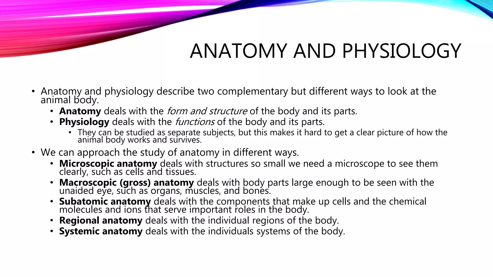
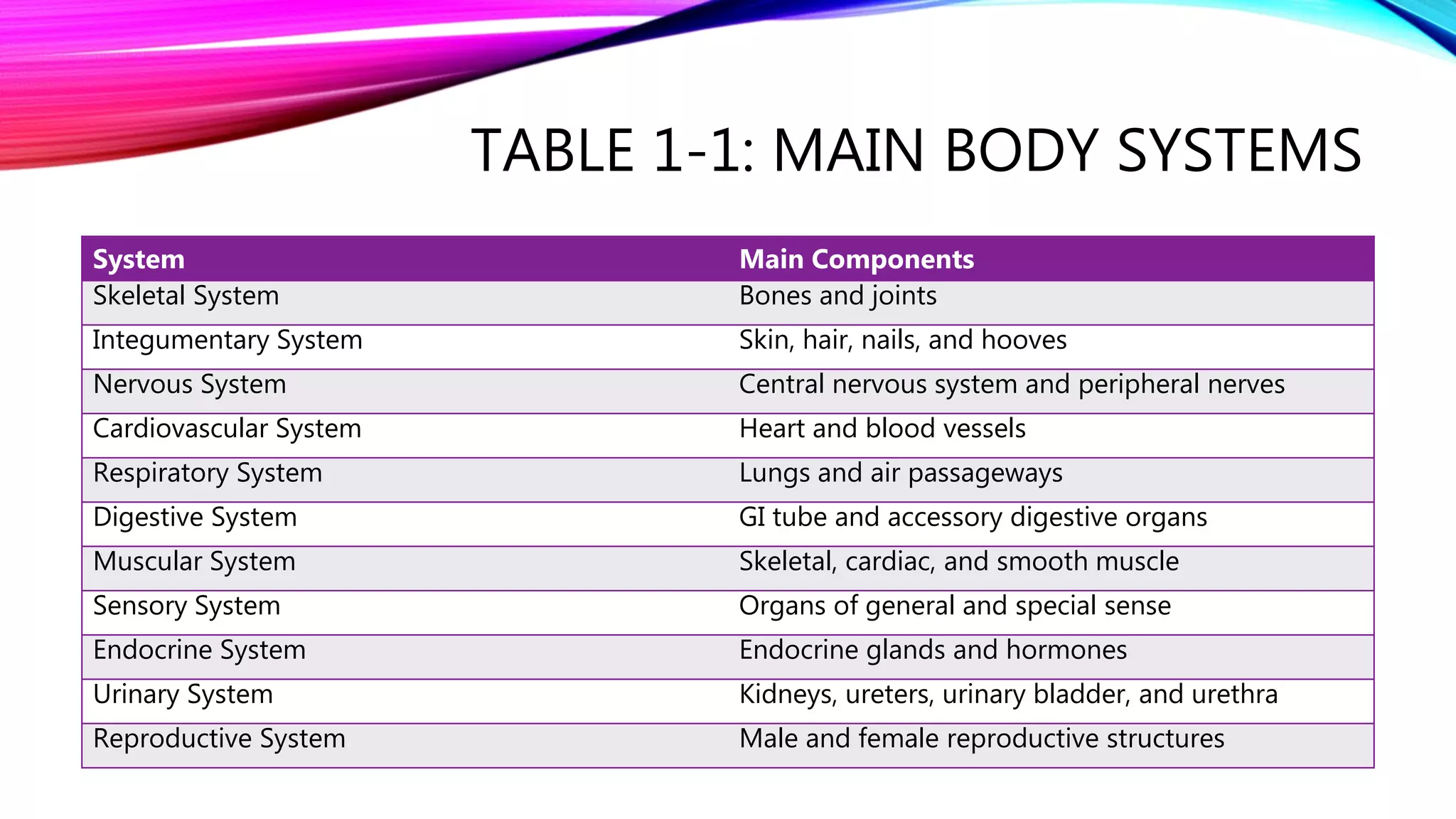
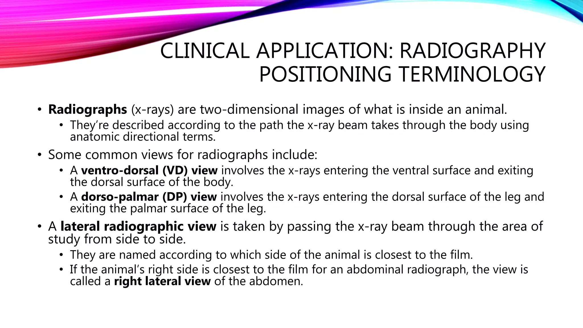
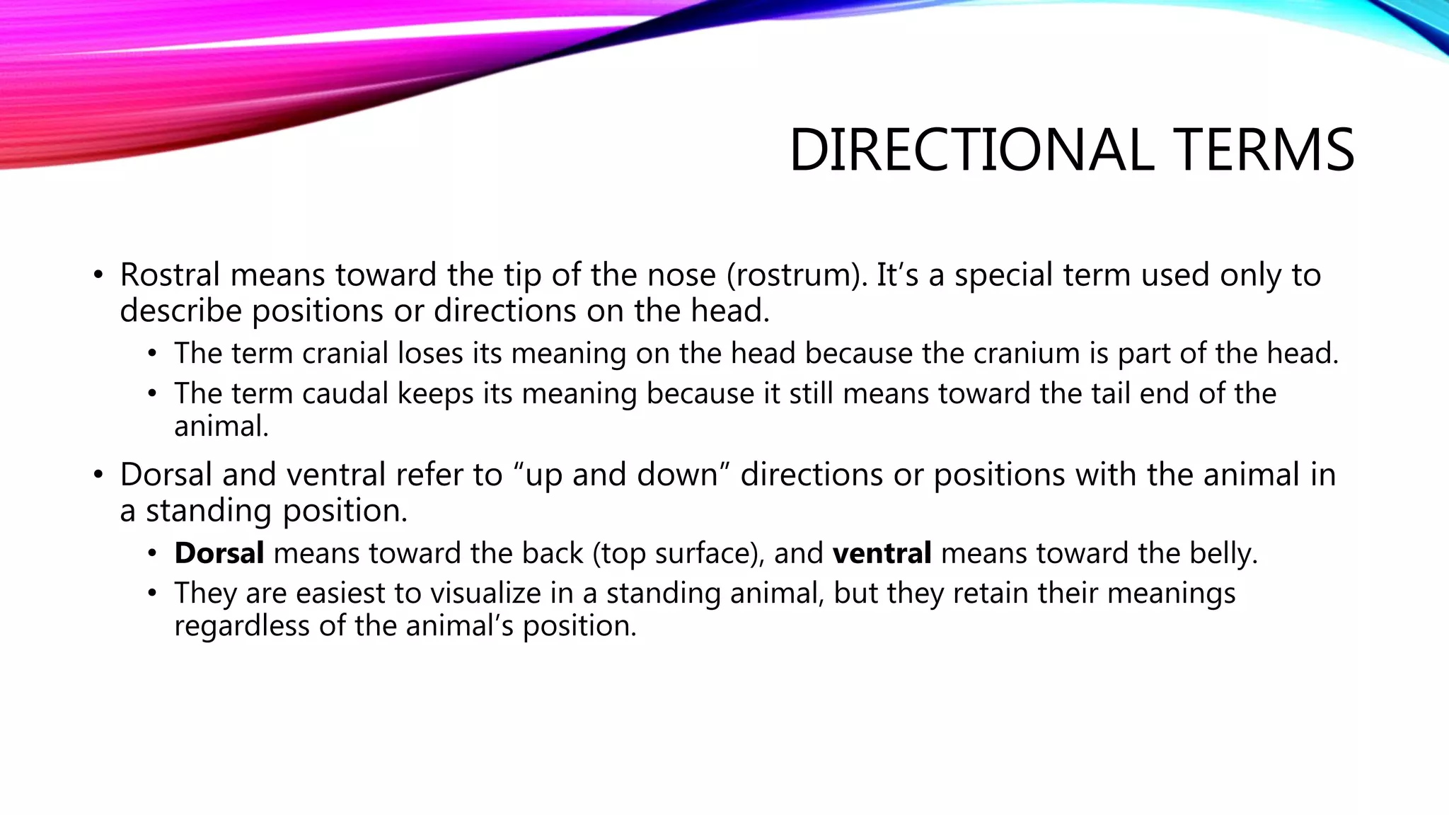
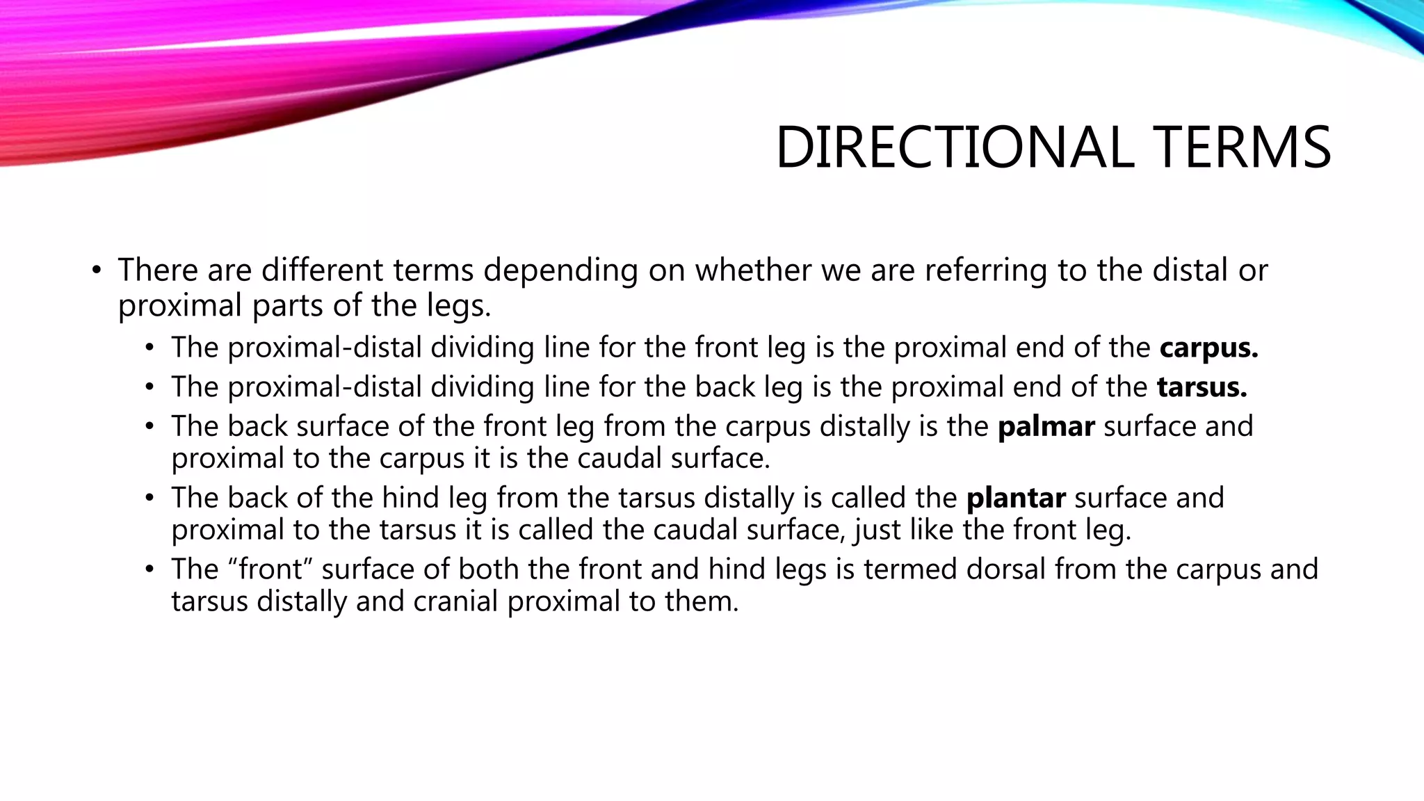
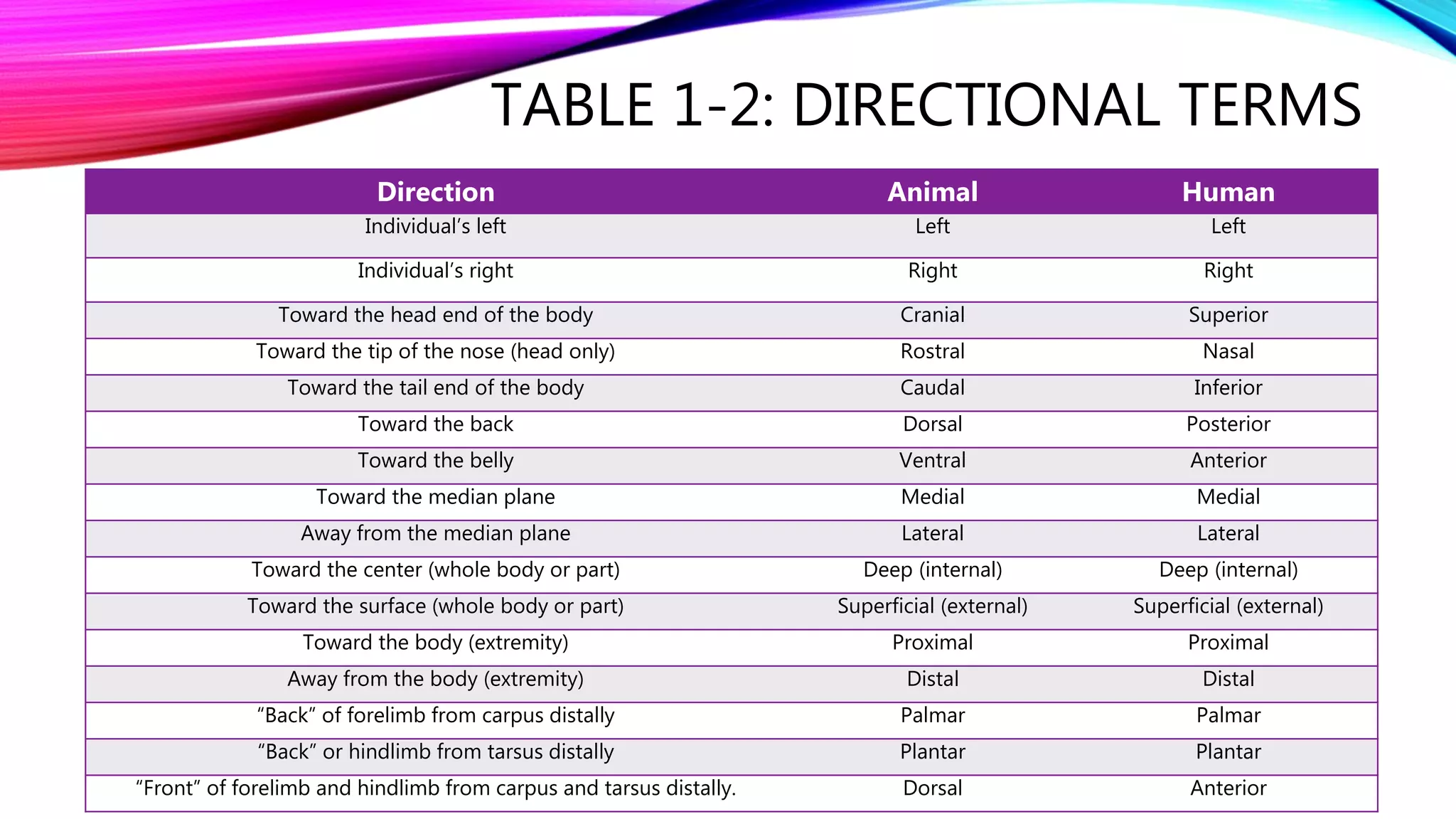
CLINICAL ANATOMY AND
PHYSIOLOGY FOR
VETERINARY TECHNICIANS
Madeline Olson | Medaille College | Semester One
CHAPTER ONE
Introduction to Anatomy and Physiology
INTRODUCTION
• As veterinary health care professionals, we must understand how animals are put together
and how their bodies work.
• The various parts of the body must work in near-perfect harmony to maintain the life and
well-being of an animal.
• The apparently simple and automatic states of life and health are not as they seem. Life is not
simple, and health is not automatic.
• Life is extraordinarily complicated, and health is the result of things going right.
• It seems as though health is the normal state of affairs, and disease and death result from
some awful outside influences attacking the body.
• Outside influences alone usually play smaller roles that we might think.
• Disease and death often result from the absence of normal body structure and functioning.
• Normal anatomy and physiology are critical to an animal’s health and survival.
• Our knowledge of them is critical to our ability to influence the animal’s health and survival in
cases of disease or injury.
ANATOMY AND PHYSIOLOGY
• Anatomy and physiology describe two complementary but different ways to look at the
animal body.
• Anatomy deals with the form and structure of the body and its parts.
• Physiology deals with the functions of the body and its parts.
• They can be studied as separate subjects, but this makes it hard to get a clear picture of how the
animal body works and survives.
• We can approach the study of anatomy in different ways.
• Microscopic anatomy deals with structures so small we need a microscope to see them
clearly, such as cells and tissues.
• Macroscopic (gross) anatomy deals with body parts large enough to be seen with the
unaided eye, such as organs, muscles, and bones.
• Subatomic anatomy deals with the components that make up cells and the chemical
molecules and ions that serve important roles in the body.
• Regional anatomy deals with the individual regions of the body.
• Systemic anatomy deals with the individuals systems of the body.
ANATOMY AND PHYSIOLOGY
• Regional anatomy versus systemic anatomy.
• In the regional approach, all the components of each region of the body are examined.
• For example, the anatomy of the cervical (neck) region would include all the cells, tissues,
blood vessels, nerves, muscles, organs, and bones present in the neck.
• The problem is that the body is not always easy to subdivide this way, and there is often an
overlap between adjacent regions.
• The systemic approach to anatomy deals with the systems of the body as separate
topics.
• The many interrelationships between the body systems can be described as the systems are
examined.
• This lets us look at the whole body by breaking it down into clear, logical components.
TABLE 1-1: MAIN BODY SYSTEMS
System Main Components
Skeletal System Bones and joints
Integumentary System Skin, hair, nails, and hooves
Nervous System Central nervous system and peripheral nerves
Cardiovascular System Heart and blood vessels
Respiratory System Lungs and air passageways
Digestive System GI tube and accessory digestive organs
Muscular System Skeletal, cardiac, and smooth muscle
Sensory System Organs of general and special sense
Endocrine System Endocrine glands and hormones
Urinary System Kidneys, ureters, urinary bladder, and urethra
Reproductive System Male and female reproductive structures
TERMINOLOGY
• To be clear and accurate with descriptions of body parts, we have to use terms that
leave no doubt about their meanings.
• Terms such as up, down, above, below, and beside are not very useful because they
depend on the orientation of the animal. Even the position of the observer can make a
difference in these kinds of terms.
• Anatomic terms must have the same meaning regardless of the orientation of the animal
or the position of the observer.
• Basic anatomic terminology is based on imaginary slices, called planes, through the
animal body that can be used as points or areas of reference and on sets of
directional terms that have opposite meanings from each other.
ANATOMIC PLANES OF REFERENCE
• There are four anatomic planes of reference, two of which are variations of each other. Each
plane is an imaginary slice through the body that is oriented at right angles to the other two.
• The sagittal plane is a plane that runs the length of the body and divides it into left and
right parts that are not necessarily equal halves.
• The median plane is a special kind of sagittal plane that runs down the center of the body
lengthwise and divides it into equal left and right halves.
• It could also be called a midsagittal plane, but that term is not commonly used.
• The transverse plane is a plane across the body that divides it into cranial (head-end) and
caudal (tail-end) parts that are not necessarily equal.
• The dorsal plane is a plane at right angles to the sagittal and transverse planes. It divides the
body into dorsal (back) and ventral (belly) parts that aren’t necessarily equal.
• If an animal stands in water with its body partially submerged, the surface of the water describes a
dorsal plane.
CLINICAL APPLICATION: RADIOGRAPHY
POSITIONING TERMINOLOGY
• Radiographs (x-rays) are two-dimensional images of what is inside an animal.
• They’re described according to the path the x-ray beam takes through the body using
anatomic directional terms.
• Some common views for radiographs include:
• A ventro-dorsal (VD) view involves the x-rays entering the ventral surface and exiting
the dorsal surface of the body.
• A dorso-palmar (DP) view involves the x-rays entering the dorsal surface of the leg and
exiting the palmar surface of the leg.
• A lateral radiographic view is taken by passing the x-ray beam through the area of
study from side to side.
• They are named according to which side of the animal is closest to the film.
• If the animal’s right side is closest to the film for an abdominal radiograph, the view is
called a right lateral view of the abdomen.
DIRECTIONAL TERMS
• Directional terms in anatomy provide a common language for accurately and clearly
describing body structures regardless of the animal’s position.
• These terms generally occur in pairs that have opposite meanings and are used chiefly
to describe relative positions of body parts.
• Since humans walk upright, there are a few differences between human directional
terms and those of nonhuman animals.
• Left and right always refer to the animal’s left and right sides.
• Cranial and caudal refer to the ends of the animal as it stands on four legs.
• Cranial means toward the head, and caudal means toward the tail.
DIRECTIONAL TERMS
• Rostral means toward the tip of the nose (rostrum). It’s a special term used only to
describe positions or directions on the head.
• The term cranial loses its meaning on the head because the cranium is part of the head.
• The term caudal keeps its meaning because it still means toward the tail end of the
animal.
• Dorsal and ventral refer to “up and down” directions or positions with the animal in
a standing position.
• Dorsal means toward the back (top surface), and ventral means toward the belly.
• They are easiest to visualize in a standing animal, but they retain their meanings
regardless of the animal’s position.
DIRECTIONAL TERMS
• Medial and lateral refer to positions relative to the median plane.
• Medial means toward and lateral means away from the median plane.
• Deep (internal) and superficial (external) refer to the position of something relative
to the center or surface of the body or a body part.
• Deep (internal) means toward the center of the body or a body part.
• Superficial (external) means toward the surface of the body or a body part.
• Proximal and distal are used to describe positions only on extremities.
• Proximal means toward the body and distal means away from the body.
• When describing the front and back surfaces of the legs, things get just a little more
complicated.
DIRECTIONAL TERMS
• There are different terms depending on whether we are referring to the distal or
proximal parts of the legs.
• The proximal-distal dividing line for the front leg is the proximal end of the carpus.
• The proximal-distal dividing line for the back leg is the proximal end of the tarsus.
• The back surface of the front leg from the carpus distally is the palmar surface and
proximal to the carpus it is the caudal surface.
• The back of the hind leg from the tarsus distally is called the plantar surface and
proximal to the tarsus it is called the caudal surface, just like the front leg.
• The “front” surface of both the front and hind legs is termed dorsal from the carpus and
tarsus distally and cranial proximal to them.
TABLE 1-2: DIRECTIONAL TERMS
Direction Animal Human
Individual’s left Left Left
Individual’s right Right Right
Toward the head end of the body Cranial Superior
Toward the tip of the nose (head only) Rostral Nasal
Toward the tail end of the body Caudal Inferior
Toward the back Dorsal Posterior
Toward the belly Ventral Anterior
Toward the median plane Medial Medial
Away from the median plane Lateral Lateral
Toward the center (whole body or part) Deep (internal) Deep (internal)
Toward the surface (whole body or part) Superficial (external) Superficial (external)
Toward the body (extremity) Proximal Proximal
Away from the body (extremity) Distal Distal
“Back” of forelimb from carpus distally Palmar Palmar
“Back” or hindlimb from tarsus distally Plantar Plantar
“Front” of forelimb and hindlimb from carpus and tarsus distally. Dorsal Anterior
TABLE 1-2: DIRECTIONAL TERMS
Direction Animal Human
Toward the body (extremity) Proximal Proximal
Away from the body (extremity) Distal Distal
“Back” of forelimb from carpus distally Palmar Palmar
“Back” of hindlimb from tarsus distally Plantar Plantar
“Front” of forelimb and hindlimb from carpus and tarsus distally Dorsal Anterior
COMMON REGIONAL TERMS
• Common regional terms give us a shorthand way of recording anatomic locations in
veterinary records.
TABLE 1-3: COMMON REGIONAL TERMS
Term Region
Barrel Trunk of the body—formed by the rib cage and the abdomen
Brisket Area at the base of the neck between the front legs that covers the cranial end of the sternum
Cannon Large metacarpal or metatarsal bone of hoofed animals
Fetlock Joint between cannon bone (large metacarpal/metatarsal) and the proximal phalanx of hoofed animals
Flank Lateral surface of the abdomen between the last rib and the hind legs
Hock Tarsus
Knee Carpus of hoofed animals
Muzzle Rostral part of the face formed mainly by the maxillary and nasal bones
Pastern Area of the proximal phalanx of hoofed animals
Poll Top of the head between the bases of the ears
Stifle Femorotibial/femoropatellar joint—equivalent to human knee
Tailhead Dorsal part of the base of the tail
Withers Area dorsal to the scapula
BILATERAL SYMMETRY
• Bilateral symmetry means that the left and right halves of an animal’s body are
essentially mirror images of each other.
• Although not absolute, the principle of bilateral symmetry accurately reflects the basic
inner and outer structure of the body.
• Paired structures and internal organs are approximately mirror images.
• Single structures in the body are generally found near the center of the body, near the
median plane. This is true of structures like the brain, heart, and GI tract.
BODY CAVITIES
DORSAL BODY CAVITY
• The dorsal body cavity contains the brain and spinal cord, that is, the central
nervous system.
• It consists of two parts: a somewhat spherical cranial cavity in the skull and a long,
narrow spinal cavity running down the spine.
• The cranial cavity is also known as the cranium. It’s formed from several bones of
the skull, and it houses and protects the brain.
• The spinal cavity is also known as the spinal canal. It’s formed from the vertebrae of
the spine, and it houses and protects the spinal cord.
VENTRAL BODY CAVITY
• The ventral body cavity contains most of the body’s soft organs (viscera).
• It’s divided by the thin diaphragm muscle into the cranial thoracic cavity, also known as the
thorax or chest, and the caudal abdominal cavity, or abdomen.
• Major structures in the thoracic cavity include the heart, lungs, esophagus, and many major
blood vessels coming to and going from the heart.
• All of its organs are covered by a thin membrane called the pleura, and even the cavity itself
is lined with this as well.
• The layer that covers the organs is called the visceral layer of pleura because it lies right on
the viscera (the organs).
• The layer that lines the whole cavity is called the parietal layer of pleura.
• The potential space between the two layers is filled with a small amount of lubricating fluid.
The smooth pleural surfaces are lubricated by the pleural fluid to ensure that the two surface
slide over each other easily during breathing.
• If the pleural surfaces become thickened and roughened by inflammation, a condition called
pleuritis or pleurisy, the surfaces scrape over each other with every breath, making breathing
very painful.
VENTRAL BODY CAVITY
• The abdomen contains the digestive, urinary, and reproductive organs.
• The cavity and its contents are lined by a thin membrane called the peritoneum.
• The visceral layer of peritoneum covers the abdominal organs, and the parietal layer
lines the abdominal cavity.
• A potential space filled with peritoneal fluid separates the two layers.
• Inflammation of the peritoneum (peritonitis) is very painful and most commonly
results either from a wound that penetrates into the abdomen from the outside or
from a rupture or perforation of the GI tract.
• When performing surgery on the digestive tract, we must take care to suture it securely
closed to prevent leakage, which could lead to peritonitis.
CELLS
• Cells are the basic functional units of animal life—the smallest subdivision of the
body that are capable of life.
• A simple, single-celled animal like an ameba has to carry out all the life functions
necessary to support itself within its one cell.
• It must do things such as: grow; respond to positive and negative stimuli; seek out, engulf, and
absorb food; eliminate wastes; and reproduce.
• It has no ability to influences its environment and has to take things as they come.
• If environmental conditions are favorable, the ameba survives. If not, it dies.
• In complex animals, the body’s cells must divide the work.
• The body must create and support an internal environment that allows all of its cells to live
and function.
• To do this, cells must specialize in some functions and eliminate others.
• The body’s cells depend on each other to ensure the survival of the animal overall.
TISSUES
• When specialized cells group together, they form tissues.
• The entire animal body is made up of only four basic tissues: epithelial, connective,
muscle, and nervous tissue.
• Epithelial tissue is composed entirely of cells, and its main jobs are to cover body
surfaces, secrete materials, and absorb materials.
• The surface of the skin is covered by epithelium, as are the linings of the mouth,
intestine, and urinary bladder.
• Epithelial tissue also forms glands, which are structures that secrete useful substances
and excrete wastes.
• The secreting units of sweat, salivary, and mammary glands are all composed of
specialized epithelial tissue.
TISSUES
• Connective tissue supports and holds the body together (connects its cells).
• Cells are very soft and cannot support themselves without outside help.
• They range from very soft (adipose tissue) to very firm (cartilage or bone).
• Connective tissues are composed of cells and a variety of nonliving intercellular substances,
such as fibers, that add strength.
• Muscle tissue moves the body inside and out, and exists as three types.
• Skeletal muscle moves the bones of the skeleton and is under conscious control.
• Cardiac muscle makes up the heart and works “automatically”.
• Smooth muscle is found in internal organs and mostly works automatically.
• Nervous tissue transmits information around the body and controls body functions.
• It transmits sensory information from the body to the brain, processes the information, and
sends instructions out to tell the body how to react to changing conditions.
ORGANS AND ORGAN SYSTEMS
• Organs are made up of groups of tissues that work together for common purposes.
• For example, the kidney is an organ composed of various tissues that function together
to eliminate wastes from the body.
• Some organs, such as the eyes, lungs, and kidneys, occur in pairs.
• Other organs, such as the brain, heart, and uterus, are single structures.
• Organ systems are groups of organs that are involved in a common set of activities.
• For example, the digestive system is concerned with obtaining, digesting, and absorbing
nutrients to fuel the rest of the body.
• It’s composed of the organs that make up the digestive tube, as well as accessory
digestive organs.
TABLE 1-4: BODY TISSUES
Tissue Characteristics Functions
Epithelial Composed only of cells Covers and protects (surfaces)
Secretes (glands)
Absorbs (intestinal lining)
Connective Composed of living cells and non-living
intercellular substances
Binds cells and structures together and
supports the body
Muscle Skeletal (voluntary) Movements
Cardiac (heart)
Smooth (involuntary)
Nervous Composed of neurons and supporting cells Transmits information around body;
coordinates and controls activities
HEALTH
• The term health has a lot of meanings.
• The simplest way to think of it is as a state of normal anatomy and physiology.
• When the structures or functions of the body become abnormal, disease results.
• Maintaining health is a complicated process.
• The health of the body as a whole depends on the health and proper functioning of
each of its systems, organs, tissues, and cells.
• Each of the body’s cells depends on the health and proper functioning of all the tissues,
organs, systems, and the body as a whole.
• All structures and functions in the body are interrelated; nothing takes place in isolation.
HOMEOSTASIS
• Homeostasis is the maintenance of a dynamic equilibrium in the body.
• The word dynamic implies activity, energy, and work, and equilibrium refers to balance.
Together they summarize all the physiological processes that actively maintain balance
in the various structures, functions, and properties of the body.
• The processes that monitor and adjust all the various essential parameters of the body
are summarized by the term homeostasis.
• An animal’s body temperature cannot vary more than a few degrees from either
side of the normal range without starting to interfere with other body functions.
• The acid-base balance, fluid balance, hormone levels, nutrient levels, and oxygen levels
cannot vary by much if the body is to operate normally. They must be kept within fairly
narrow operational ranges.
HOMEOSTASIS
• The whole body is responsible for homeostasis.
• All the body systems are involved in the many mechanisms of homeostasis, which require
lots of energy and work.
• The various homeostatic mechanisms in the body keep it functioning amid the twists and
turns of life.
• The processes of homeostasis help maintain a fairly constant internal environment in
the body as conditions inside and outside the animal change.
• Along with normal functioning of the body’s cells, tissues, organs, and systems, the
processes of homeostasis make life possible.
CLINICAL APPLICATION:
HOMEOSTASIS AND CONGESTIVE HEART FAILURE
• Congestive heart failure is a clinical term used to describe a hear that is not
pumping adequate amounts of blood.
• This results in blood “backing up” in the body, which produces congestion, or abnormal
fluid accumulation, upstream from the failing heart.
• There are many causes and forms of congestive heart failure, but the overall
homeostatic mechanisms that attempt to maintain normal blood circulation in the body
are basically the same.
• The first indication that the heart is starting to fail is a drop in the cardiac output,
that is, the amount of blood the heart pumps out per minute.
• The decreased blood flow and pressure are picked up by receptors in the vascular
system and relayed to the central nervous system.
• Signals then go out to activate the sympathetic portion of the nervous system, or the
fight-or-flight system.
CLINICAL APPLICATION:
HOMEOSTASIS AND CONGESTIVE HEART FAILURE
• The fight-or-flight system helps prepare the body for intense physical activity.
• Its effect on the cardiovascular system is to increase blood flow and pressure by stimulating
the heart to beat harder and faster and by constricting blood vessels.
• In short term, these mechanisms help bring blood flow and pressure back to normal.
• These compensatory mechanisms cause the weak heart to work harder.
• The result is a further weakening of the heart and further decreases in cardiac output.
• This causes more sympathetic nervous system stimulation.
• The cycle continues to repeat until either the heart gives up completely or we intervene with
medical therapy.
• Homeostatic mechanisms cannot change the basic defects that are causing the heart to
fail, but they help the damaged heart to maintain vital blood flow to the rest of the body
for as long as possible.
• By adding good medical care to the body’s natural homeostatic mechanisms, we can often
extend the length and quality of life of an animal with this heart failure.

Clinical Anatomy and Physiology for Veterinary Science: Chapter One

  • 1.
    CLINICAL ANATOMY AND PHYSIOLOGYFOR VETERINARY TECHNICIANS Madeline Olson | Medaille College | Semester One
  • 2.
    CHAPTER ONE Introduction toAnatomy and Physiology
  • 3.
    INTRODUCTION • As veterinaryhealth care professionals, we must understand how animals are put together and how their bodies work. • The various parts of the body must work in near-perfect harmony to maintain the life and well-being of an animal. • The apparently simple and automatic states of life and health are not as they seem. Life is not simple, and health is not automatic. • Life is extraordinarily complicated, and health is the result of things going right. • It seems as though health is the normal state of affairs, and disease and death result from some awful outside influences attacking the body. • Outside influences alone usually play smaller roles that we might think. • Disease and death often result from the absence of normal body structure and functioning. • Normal anatomy and physiology are critical to an animal’s health and survival. • Our knowledge of them is critical to our ability to influence the animal’s health and survival in cases of disease or injury.
  • 4.
    ANATOMY AND PHYSIOLOGY •Anatomy and physiology describe two complementary but different ways to look at the animal body. • Anatomy deals with the form and structure of the body and its parts. • Physiology deals with the functions of the body and its parts. • They can be studied as separate subjects, but this makes it hard to get a clear picture of how the animal body works and survives. • We can approach the study of anatomy in different ways. • Microscopic anatomy deals with structures so small we need a microscope to see them clearly, such as cells and tissues. • Macroscopic (gross) anatomy deals with body parts large enough to be seen with the unaided eye, such as organs, muscles, and bones. • Subatomic anatomy deals with the components that make up cells and the chemical molecules and ions that serve important roles in the body. • Regional anatomy deals with the individual regions of the body. • Systemic anatomy deals with the individuals systems of the body.
  • 5.
    ANATOMY AND PHYSIOLOGY •Regional anatomy versus systemic anatomy. • In the regional approach, all the components of each region of the body are examined. • For example, the anatomy of the cervical (neck) region would include all the cells, tissues, blood vessels, nerves, muscles, organs, and bones present in the neck. • The problem is that the body is not always easy to subdivide this way, and there is often an overlap between adjacent regions. • The systemic approach to anatomy deals with the systems of the body as separate topics. • The many interrelationships between the body systems can be described as the systems are examined. • This lets us look at the whole body by breaking it down into clear, logical components.
  • 6.
    TABLE 1-1: MAINBODY SYSTEMS System Main Components Skeletal System Bones and joints Integumentary System Skin, hair, nails, and hooves Nervous System Central nervous system and peripheral nerves Cardiovascular System Heart and blood vessels Respiratory System Lungs and air passageways Digestive System GI tube and accessory digestive organs Muscular System Skeletal, cardiac, and smooth muscle Sensory System Organs of general and special sense Endocrine System Endocrine glands and hormones Urinary System Kidneys, ureters, urinary bladder, and urethra Reproductive System Male and female reproductive structures
  • 7.
    TERMINOLOGY • To beclear and accurate with descriptions of body parts, we have to use terms that leave no doubt about their meanings. • Terms such as up, down, above, below, and beside are not very useful because they depend on the orientation of the animal. Even the position of the observer can make a difference in these kinds of terms. • Anatomic terms must have the same meaning regardless of the orientation of the animal or the position of the observer. • Basic anatomic terminology is based on imaginary slices, called planes, through the animal body that can be used as points or areas of reference and on sets of directional terms that have opposite meanings from each other.
  • 8.
    ANATOMIC PLANES OFREFERENCE • There are four anatomic planes of reference, two of which are variations of each other. Each plane is an imaginary slice through the body that is oriented at right angles to the other two. • The sagittal plane is a plane that runs the length of the body and divides it into left and right parts that are not necessarily equal halves. • The median plane is a special kind of sagittal plane that runs down the center of the body lengthwise and divides it into equal left and right halves. • It could also be called a midsagittal plane, but that term is not commonly used. • The transverse plane is a plane across the body that divides it into cranial (head-end) and caudal (tail-end) parts that are not necessarily equal. • The dorsal plane is a plane at right angles to the sagittal and transverse planes. It divides the body into dorsal (back) and ventral (belly) parts that aren’t necessarily equal. • If an animal stands in water with its body partially submerged, the surface of the water describes a dorsal plane.
  • 9.
    CLINICAL APPLICATION: RADIOGRAPHY POSITIONINGTERMINOLOGY • Radiographs (x-rays) are two-dimensional images of what is inside an animal. • They’re described according to the path the x-ray beam takes through the body using anatomic directional terms. • Some common views for radiographs include: • A ventro-dorsal (VD) view involves the x-rays entering the ventral surface and exiting the dorsal surface of the body. • A dorso-palmar (DP) view involves the x-rays entering the dorsal surface of the leg and exiting the palmar surface of the leg. • A lateral radiographic view is taken by passing the x-ray beam through the area of study from side to side. • They are named according to which side of the animal is closest to the film. • If the animal’s right side is closest to the film for an abdominal radiograph, the view is called a right lateral view of the abdomen.
  • 11.
    DIRECTIONAL TERMS • Directionalterms in anatomy provide a common language for accurately and clearly describing body structures regardless of the animal’s position. • These terms generally occur in pairs that have opposite meanings and are used chiefly to describe relative positions of body parts. • Since humans walk upright, there are a few differences between human directional terms and those of nonhuman animals. • Left and right always refer to the animal’s left and right sides. • Cranial and caudal refer to the ends of the animal as it stands on four legs. • Cranial means toward the head, and caudal means toward the tail.
  • 12.
    DIRECTIONAL TERMS • Rostralmeans toward the tip of the nose (rostrum). It’s a special term used only to describe positions or directions on the head. • The term cranial loses its meaning on the head because the cranium is part of the head. • The term caudal keeps its meaning because it still means toward the tail end of the animal. • Dorsal and ventral refer to “up and down” directions or positions with the animal in a standing position. • Dorsal means toward the back (top surface), and ventral means toward the belly. • They are easiest to visualize in a standing animal, but they retain their meanings regardless of the animal’s position.
  • 13.
    DIRECTIONAL TERMS • Medialand lateral refer to positions relative to the median plane. • Medial means toward and lateral means away from the median plane. • Deep (internal) and superficial (external) refer to the position of something relative to the center or surface of the body or a body part. • Deep (internal) means toward the center of the body or a body part. • Superficial (external) means toward the surface of the body or a body part. • Proximal and distal are used to describe positions only on extremities. • Proximal means toward the body and distal means away from the body. • When describing the front and back surfaces of the legs, things get just a little more complicated.
  • 14.
    DIRECTIONAL TERMS • Thereare different terms depending on whether we are referring to the distal or proximal parts of the legs. • The proximal-distal dividing line for the front leg is the proximal end of the carpus. • The proximal-distal dividing line for the back leg is the proximal end of the tarsus. • The back surface of the front leg from the carpus distally is the palmar surface and proximal to the carpus it is the caudal surface. • The back of the hind leg from the tarsus distally is called the plantar surface and proximal to the tarsus it is called the caudal surface, just like the front leg. • The “front” surface of both the front and hind legs is termed dorsal from the carpus and tarsus distally and cranial proximal to them.
  • 15.
    TABLE 1-2: DIRECTIONALTERMS Direction Animal Human Individual’s left Left Left Individual’s right Right Right Toward the head end of the body Cranial Superior Toward the tip of the nose (head only) Rostral Nasal Toward the tail end of the body Caudal Inferior Toward the back Dorsal Posterior Toward the belly Ventral Anterior Toward the median plane Medial Medial Away from the median plane Lateral Lateral Toward the center (whole body or part) Deep (internal) Deep (internal) Toward the surface (whole body or part) Superficial (external) Superficial (external) Toward the body (extremity) Proximal Proximal Away from the body (extremity) Distal Distal “Back” of forelimb from carpus distally Palmar Palmar “Back” or hindlimb from tarsus distally Plantar Plantar “Front” of forelimb and hindlimb from carpus and tarsus distally. Dorsal Anterior
  • 16.
    TABLE 1-2: DIRECTIONALTERMS Direction Animal Human Toward the body (extremity) Proximal Proximal Away from the body (extremity) Distal Distal “Back” of forelimb from carpus distally Palmar Palmar “Back” of hindlimb from tarsus distally Plantar Plantar “Front” of forelimb and hindlimb from carpus and tarsus distally Dorsal Anterior
  • 17.
    COMMON REGIONAL TERMS •Common regional terms give us a shorthand way of recording anatomic locations in veterinary records.
  • 18.
    TABLE 1-3: COMMONREGIONAL TERMS Term Region Barrel Trunk of the body—formed by the rib cage and the abdomen Brisket Area at the base of the neck between the front legs that covers the cranial end of the sternum Cannon Large metacarpal or metatarsal bone of hoofed animals Fetlock Joint between cannon bone (large metacarpal/metatarsal) and the proximal phalanx of hoofed animals Flank Lateral surface of the abdomen between the last rib and the hind legs Hock Tarsus Knee Carpus of hoofed animals Muzzle Rostral part of the face formed mainly by the maxillary and nasal bones Pastern Area of the proximal phalanx of hoofed animals Poll Top of the head between the bases of the ears Stifle Femorotibial/femoropatellar joint—equivalent to human knee Tailhead Dorsal part of the base of the tail Withers Area dorsal to the scapula
  • 19.
    BILATERAL SYMMETRY • Bilateralsymmetry means that the left and right halves of an animal’s body are essentially mirror images of each other. • Although not absolute, the principle of bilateral symmetry accurately reflects the basic inner and outer structure of the body. • Paired structures and internal organs are approximately mirror images. • Single structures in the body are generally found near the center of the body, near the median plane. This is true of structures like the brain, heart, and GI tract.
  • 20.
  • 21.
    DORSAL BODY CAVITY •The dorsal body cavity contains the brain and spinal cord, that is, the central nervous system. • It consists of two parts: a somewhat spherical cranial cavity in the skull and a long, narrow spinal cavity running down the spine. • The cranial cavity is also known as the cranium. It’s formed from several bones of the skull, and it houses and protects the brain. • The spinal cavity is also known as the spinal canal. It’s formed from the vertebrae of the spine, and it houses and protects the spinal cord.
  • 22.
    VENTRAL BODY CAVITY •The ventral body cavity contains most of the body’s soft organs (viscera). • It’s divided by the thin diaphragm muscle into the cranial thoracic cavity, also known as the thorax or chest, and the caudal abdominal cavity, or abdomen. • Major structures in the thoracic cavity include the heart, lungs, esophagus, and many major blood vessels coming to and going from the heart. • All of its organs are covered by a thin membrane called the pleura, and even the cavity itself is lined with this as well. • The layer that covers the organs is called the visceral layer of pleura because it lies right on the viscera (the organs). • The layer that lines the whole cavity is called the parietal layer of pleura. • The potential space between the two layers is filled with a small amount of lubricating fluid. The smooth pleural surfaces are lubricated by the pleural fluid to ensure that the two surface slide over each other easily during breathing. • If the pleural surfaces become thickened and roughened by inflammation, a condition called pleuritis or pleurisy, the surfaces scrape over each other with every breath, making breathing very painful.
  • 23.
    VENTRAL BODY CAVITY •The abdomen contains the digestive, urinary, and reproductive organs. • The cavity and its contents are lined by a thin membrane called the peritoneum. • The visceral layer of peritoneum covers the abdominal organs, and the parietal layer lines the abdominal cavity. • A potential space filled with peritoneal fluid separates the two layers. • Inflammation of the peritoneum (peritonitis) is very painful and most commonly results either from a wound that penetrates into the abdomen from the outside or from a rupture or perforation of the GI tract. • When performing surgery on the digestive tract, we must take care to suture it securely closed to prevent leakage, which could lead to peritonitis.
  • 24.
    CELLS • Cells arethe basic functional units of animal life—the smallest subdivision of the body that are capable of life. • A simple, single-celled animal like an ameba has to carry out all the life functions necessary to support itself within its one cell. • It must do things such as: grow; respond to positive and negative stimuli; seek out, engulf, and absorb food; eliminate wastes; and reproduce. • It has no ability to influences its environment and has to take things as they come. • If environmental conditions are favorable, the ameba survives. If not, it dies. • In complex animals, the body’s cells must divide the work. • The body must create and support an internal environment that allows all of its cells to live and function. • To do this, cells must specialize in some functions and eliminate others. • The body’s cells depend on each other to ensure the survival of the animal overall.
  • 25.
    TISSUES • When specializedcells group together, they form tissues. • The entire animal body is made up of only four basic tissues: epithelial, connective, muscle, and nervous tissue. • Epithelial tissue is composed entirely of cells, and its main jobs are to cover body surfaces, secrete materials, and absorb materials. • The surface of the skin is covered by epithelium, as are the linings of the mouth, intestine, and urinary bladder. • Epithelial tissue also forms glands, which are structures that secrete useful substances and excrete wastes. • The secreting units of sweat, salivary, and mammary glands are all composed of specialized epithelial tissue.
  • 26.
    TISSUES • Connective tissuesupports and holds the body together (connects its cells). • Cells are very soft and cannot support themselves without outside help. • They range from very soft (adipose tissue) to very firm (cartilage or bone). • Connective tissues are composed of cells and a variety of nonliving intercellular substances, such as fibers, that add strength. • Muscle tissue moves the body inside and out, and exists as three types. • Skeletal muscle moves the bones of the skeleton and is under conscious control. • Cardiac muscle makes up the heart and works “automatically”. • Smooth muscle is found in internal organs and mostly works automatically. • Nervous tissue transmits information around the body and controls body functions. • It transmits sensory information from the body to the brain, processes the information, and sends instructions out to tell the body how to react to changing conditions.
  • 27.
    ORGANS AND ORGANSYSTEMS • Organs are made up of groups of tissues that work together for common purposes. • For example, the kidney is an organ composed of various tissues that function together to eliminate wastes from the body. • Some organs, such as the eyes, lungs, and kidneys, occur in pairs. • Other organs, such as the brain, heart, and uterus, are single structures. • Organ systems are groups of organs that are involved in a common set of activities. • For example, the digestive system is concerned with obtaining, digesting, and absorbing nutrients to fuel the rest of the body. • It’s composed of the organs that make up the digestive tube, as well as accessory digestive organs.
  • 28.
    TABLE 1-4: BODYTISSUES Tissue Characteristics Functions Epithelial Composed only of cells Covers and protects (surfaces) Secretes (glands) Absorbs (intestinal lining) Connective Composed of living cells and non-living intercellular substances Binds cells and structures together and supports the body Muscle Skeletal (voluntary) Movements Cardiac (heart) Smooth (involuntary) Nervous Composed of neurons and supporting cells Transmits information around body; coordinates and controls activities
  • 29.
    HEALTH • The termhealth has a lot of meanings. • The simplest way to think of it is as a state of normal anatomy and physiology. • When the structures or functions of the body become abnormal, disease results. • Maintaining health is a complicated process. • The health of the body as a whole depends on the health and proper functioning of each of its systems, organs, tissues, and cells. • Each of the body’s cells depends on the health and proper functioning of all the tissues, organs, systems, and the body as a whole. • All structures and functions in the body are interrelated; nothing takes place in isolation.
  • 30.
    HOMEOSTASIS • Homeostasis isthe maintenance of a dynamic equilibrium in the body. • The word dynamic implies activity, energy, and work, and equilibrium refers to balance. Together they summarize all the physiological processes that actively maintain balance in the various structures, functions, and properties of the body. • The processes that monitor and adjust all the various essential parameters of the body are summarized by the term homeostasis. • An animal’s body temperature cannot vary more than a few degrees from either side of the normal range without starting to interfere with other body functions. • The acid-base balance, fluid balance, hormone levels, nutrient levels, and oxygen levels cannot vary by much if the body is to operate normally. They must be kept within fairly narrow operational ranges.
  • 31.
    HOMEOSTASIS • The wholebody is responsible for homeostasis. • All the body systems are involved in the many mechanisms of homeostasis, which require lots of energy and work. • The various homeostatic mechanisms in the body keep it functioning amid the twists and turns of life. • The processes of homeostasis help maintain a fairly constant internal environment in the body as conditions inside and outside the animal change. • Along with normal functioning of the body’s cells, tissues, organs, and systems, the processes of homeostasis make life possible.
  • 32.
    CLINICAL APPLICATION: HOMEOSTASIS ANDCONGESTIVE HEART FAILURE • Congestive heart failure is a clinical term used to describe a hear that is not pumping adequate amounts of blood. • This results in blood “backing up” in the body, which produces congestion, or abnormal fluid accumulation, upstream from the failing heart. • There are many causes and forms of congestive heart failure, but the overall homeostatic mechanisms that attempt to maintain normal blood circulation in the body are basically the same. • The first indication that the heart is starting to fail is a drop in the cardiac output, that is, the amount of blood the heart pumps out per minute. • The decreased blood flow and pressure are picked up by receptors in the vascular system and relayed to the central nervous system. • Signals then go out to activate the sympathetic portion of the nervous system, or the fight-or-flight system.
  • 33.
    CLINICAL APPLICATION: HOMEOSTASIS ANDCONGESTIVE HEART FAILURE • The fight-or-flight system helps prepare the body for intense physical activity. • Its effect on the cardiovascular system is to increase blood flow and pressure by stimulating the heart to beat harder and faster and by constricting blood vessels. • In short term, these mechanisms help bring blood flow and pressure back to normal. • These compensatory mechanisms cause the weak heart to work harder. • The result is a further weakening of the heart and further decreases in cardiac output. • This causes more sympathetic nervous system stimulation. • The cycle continues to repeat until either the heart gives up completely or we intervene with medical therapy. • Homeostatic mechanisms cannot change the basic defects that are causing the heart to fail, but they help the damaged heart to maintain vital blood flow to the rest of the body for as long as possible. • By adding good medical care to the body’s natural homeostatic mechanisms, we can often extend the length and quality of life of an animal with this heart failure.