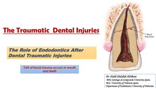
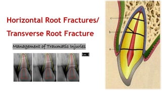
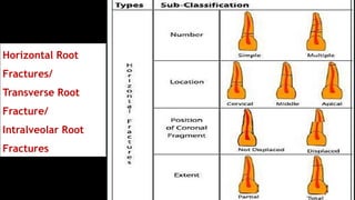
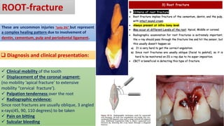
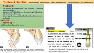
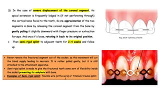
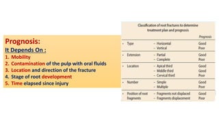
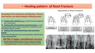
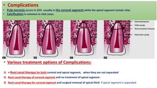
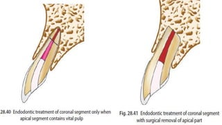
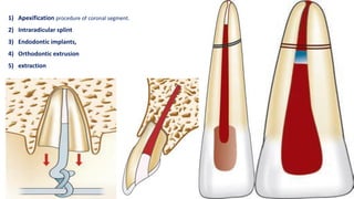
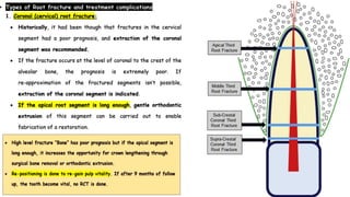
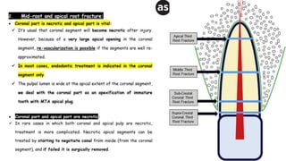
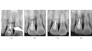
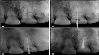
The Traumatic Dental Injuries
The Role of Endodontics After
Dental Traumatic Injuries
Dr. Hadil Abdallah Altilbani
BDS Santiago de Compostela University Spain.
MSc. University of Valencia Spain.
Department of Endodontics University of Palestine .
%50 of facial trauma occurs in mouth
and teeth
Horizontal Root Fractures/
Transverse Root Fracture
Horizontal Root
Fractures/
Transverse Root
Fracture/
Intralveolar Root
Fractures
ROOT-fracture
These are uncommon injuries “only 3%” but represent
a complex healing pattern due to involvement of
dentin, cementum, pulp and periodontal ligament.
❑ Diagnosis and clinical presentation:
✓ Clinical mobility of the tooth
✓ Displacement of the coronal segment:
(no mobility ‘apical fracture’ to extensive
mobility ‘’cervical fracture’).
✓ Palpation tenderness over the root
✓ Radiographic evidence:
Since root fractures are usually oblique, 3 angled
x-rays(45, 90, 110 degrees) to be taken
✓ Pain on bitting
✓ Sulcular bleeding
Palpating the facial mucosa with one finger and moving
crown with other finger;
Root fracture can be felt by placing finger on mucosa
over tooth and moving the crown
Arc of mobility of incisal segment of tooth with root. As fractur
e moves incisally, arc of mobility increases
• Treatment objectives : Repositioning And Splinting Of Root Segments Into Close Proximity As Much As Possible
• Treatment:
• No Mobility, No Displacement …..No Treatment ……spontane
ous Healing 80%
• Displacement And Mobility ……. Repositioning And Splinting.
• Splinting period: 2-4 weeks
• Splint type : flexible splint to allow functional movement
and avoid
ANKYLOSIS
• follow up :
3,6,12 months and yearly for 5 years
Prognosis:
It Depends On :
1. Mobility
2. Contamination of the pulp with oral fluids
3. Location and direction of the fracture
4. Stage of root development
5. Time elapsed since injury
According to the Andreasen and Hjorting—Hansen,
root fracture can show healing in following ways:
1) Healing with calcified tissue in which fractured fragments
are in close contact
2) Healing with interproximal connective tissue in which
radiographically fragments appear separated by a radiolucent line and
the fragment edges are rounded.
1) Healing with interproximal bone and connective
tissues.
Here fractured fragments are seen separated by a distinct bony
bridge radiographically.
All the 3 types considered success
✓ Interproximal inflammatory tissue without healing,
radiographically it shows widening of fracture line
• Healing pattern of Root Fracture
• Complications
• Pulp necrosis occurs in 25% usually in the coronal segment while the apical segment remain vital.
• Calcification is common in vital cases.
• Various treatment options of Complications:
1) • Root canal therapy for both coronal and apical segment, when they are not separated
2) Root canal therapy of coronal segment and no treatment of apical segment .
3) Root canal therapy for coronal segment and surgical removal of apical third if apical segment is separated
1) Apexification procedure of coronal segment.
2) Intraradicular splint
3) Endodontic implants,
4) Orthodontic extrusion
5) extraction
horizontal fracture .pdf

horizontal fracture .pdf

  • 1.
    The Traumatic DentalInjuries The Role of Endodontics After Dental Traumatic Injuries Dr. Hadil Abdallah Altilbani BDS Santiago de Compostela University Spain. MSc. University of Valencia Spain. Department of Endodontics University of Palestine . %50 of facial trauma occurs in mouth and teeth
  • 2.
  • 3.
  • 4.
    ROOT-fracture These are uncommoninjuries “only 3%” but represent a complex healing pattern due to involvement of dentin, cementum, pulp and periodontal ligament. ❑ Diagnosis and clinical presentation: ✓ Clinical mobility of the tooth ✓ Displacement of the coronal segment: (no mobility ‘apical fracture’ to extensive mobility ‘’cervical fracture’). ✓ Palpation tenderness over the root ✓ Radiographic evidence: Since root fractures are usually oblique, 3 angled x-rays(45, 90, 110 degrees) to be taken ✓ Pain on bitting ✓ Sulcular bleeding
  • 5.
    Palpating the facialmucosa with one finger and moving crown with other finger; Root fracture can be felt by placing finger on mucosa over tooth and moving the crown
  • 6.
    Arc of mobilityof incisal segment of tooth with root. As fractur e moves incisally, arc of mobility increases
  • 8.
    • Treatment objectives: Repositioning And Splinting Of Root Segments Into Close Proximity As Much As Possible • Treatment: • No Mobility, No Displacement …..No Treatment ……spontane ous Healing 80% • Displacement And Mobility ……. Repositioning And Splinting. • Splinting period: 2-4 weeks • Splint type : flexible splint to allow functional movement and avoid ANKYLOSIS • follow up : 3,6,12 months and yearly for 5 years
  • 10.
    Prognosis: It Depends On: 1. Mobility 2. Contamination of the pulp with oral fluids 3. Location and direction of the fracture 4. Stage of root development 5. Time elapsed since injury
  • 11.
    According to theAndreasen and Hjorting—Hansen, root fracture can show healing in following ways: 1) Healing with calcified tissue in which fractured fragments are in close contact 2) Healing with interproximal connective tissue in which radiographically fragments appear separated by a radiolucent line and the fragment edges are rounded. 1) Healing with interproximal bone and connective tissues. Here fractured fragments are seen separated by a distinct bony bridge radiographically. All the 3 types considered success ✓ Interproximal inflammatory tissue without healing, radiographically it shows widening of fracture line • Healing pattern of Root Fracture
  • 12.
    • Complications • Pulpnecrosis occurs in 25% usually in the coronal segment while the apical segment remain vital. • Calcification is common in vital cases. • Various treatment options of Complications: 1) • Root canal therapy for both coronal and apical segment, when they are not separated 2) Root canal therapy of coronal segment and no treatment of apical segment . 3) Root canal therapy for coronal segment and surgical removal of apical third if apical segment is separated
  • 14.
    1) Apexification procedureof coronal segment. 2) Intraradicular splint 3) Endodontic implants, 4) Orthodontic extrusion 5) extraction