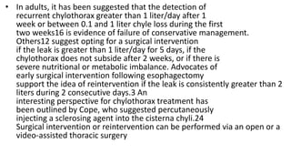
Downloaded 20 times

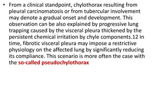


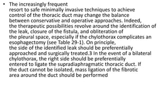
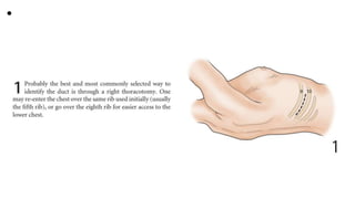
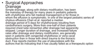
![• Complications of pleurovenous shunts
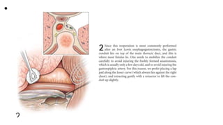
• Pulmonary edema and development of respiratory distress syndrome is reported as an
early complication due to fluid overload in the early post shunt replacement period.[6]
• Post shunt coagulopathy is reported in up to 5% of patients following shunt placement.
Coagulopathy is presumed to be initiated by a large amount of tissue plasminogen factors
being introduced to the blood from the peritoneal or the pleural fluid.[6]
• Infection is reported to have a lower incidence due to use of prophylactic antibiotics and
wound irrigation with aminoglycosides.[6]
• Deep vein thrombosis involving the upper extremity, ipsilateral to the shunt insertion has
also been reported.
• Shunt failure is the most common complication and is reported in 10% to 15% of the
patients. Failure can be due to mal-positioning of the venous tip of the shunt or
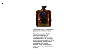
mechanical obstruction such as a kink or a thrombus.[6] The main cause of Denver shunt
occlusion is the accumulation of fibrin and cellular debris that often get impacted after
each manual compression.[10]
• Air embolism as a complication is uncommon and has been reported in the literature. To
prevent air embolism, pleural or peritoneal fluid should not be aspirated to its fullest
extent and a residue amount should always be left in pleural or peritoneal cavity.[9]
• Although catheter leakage is a known entity, the leakage encountered in our patient led to
the formation of a collection within the soft tissue that presented as a neck mass.](https://image.slidesharecdn.com/chylothorax-220309201650/85/Chylothorax-42-320.jpg)
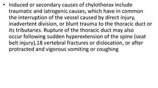
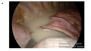
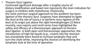
![• Clinical features
• The first sign of development of post-operative chylothorax is pleural fluid
turning milky white in the chest tube. Sometimes chylothorax is serous,
sanguineous or bloody [12]. It can develop from the 1st day up to 24th
day after surgery [4] and can be unilateral or bilateral. However, in
patients who are fasting post-operatively, effusions may appear serious.
Pleural empyema can also produce opaque pleural fluid, as can pseudo-
chylothorax (long-standing pleural effusion in which transudate becomes
turbid due to accumulation of cholesterol and lecithin). The latter two can
be distinguished by clinical features and laboratory investigations (see
below) [8,14].
• Without the chest tube, low volume chylothorax can be clinically silent.
High volume collections can lead to dyspnea, cough, hypovolemic
symptoms, and rarely, with rapid accumulation of fluid, may cause tension
chylothorax. Since the accumulation is non-inflammatory, fever and
pleuritic chest pain are not present](https://image.slidesharecdn.com/chylothorax-220309201650/85/Chylothorax-47-320.jpg)
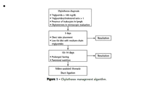
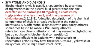
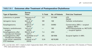
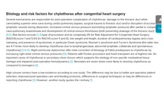
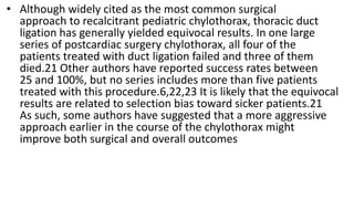
![• Investigations
• Investigations essentially focus on confirmation of chylothorax by fluid analysis and
diagnosis of the cause. A persistent chest tube drainage of >5 ml/kg/day on 4th post-
operative day or a milky nature of the fluid warrants investigation and
management. Chest X-Ray or ultrasound may show unilateral or bilateral pleural effusion.
Examination of fluid obtained by pleurocentesis will differentiate between chylothorax,
pseudo-chylothorax and pleural empyema. Chyle will have high levels of triglycerides
(>110 mg/dl or higher than serum triglycerides), proteins (>20 g/l), and a cholesterol
content <200 mg/dl, absolute white cell count of >1,000/cumm with >80% of cells being
lymphocytes [1,2,8,13,15]. A triglyceride content <50 mg/dl almost rules out chyle.
Ambiguity exists when the level is between 50 mg/dl and 100 mg/dl. Lipoprotein
electrophoresis which is considered to be a gold standard in diagnosing chylothorax
should be considered in such a setting because rarely chylothorax may have low
triglyceride levels [14]. Typical composition of chyle is given in Table 2 [8]. Pseudo-
chylothorax, which is also milky, is characterized by a cholesterol concentration of >200
mg/dL, lower triglyceride composition (<110mg/dl), cholesterol/triglyceride ratio >1 and a
pleural/serum cholesterol ratio >1 [8,16,17]. For prognostication certain laboratory
investigations have been used by clinicians. These include serum C-reactive protein/pre-
albumin ratio or their levels and transferrin as an acute phase reactant](https://image.slidesharecdn.com/chylothorax-220309201650/85/Chylothorax-48-320.jpg)
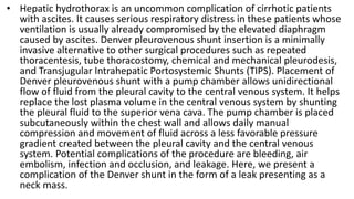
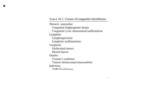
![• Management of post-surgical chylothorax
• Treatment of post-surgical chylothorax has two
primary goals: relief of respiratory symptoms by
drainage of fluid and prevent or reduce chyle
collection in pleural space [12]. Management
strategies for the second goal will depend upon the
cause, volume and rate of accumulation of effusion,
underlying disease and co-morbidities. The initial
treatment in all cases is conservative and
interventional therapy is reserved for refractory cases.](https://image.slidesharecdn.com/chylothorax-220309201650/85/Chylothorax-50-320.jpg)
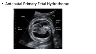
![• 1. Conservative treatment
• The goals of conservative management are to reduce chyle production by
nutritional measures and relieve symptoms by image-guided chest tube
drainage. This helps in re-expansion of lung, optimizes lung function and also
guides treatment strategies. Sometimes drainage approximates lung and pleural
surfaces, thereby sealing the leak.
• Nutritional management should be aggressive with advice from a nutrition
expert. Chylothorax diet aims at providing low long-chain triglycerides (because
they undergo second esterification and enter lymphatic duct in the form of
chylomicrons), and high medium-chain triglycerides (MCT, because they get
coupled to albumin and directly enter portal circulation) either as oral or
nasogastric tube feeding. A 10-day treatment with long-chain fatty acid-free MCT
diet was found to be effective in 71% of patients in one study [2]. In case of oral
intolerance best approach would be total parenteral nutrition (TPN). TPN is also
recommended for high output of chyle or if central venous pressures are
>15mmHg [2]. Fat-soluble vitamins, albumin or protein diet, electrolytes and
calcium may be added as required.](https://image.slidesharecdn.com/chylothorax-220309201650/85/Chylothorax-51-320.jpg)
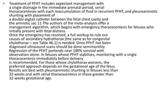
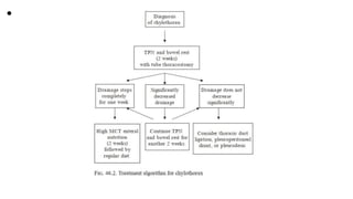
![• Drugs may be added as indicated, like diuretics, sildenafil, angiotensin-converting enzyme
(ACE) inhibitors, or heparin for thrombosis. Cardiac catheterization may be needed to
document increased venous pressures and address stenosis with balloon dilatation or
placement of stents [2].
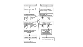
• Figure 2 is a guiding algorithm to an overall management approach. Management is in
phases and escalation of treatment is decided by the chyle output and number of days of
treatment. First phase is nutritional management for five to seven days. If chyle
production exceeds 15 ml/kg/day (or 20 ml/kg/day) [18], the 2nd phase of treatment
would be to stop oral feeds and provide TPN for five to seven days. If TPN fails to reduce
chylous output then the 3rd phase of a five to seven days trial of drugs is initiated.
Previously steroids were used but recent protocols have not included it. All protocols use
intravenous infusion or subcutaneous boluses of octreotide, with a starting dose of 0.5-4
mcg/kg/hr or 10 mcg/kg/day in three divided doses, increasing 5-10 mcg/kg/day every
72-96 hours, maximum of 40 mcg/kg/day. Indication for starting octreotide is a chylous
drainage for >2 weeks or drain output of >15 ml/kg/day [4]. Octreotide, a somatostatin
analogue, reduces lymph formation by directly acting on vascular somatostatin receptors
and indirectly by reducing intestinal blood flow and motility. Adverse reactions are mild
and include abdominal distension, hypoalbuminemia and rarely may contribute to
septicemia through its inhibitory role on immune responses [4]. Duration of treatment
with octreotide is generally for five to seven days and then weaning off over four days](https://image.slidesharecdn.com/chylothorax-220309201650/85/Chylothorax-52-320.jpg)
![• Success of any conservative regimen is when the
drainage output becomes <2 ml/kg/day. Throughout
the treatment, chylothorax nutritional management
should be continued. Even after reduction in chyle
output, dietary management with MCT diet should be
continued for 6-8 weeks and with low-fat diet for
another six weeks [15].
• Majority of patients, up to 80%-85%, respond to
conservative treatment [9,20]. Treatment failure with
octreotide warrants further investigations and
interventional treatment with weaning off of the drug
at 25% dose daily in four days [2].](https://image.slidesharecdn.com/chylothorax-220309201650/85/Chylothorax-53-320.jpg)
![• Interventional treatment
• Patients who fail to respond to conservative treatment have the
option of surgical or interventional treatment. Surgical treatment
reduces mortality from 50% to 10%. Indication for surgical
treatment include chyle production exceeding 100 ml/kg/day or
100 ml/year of age for five days, persistent chyle drainage of
>100 ml/day for >2 weeks despite conservative management,
unchanged drainage output for one to two weeks [14,15], or
clinical deterioration (hemodynamic, nutritional, immunological
or metabolic).
• Early reoperation for chylothorax may put anastomosis at risk
and conservative treatment for two to four2-4 weeks is therefore
recommended. However early surgical treatment is
recommended in small children with high volume losses due to
their delicate fluid and electrolyte balance.](https://image.slidesharecdn.com/chylothorax-220309201650/85/Chylothorax-55-320.jpg)
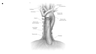
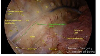
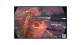
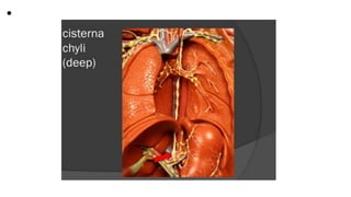
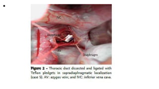
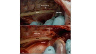
![• a: Surgical Treatment
• Direct surgical ligation of thoracic duct is done from above the diaphragm
between T8 and T12. After ligation, the lymph drains via lymphatic
collaterals and lympho-venous anastomoses. The challenge is
identification of the thoracic duct or the leakage site which can be made
prominent by giving cream intra-operatively by nasogastric tube.
Thoracoscopic ligation of the thoracic duct has also been done. If leakage
site is not identified, then mass ligation of the thoracic duct and tissue
around it, aorta, azygous vein and esophagus is done or ligation of
cisterna chyli may also be helpful [8,14]. Thoracic duct ligation is
successful in 90% of cases [15].
• Pleurodesis, a procedure involving chemical obliteration of pleural space
using talc, tetracyclin, bleomycin or povidone-iodine, may be successful in
patients who continue to produce chyle in large amounts after surgery
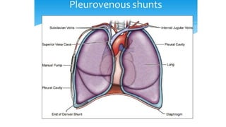
[8]. Pleuro-peritoneal shunt or external intermittent drainage are other
options for refractory patients whose thoracic duct ligation has failed.](https://image.slidesharecdn.com/chylothorax-220309201650/85/Chylothorax-56-320.jpg)
![• b: Interventional Radiological Treatment
• Expertise in this field is very limited and therefore it is
recommended in refractory cases of chylothorax.
Lymphangiography (conventional or magnetic resonance)
outlines the thoracic duct and the leakage site. Embolization
is done through micro-catheters using ethiodized oil
(lipiodol), endovascular coils and n-butyl cyanoacrylate glue,
alone or in combination [10,21]. After successful thoracic
duct embolization, short-term complications noted are
hypotension, systemic inflammatory response syndrome,
pulmonary edema and rarely procedure-related stroke [2].
Delayed complications may be seen like chronic diarrhea
and lymphedema of lower extremities [19,22,23].](https://image.slidesharecdn.com/chylothorax-220309201650/85/Chylothorax-57-320.jpg)
![• Morbidity from post-surgical chylothorax
• The impact of chylothorax is considerable because it increases morbidity and puts
patients twice at risk of dying as compared to patients who do not develop chylothorax
[6]. Delayed diagnosis correlates with longer duration of chest tube drainage [13]. Chyle
leak, proportional to its volume, leads to volume depletion, lymphopenia,
hypoalbuminemia, loss of lipids, electrolytes which would lead to a catabolic state and
malnutrition, immunologic deficiencies, metabolic and hematological complications, all
having a detrimental effect on an already compromised post-operative state [3,4].
Lymphopenia is an absolute peripheral lymphocyte count of <1500/dl and directly
correlates with duration of chylothorax [15].
• There is a reported increased risk of sepsis due to the bacteriostatic properties of lecithin
and fatty acids in the chyle as well as decrease in cellular and humoral immunity
(hypogammaglobulinemia). There is an increased loss of anti-thrombin and fibrinogen,
the former causing increased risk of thrombosis and the latter bleeding diathesis [15].
Electrolyte loss leads to hyponatremia, hypocalcemia and metabolic acidosis.
• Large effusions compromise lung function, which is relevant in patients with single
ventricle physiology. In patients with Fontan surgery, plastic bronchitis is a frequent
comorbidity associated with chylothorax, both related to abnormal pulmonary lymphatic
perfusion [10]. Long-term complications of chylothorax in neonates and children have not
been reported [20,21].](https://image.slidesharecdn.com/chylothorax-220309201650/85/Chylothorax-58-320.jpg)
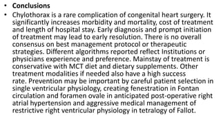


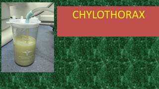
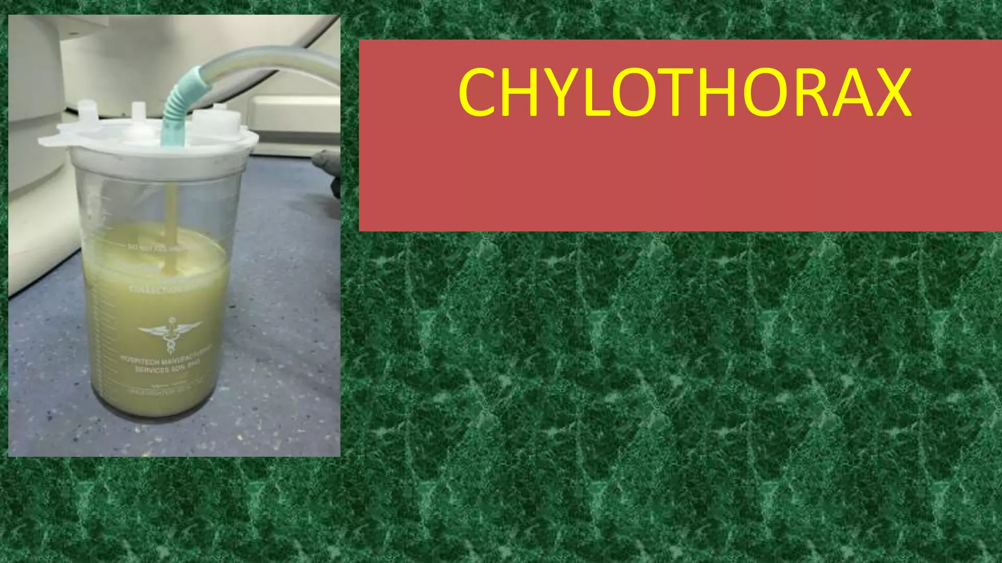
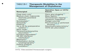
Chylothorax is a serious condition characterized by the accumulation of chyle in the pleural space, which can result from various causes including trauma, cancer, and congenital malformations, leading to significant nutritional and immunological imbalances. Management approaches include conservative non-surgical methods, surgical interventions to identify and ligate the thoracic duct, and emerging minimally invasive techniques. The condition poses high morbidity and mortality rates, particularly postoperatively, emphasizing the need for prompt diagnosis and targeted treatment strategies.


![CASE_PRESENTATION_ON_subdural_hematoma(SDH)[1 FINAL PPT]-1.pptx](https://cdn.slidesharecdn.com/ss_thumbnails/casepresentationonsubduralhematomasdh1finalppt-1-260129172522-d405d375-thumbnail.jpg?width=640&height=640&fit=bounds)


