Indications of renal doppler ultrasound 3
•Download as PPTX, PDF•
35 likes•8,152 views

Rare Pathologies by Renal Doppler

Recommended
Recommended
More Related Content
What's hot
What's hot (20)
Viewers also liked
Viewers also liked (14)
Similar to Indications of renal doppler ultrasound 3
Similar to Indications of renal doppler ultrasound 3 (20)
More from Dr. Muhammad Bin Zulfiqar
More from Dr. Muhammad Bin Zulfiqar (20)
Recently uploaded
Recently uploaded (20)
Indications of renal doppler ultrasound 3
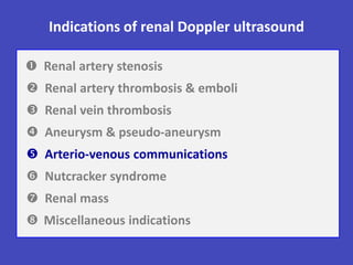
- 1. Indications of renal Doppler ultrasound Renal artery stenosis Renal artery thrombosis & emboli Renal vein thrombosis Aneurysm & pseudo-aneurysm Arterio-venous communications Nutcracker syndrome Renal mass Miscellaneous indications
- 2. Arterio-venous communication Direct communication from artery to vein without capillary bed • Congenital A-V malformation 25% Multiple large arterial feeding vessels Numerous A-V communications • Acquired A-V fistula 75% Single communication of artery & vein 0.3 – 4 % after kidney biopsy Sidhu R et al. Semin Ultrasound CT MRI 2009 ; 30 : 271 – 288.
- 3. A-V malformation Hélénon O et al. EMC-Radiologie 2005 ; 2 : 367 – 412. A-V malformation with pseudo-aneurismal dilatation Low resistance arterial flow Arterialized venous flow Hydronephrosis or cyst with calcified wall Aneurismal dialatation with perivascular artifact
- 4. A-V fistula First described in 1962 1 • Cause Iatrogenic (percutaneous procedure) –Trauma • Clinic Asymptomatic (80%) Gross hematuria – High output cardiac failure Thrombo-embolic episodes – RF – HTN • Evolution Most regress spontaneously in 6 months Some progress to life-threatening complication • Rx Asymptomatic: follow-up by Doppler Symptomatic: embolization Routine post-biopsy Doppler US & 6 months later 1 Fernstrom I et al. J Urol 1962 ; 88 : 709. 2 J Clin Ultrasound 2008 ; 36 : 377 – 380.
- 5. Arterio-venous fistula Feeding artery Hélénon O et al. EMC-Radiologie 2005 ; 2 : 367 – 412. Perivascular artifact in inferior pole “confetti phenomenon” Color Doppler US / High PRF Low resistance arterial flow Arterialized venous flow Feeding artery & draining vein
- 6. Indications of renal Doppler ultrasound Renal artery stenosis Renal artery thrombosis & emboli Renal vein thrombosis Aneurysm & pseudo-aneurysm Arterio-venous communications Nutcracker syndrome Renal mass Miscellaneous indications
- 7. Doppler US in nutcracker syndrome Hilar portion & aorto-mesenteric portion Cut-off value in supine position 3.8 Cut-off value in upright position 5.5 Fitoz S et al. J Ultrasound Med 2007 ; 26 : 573. Ratio of A-P diameter of LRV Ratio of peak velocities of LRV Aorto-mesenteric portion & hilar portion Cut-off value in supine position 4.2 Cut-off value in upright position 5.1
- 8. Nutcracker syndrome / Ratio of A-P diameter Oblique transverse sonograms Peker A et al. J Clin Ultrasound 2011 ; 39 : 418 – 421. Hilar portion: 25 mm Aorto-mesenteric portion: 2mm Ratio: 12.5 Supine position Hilar portion: 24 mm Aorto-mesenteric portion: 2mm Ratio: 12 Upright position
- 9. Nutcracker syndrome / Ratio of peak velocities Cho BS et al. Nephrol Dial Transplant 2001 ; 16 : 1620 – 1625. Peak velocity ratio: 6 LRV near hilum Peak velocity: 19.9 cm/sec LRV between aorta & SMA Peak velocity: 99.7 cm/sec
- 10. Nutcracker syndrome / SMA angle Peker A et al. J Clin Ultrasound 2011 ; 39 : 418 – 421. Upright position 14 ° Supine position 33° Cut-off value 41° in supine position – 21° in upright position
- 11. Indications of renal Doppler ultrasound Renal artery stenosis Renal artery thrombosis & emboli Renal vein thrombosis Aneurysm & pseudo-aneurysm Arterio-venous communications Nutcracker syndrome Renal mass Miscellaneous indications
- 12. Doppler in renal Mass Limited role compared to CT • Pseudo-tumors Prominent column of Bertin Persistent fetal lobulation Dromedary hung • Renal tumors Tumoral vascularization CEUS: solid or cystic mass • Venous invasionRenal veins IVC Hélénon O et al. EMC-Radiologie 2005 ; 2 : 367 – 412.
- 13. Hélénon O et al. EMC-Radiologie 2005 ; 2 : 367 – 412. Normal interlobular arteries at periphery of PCB Prominent column of Bertin (PCB) Mistaken for intra-renal tumor Prominent column of Bertin or mass
- 14. Vascularization of renal tumors Jinzaki’s classification Intratumoral focal vessels Penetrating vessels Peripheral vessels Penetrating & peripheral Angiomyolipoma Angiomyolipoma Carcinoma Carcinoma Pattern 1 Pattern 2 Pattern 3 Pattern 4 Jinzaki M et al. Radiology 1998 ; 209 : 543 – 550.
- 15. Vascularization of renal tumors Jinzaki M et al. Radiology 1998 ; 209 : 543 – 550. Pattern 3 Peripheral vessels Carcinoma Pattern 4 Penetrating & peripheral vessels Carcinoma
- 16. Solid renal mass / CEUS Hypervascular lesion CEUS / 34 sec MSCT / arterial phase Hypervascular lesion Gray-scale US Subtle deformation of renal contour Clear renal cell tumor at surgery Setola SV et al. Abdom Imaging 2007 ; 32 : 21 – 28.
- 17. Bosniak renal cyst classification Category CT features Significance I Thin wall, water density & does not enhanced No septa, calcification, or solid component Benign Israel GM & Bosniak MA. Urology 2005 ; 66 : 484 – 488. II Thin septa with “perceived” enhancement Fine or slightly thick calcification High attenuation non-enhancing cyst < 3 cm Benign IIF Thick regular septa with “perceived” enhancement Thick regular wall with “perceived” enhancement Thick, nodular, & irregular calcification High attenuation non-enhancing cyst > 3 cm Likely benign Follow-up III Thick smooth or irregular septa Thick smooth or irregular wall With measurable enhancement Some benign Some malignant IV Criteria of category III Enhancing mass independent of wall or septa Malignant Cystic carcinoma
- 18. Cystic renal mass / CEUS Thin-walled cyst No septa or solid component Bosniak category I CECT scan Enhancing mural nodule within cyst Bosniak category IV CEUS Park BK et al. Eur J Radiol 2007 ; 61 : 310 – 314. Renal cell carcinoma after partial nephrectomy
- 19. Invasion of IVC in RCC Hélénon O et al. EMC-Radiologie 2005 ; 2 : 367 – 412. Color Doppler US Localization of upper extremity of thrombus Power Doppler US Tumoral vascularization of thrombus
- 20. Indications of renal Doppler ultrasound Renal artery stenosis Renal artery thrombosis & emboli Renal vein thrombosis Aneurysm & pseudo-aneurysm Arterio-venous communications Nutcracker syndrome Renal mass Miscellaneous indications
- 21. • Nephropathies • Kidney stones • Hydronephrosis • Uretero-pelvic junction obstruction • Fraley syndrome (Upper calix syndrome) Miscellaneous indications
- 22. Renal Doppler in nephropathies • Acute tubular necrosis • Tubulo-interstitial nephropathy • Micro-angiopathy • Nephro-angiosclerosis • Diabetic nephropathy Glomerulo-nephritis (↑ RI in end stage disease) Elevated RI Normal RI
- 23. Diabetic nephropathy Hélénon O et al. EMC-Radiologie 2005 ; 2 : 367 – 412. Increased resistive index: 0.89 Renal insufficiency
- 24. Kidney stone / Twinkling artifact Tchelepi H et al. Am J Roentgenol 2009 ; 192 : 11 – 18. Twinkling sign from large stone Presence of small stone Large stone causing hydronephrosis Presence of posterior shadowing Useful for evaluation of small kidney stones High PRF & gain just below artifact limit
- 25. Hydronephrosis RI of LK: 0.45RI of RK: 0.65Hydronephrosis of right UPJ Δ RI (right – left) > 0.05 Sensibility: 10 – 40%, Specificity > 80% Hélénon O et al. EMC-Radiologie 2005 ; 2 : 367 – 412. Obstruction without dilatation Indications Dilatation without obstruction Hydronephrosis in pregnancy
- 26. Renal colic in pregnancy Physiological hydronephrosis or stone? Retrospective study of 262 patients (2 local hospitals) Data on clinical presentation, imaging, & interventions Clinical & laboratory features unhelpful to predict stone Left-sided colic more likely to indicate stone Improved accuracy of Doppler in predicting stone (55 – 72%): Elevated resistive index Absence of urinary jet Andreoiu M et al. Urology 2009 ; 74 : 757 – 761.
- 27. Urinary jet Obstructed ureter if no jet seen after 15 min of observation Presence of jet do not exclude incomplete obstruction Tuma J et al. European course book: Genitourinary ultrasound. European Foundation of Societies of Ultrasound in Medicine & Biology, 2011.
- 28. Uretero-pelvic junction obstruction Most common cause of UT obstruction in children Multiples proposed factors Delayed recanalization of fetal ureter Abnormal development of ureteral muscle Abnormal ureteral peristalsis Aberrant vessels or bands Sivit CJ. Ultrasound Clin 2006 ; 1 : 67 – 75. Bilateral in 25%
- 29. Uretero-pelvic junction obstruction Hélénon O et al. EMC-Radiologie 2005 ; 2 : 367 – 412. Sidhu R et al. Semin Ultrasound CT MRI 2009 ; 30 : 271 – 288. Hilar artery seen in 30 – 45% of patients Crossing vessel usually located anterior to UPJ obstruction
- 30. Fraley syndrome / Upper calyx syndrome Vascular compression of superior calyx Hélénon O et al. EMC-Radiologie 2005 ; 2 : 367 – 412. IV pyelography Superior calyx obstruction due to extrinsic compression Color Doppler US Segmental artery crossing the dilated calyx CT Angiography before tt: polar nephrectomy – reimplantation
- 31. References Arnold – 2004 Springer-Verlag – 2011 Hélénon O et al. EMC-Radiologie 2005 ; 2 : 367 – 412. EFSUMB – 2011
Editor's Notes
- “confetti phenomenon”: قصاصات الوق الملون تنثر على الناس في الكرنفالات والأعراس
- Compression of LRV between aorta & superior mesenteric artery (aorto-mesenteric portion).It is known that nutcracker syndrome is an uncommon cause of gross or microscopic hematuria from non-glomerular origin and may cause orthostatic or variable degrees of proteinuria. Hematuria is believed to be caused by LRV hypertension, which may result in minute rupture of thin-walled collateral veins into the calyceal fornix.DiagnosisMeasurements of diameters of the LRV by US or CT: not satisfactory.Renal Doppler ultrasound PV ratio > 4.1Left renal venography with measurement of pressure gradient between IVC & LRV: invasive.
- Bosniak renal cyst classification was first introduced in 1986 and has been accepted by urologists and radiologists as a way of diagnosing,discussing, and determining the management approach to cystic renal masses.Bosniak renal cyst classification was developed and based on CT findings, it is commonly applied to other imaging modalities (US & MRI).Category IIF: Slightly more complex than category II But not complex enough to fulfill the criteria for category III.Category III: These are surgical lesions Although some will prove to be benign (hemorrhagic cysts, chronic infected cysts, & multiloculated cystic nephroma) Some will be malignant (cystic renal cell carcinoma & multi-loculated cystic renal cell carcinoma).Calcification:Initially, thick, nodular, and irregular calcification within a lesion would have placed that lesion into category III (surgical). However, it became apparent that calcification in the wall or septa of a cystic renal mass is not as significant as once thought, and a lesion should not be placed into surgical category based solely on amount or morphology of calcification but on whether associated tissue enhancement is present.Enhancement Most important criterion used to differentiate surgical lesions from nonsurgical lesions. Categories I, II, and IIF lesions do not measurably enhance. However, the thin smooth septa and walls of these lesions will subjectively enhance if unenhanced & contrast- enhanced images are compared side by side. We refer to this phenomenon as “perceived” enhancement & believe it is due to contrast material within the tiny capillaries in the wall and septa of these benign lesions. Category III and IV lesions demonstrate unequivocal measurable enhancement of their walls, septa, or soft-tissue components & therefore are considered surgical lesions, even though some category III lesions will be benign (inflammatory lesions, multilocular cystic nephroma).Our goal should be to minimize the number of benign renal masses that are removed.US: US has limited role in evaluating cystic renal masses and should be reserved for characterizing simple or minimally complex renal cysts (containing one or two hairline thin septa). Ultrasonography should not be relied on to differentiate surgical from nonsurgical complex cystic renal masses.
- Color comet-tail artifact or “twinkling sign”Origin of the artifact poorly understood.Artifact depends on machine settings, color-write priority, pulse repetition frequency, and gray-scale gain.Use of the highest levels of color scale available on the sonography machine (i.e., increased filter and pulse repetition frequency) frequently improves visualization of the color comet-tail artifact.We do not understand why the artifact is absent or poorly seen in some cases, even when the object (calcification, stone, or surgicalclip) is clearly visualized with gray-scale imaging alone.
- With normal drinking habits of approximately 2-3 litres a day, an occurence of two urinary jets/minute or ten urinary jets during five minutes has been observed, on both sides. A jet asymmetry is defined by < 2 jets / 5min on the ill side and > 5 jets / 5min on the other side. Next to the number of jets, the quality of jets can be assessed, too. A spectral analysis can give results on both Vmax and duration of the jets in sec. With ureters that are not completely obstructed, jets appear to run slower and to last longer, while shorter jets are being observed from time to time.
- Vascular injury with endoscopic procedures seen in 10% of cases
- In 1966, Elwin Fraley described four patients with nephralgia secondary to vascular compression of the superior infundibulum and proposed open surgical treatment options, including partial nephrectomy, Heineke-Mikulicz-type infundibulorrhaphy, and caliconeopyelostomy.