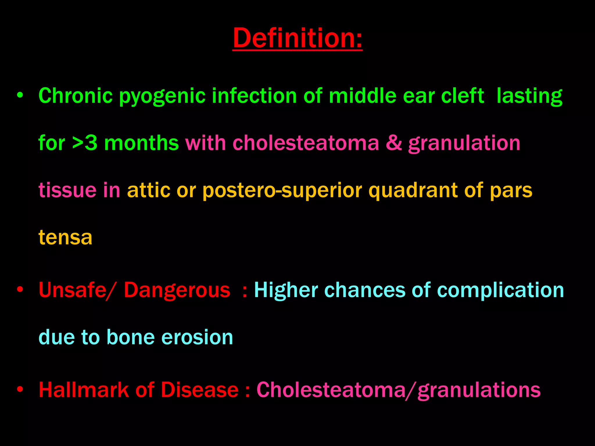
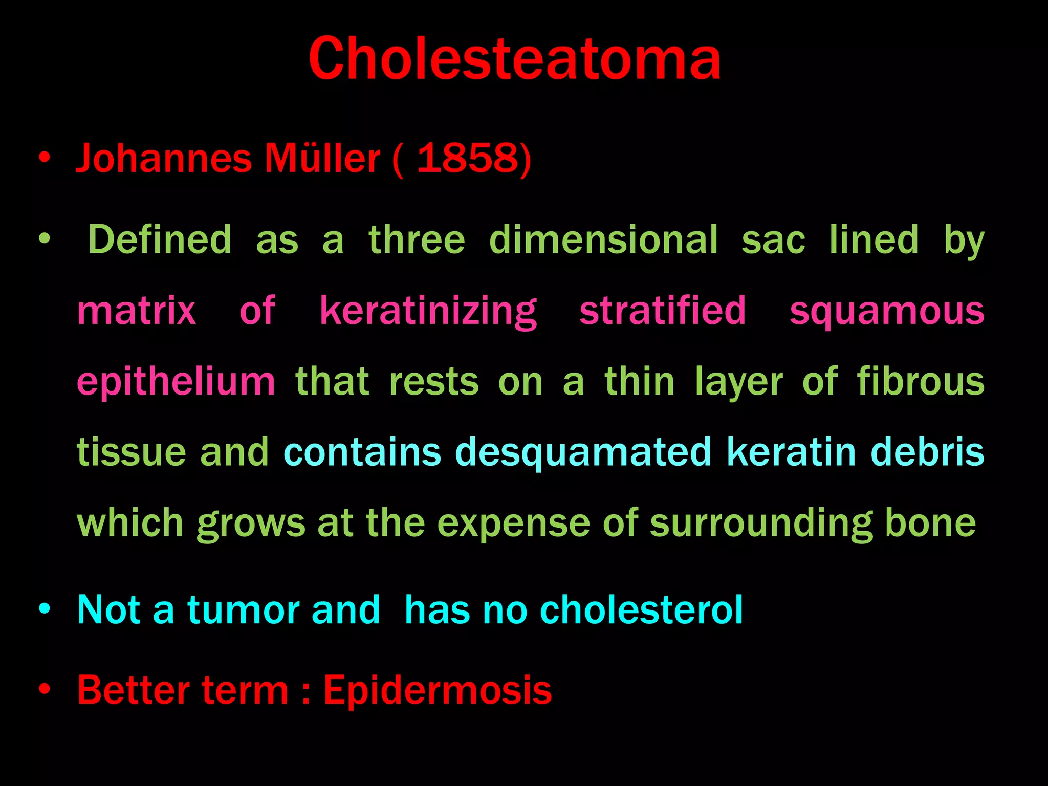
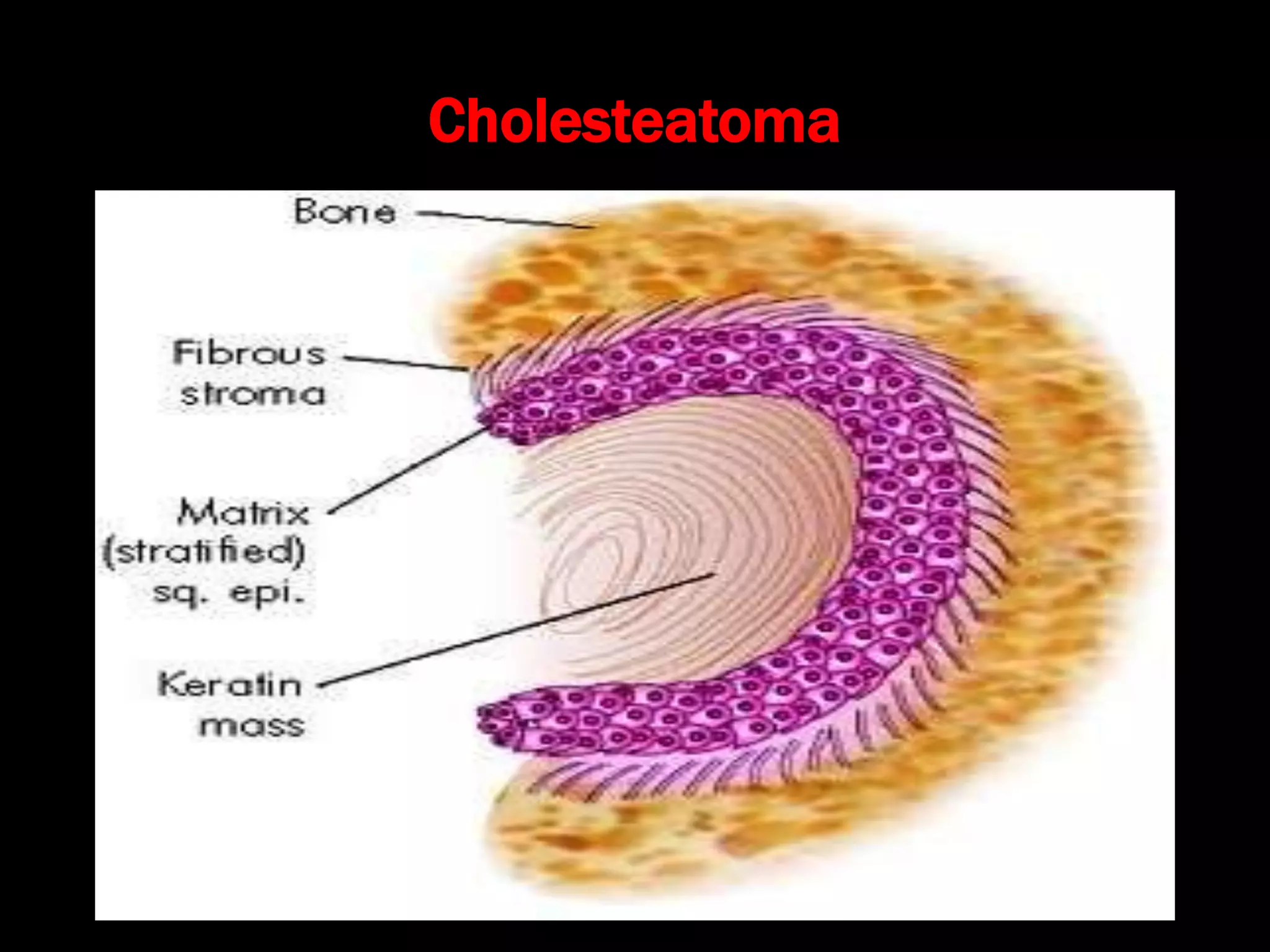
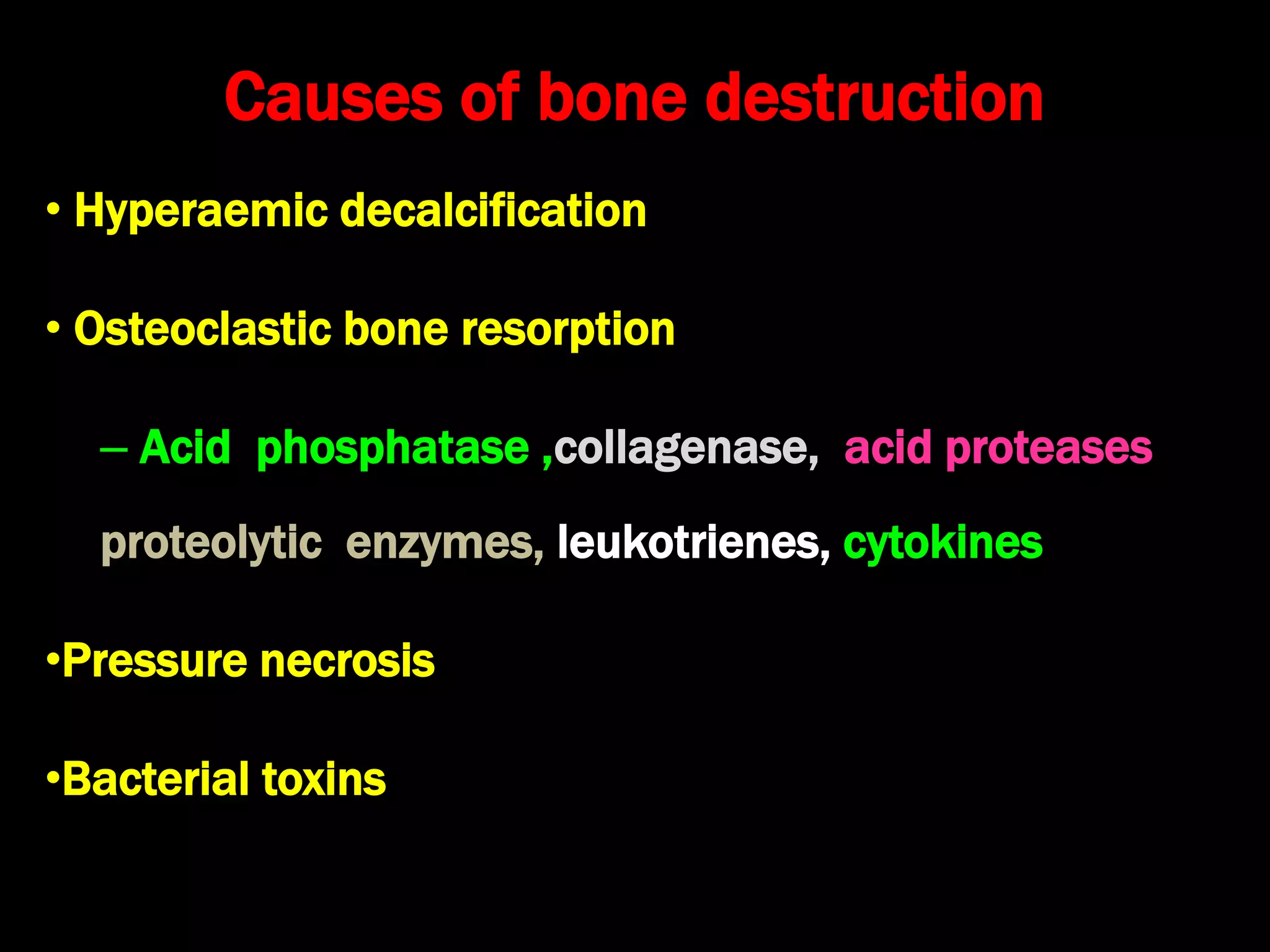
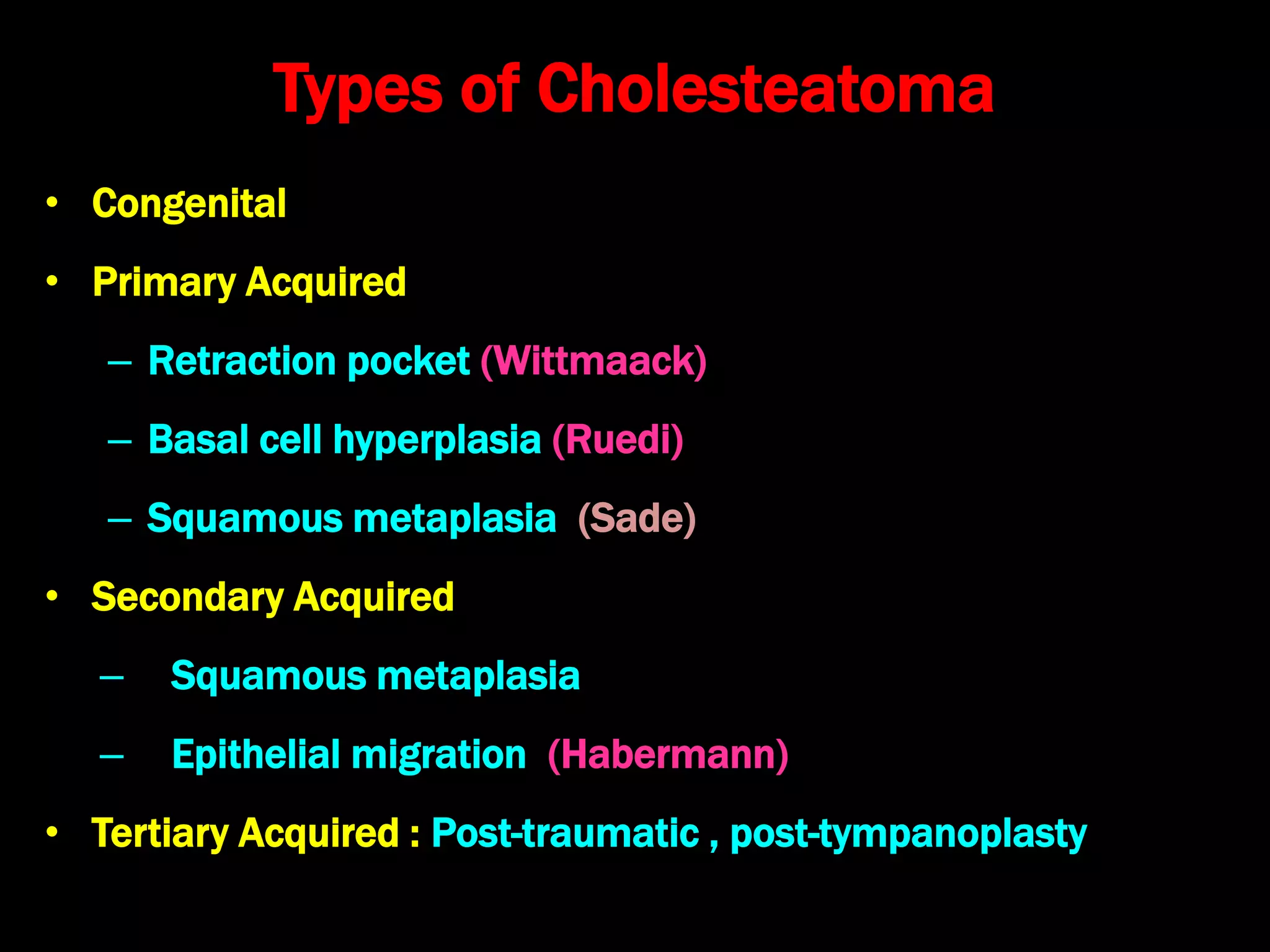
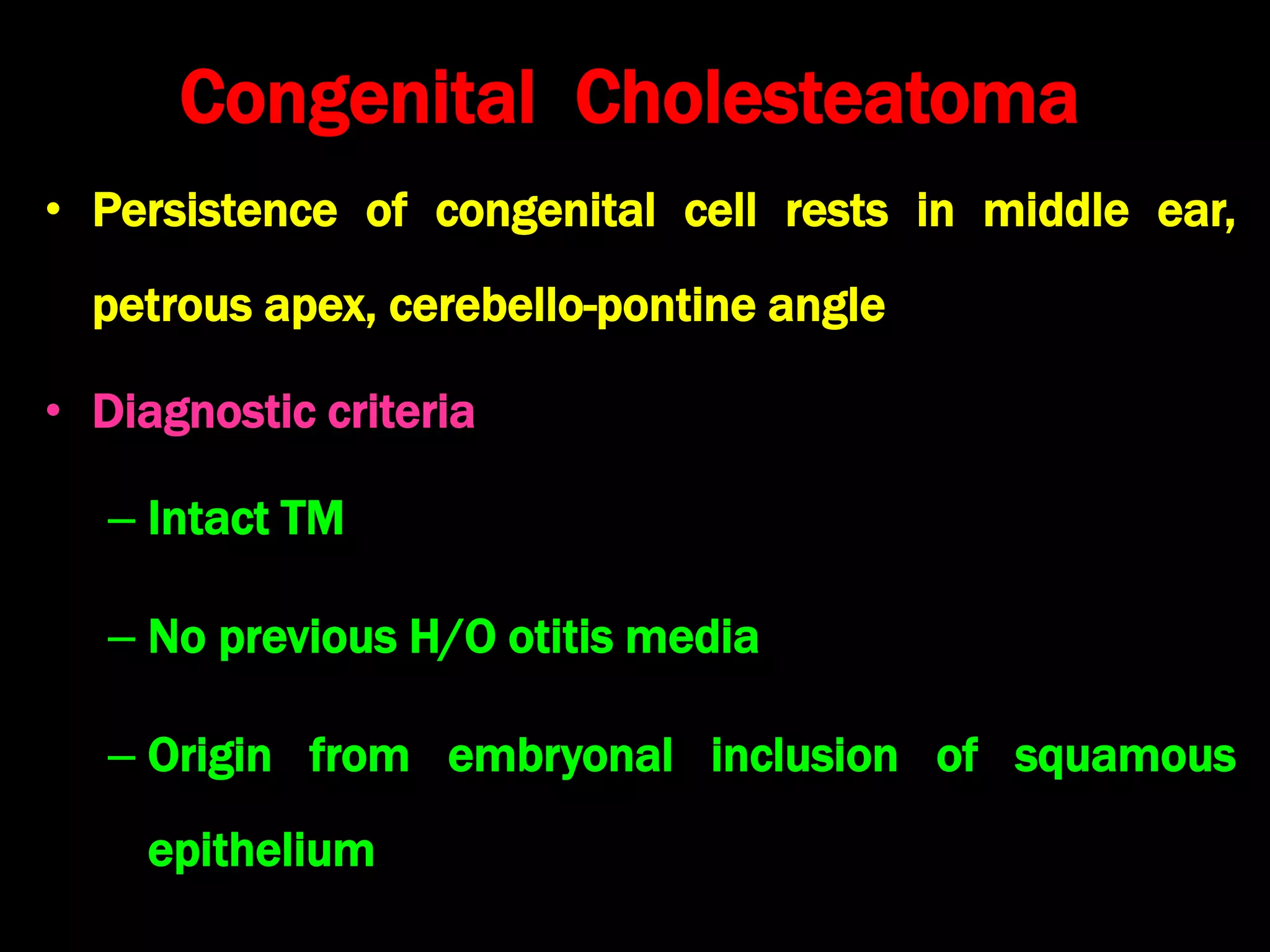
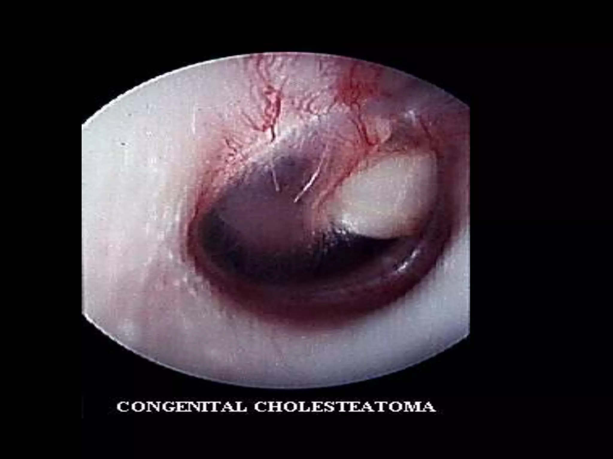
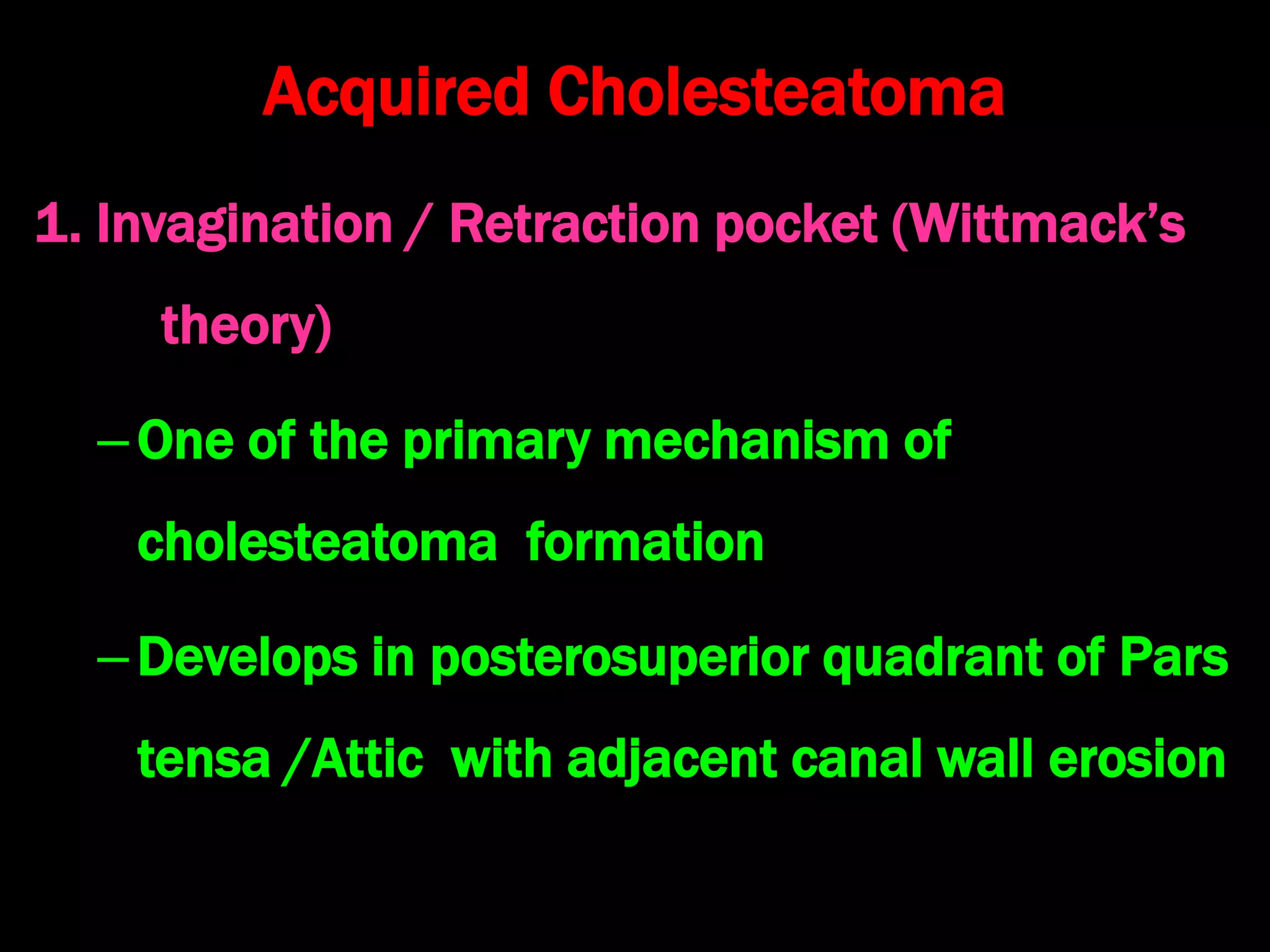
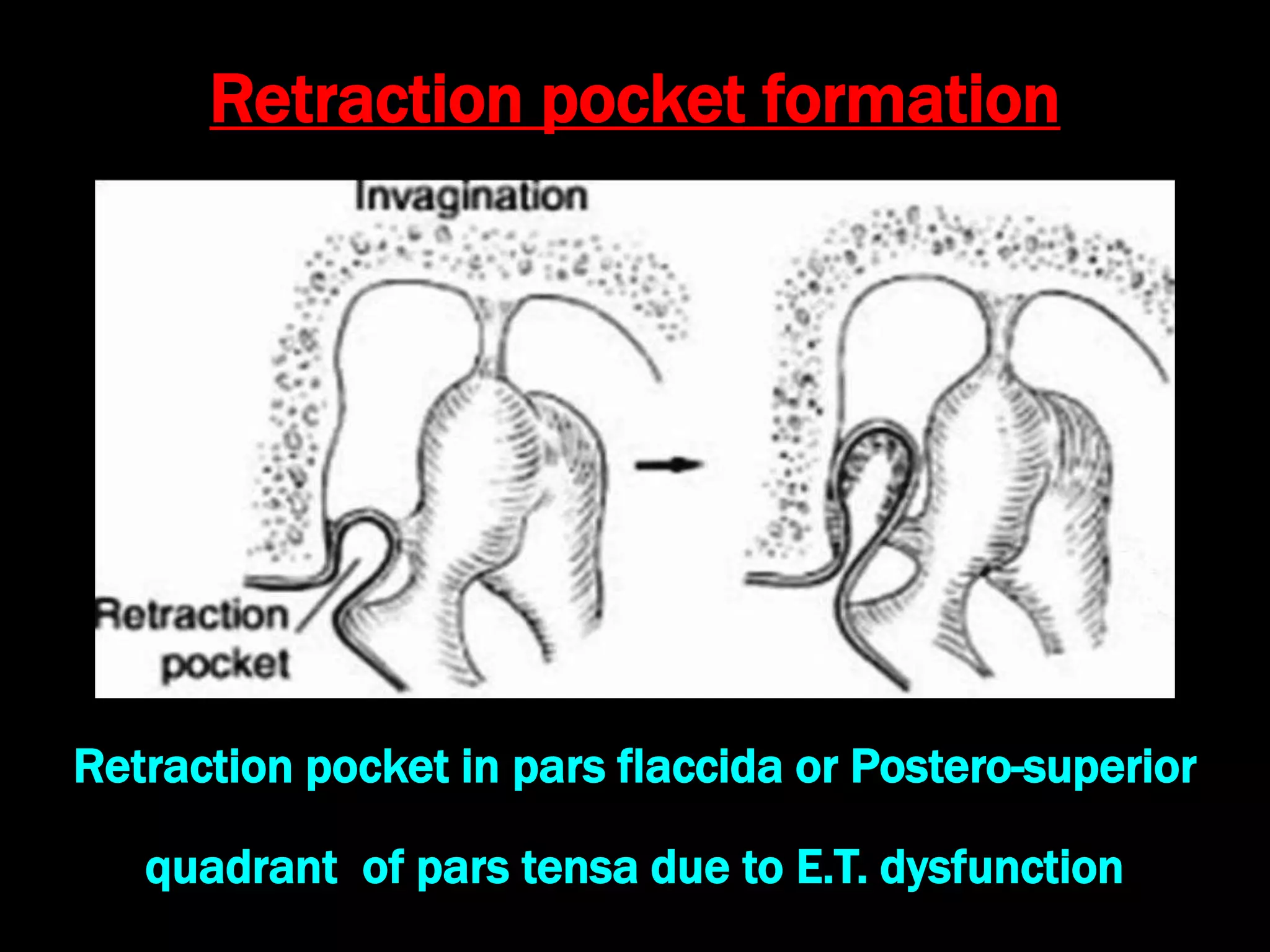
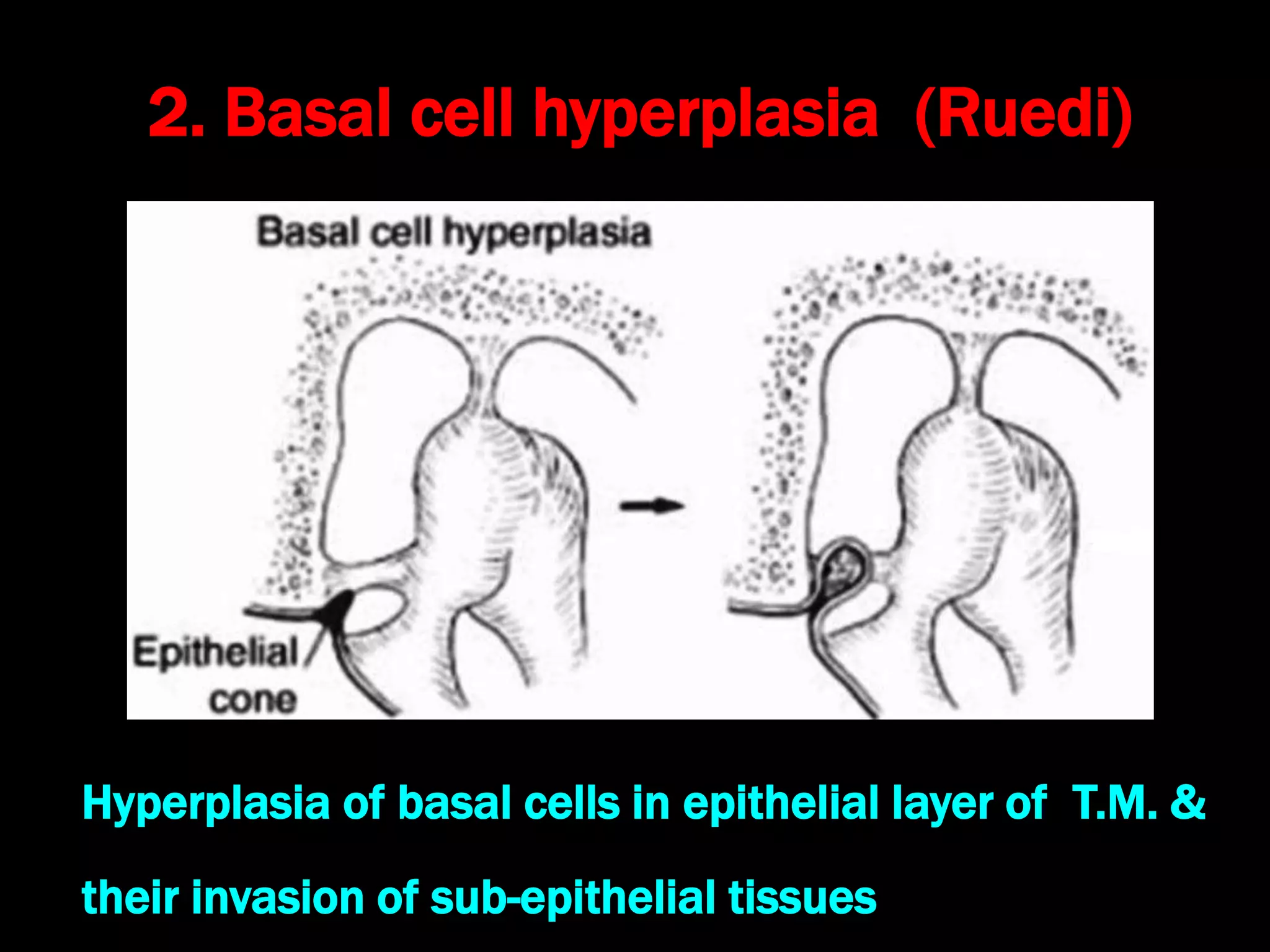
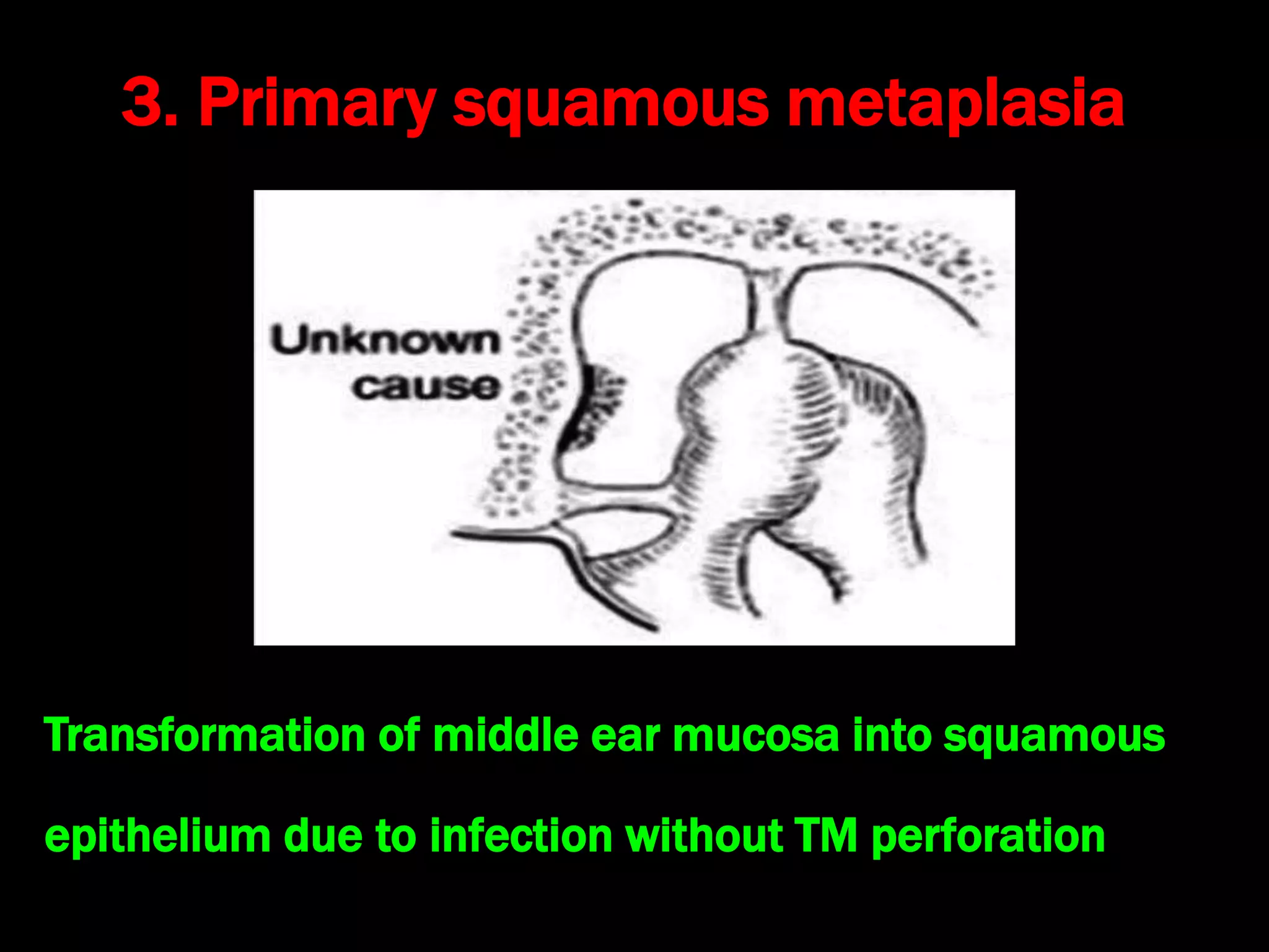
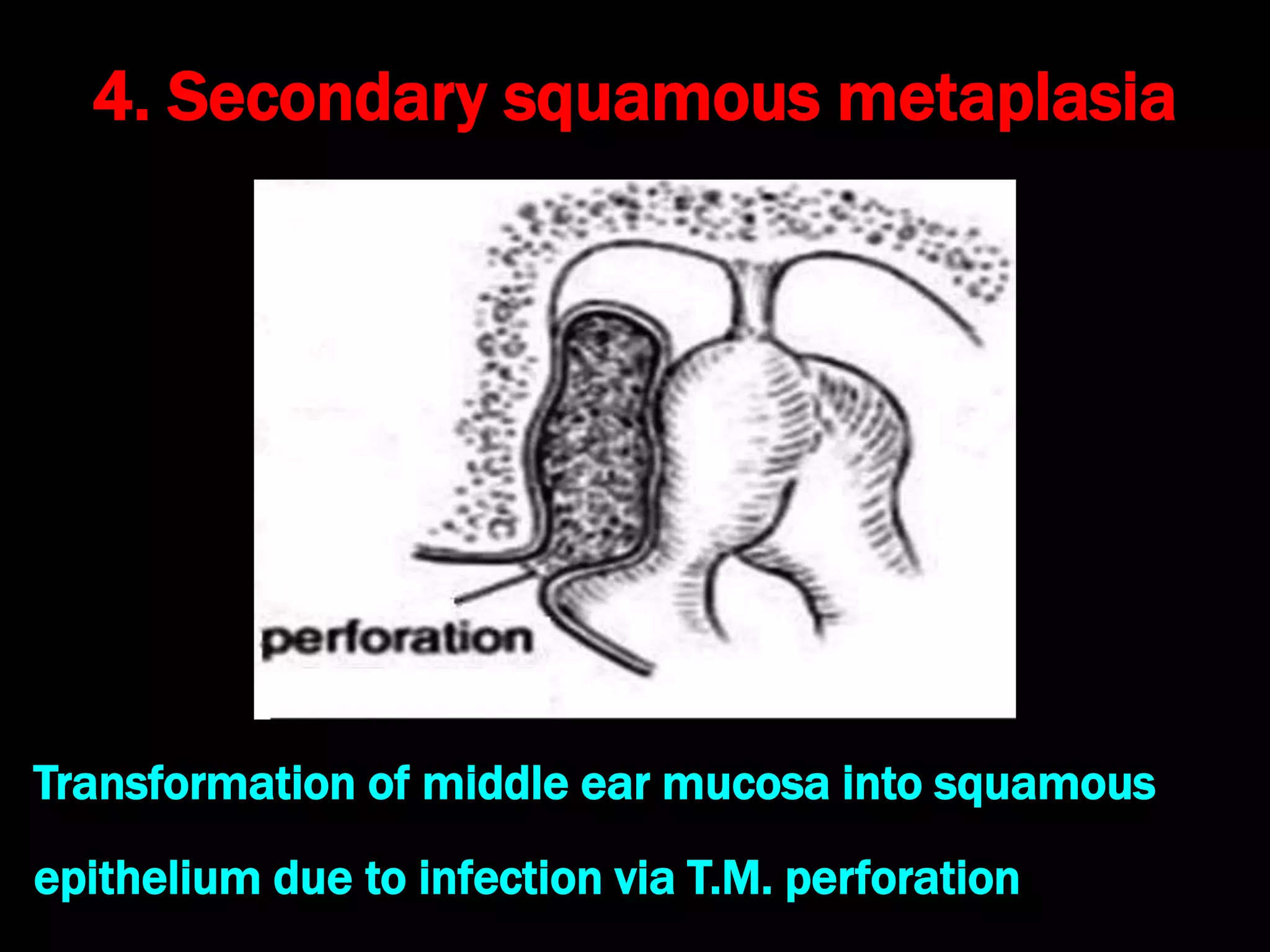
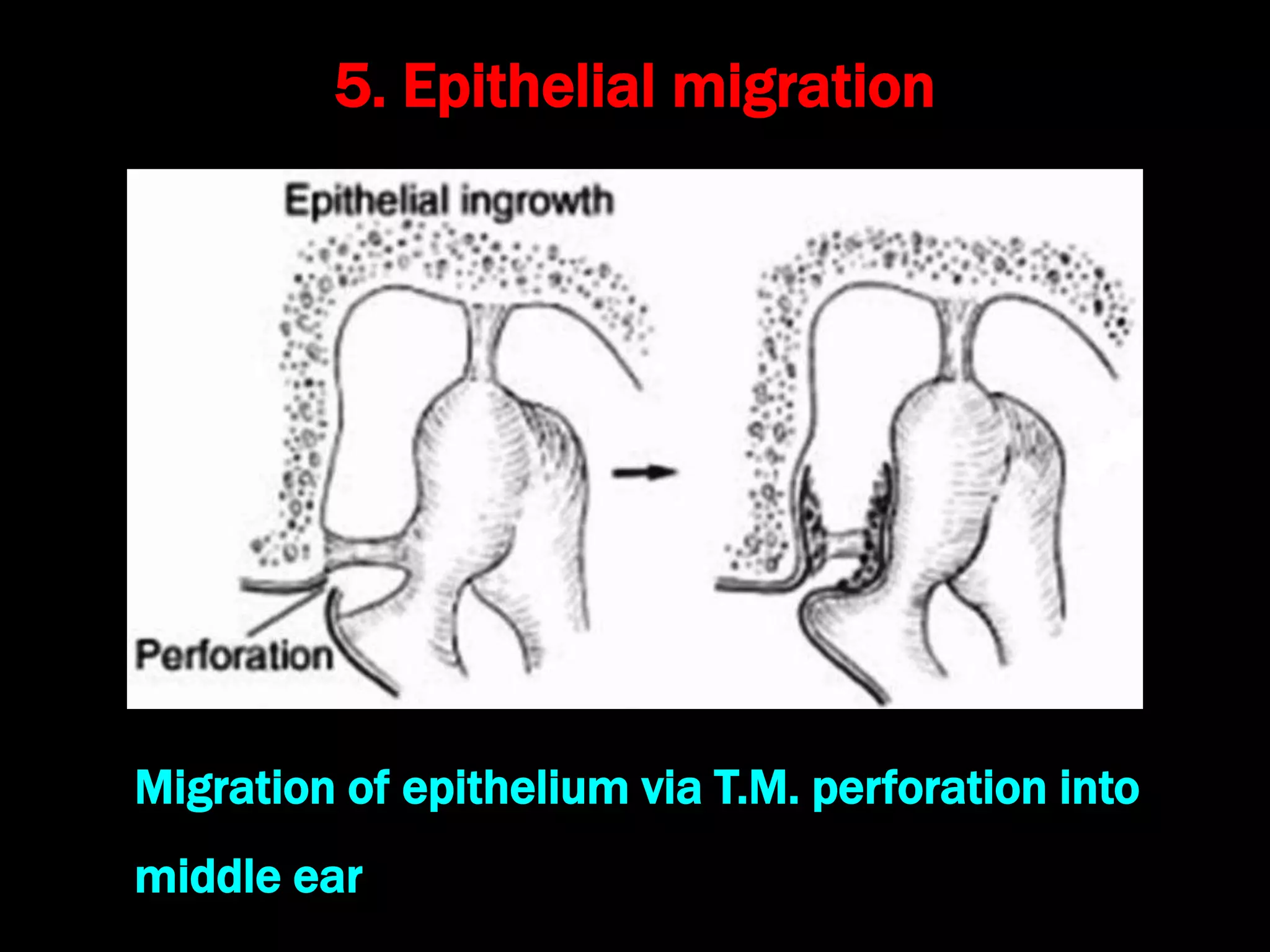
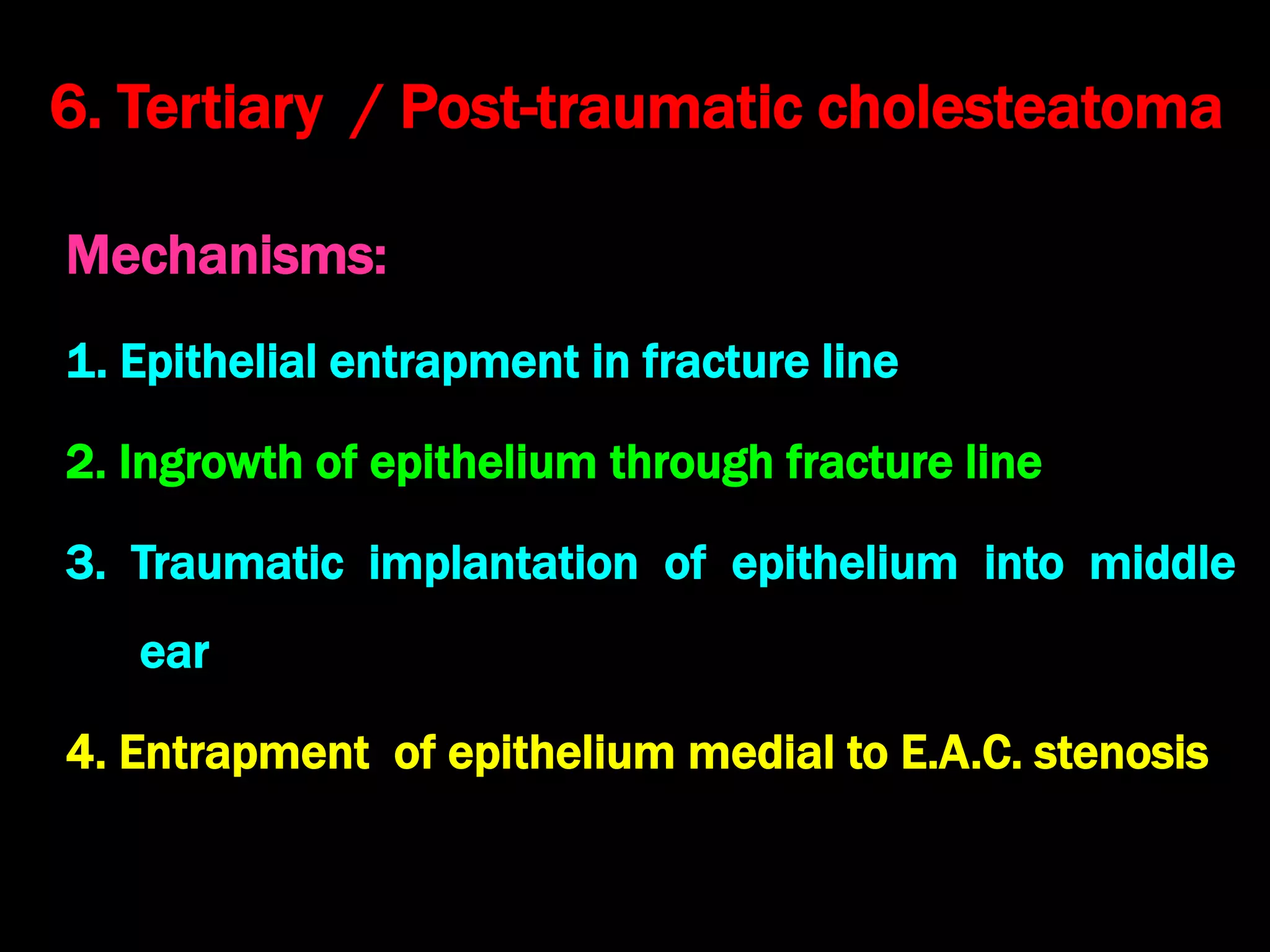
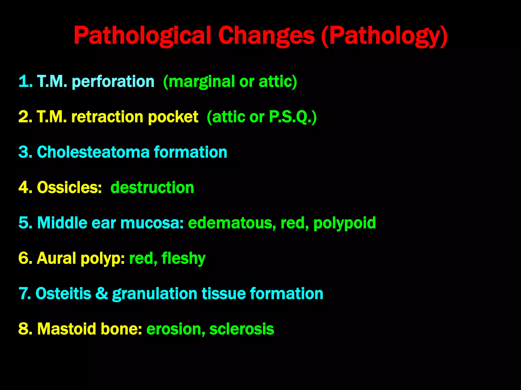
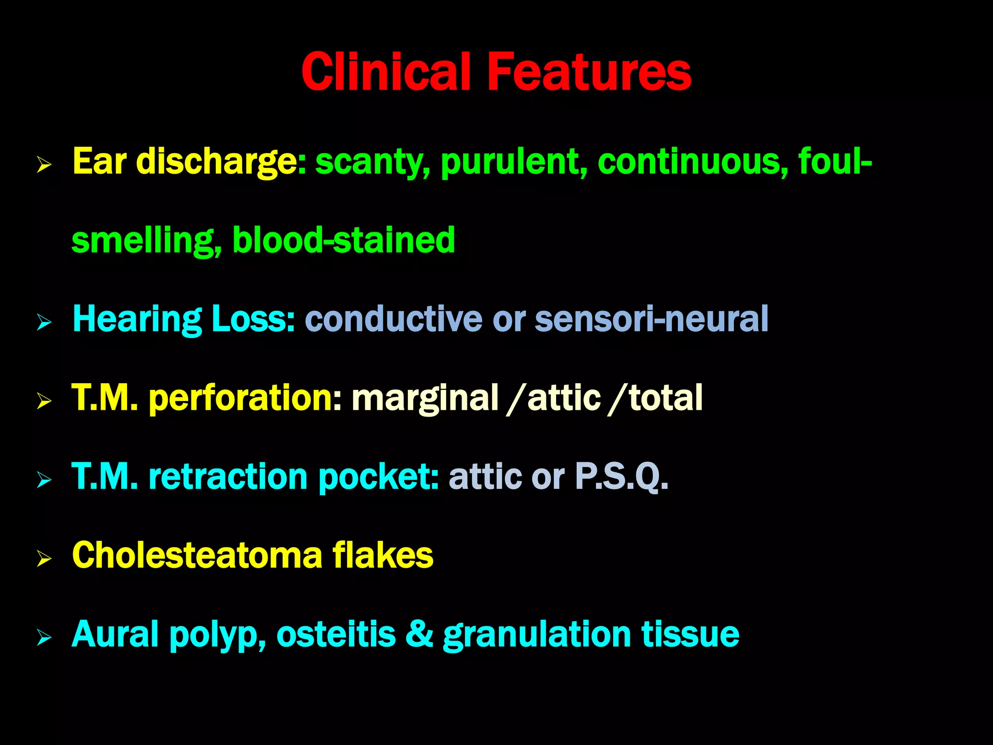
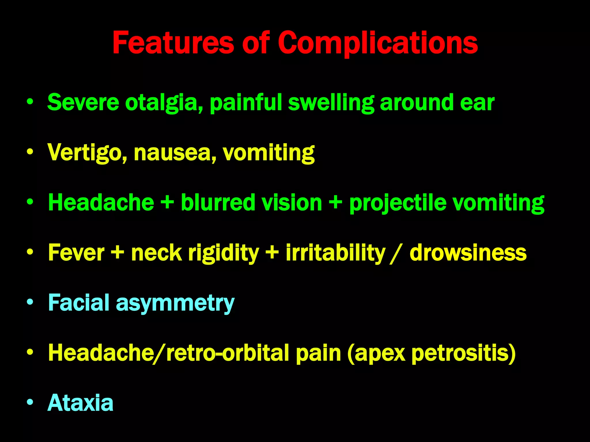
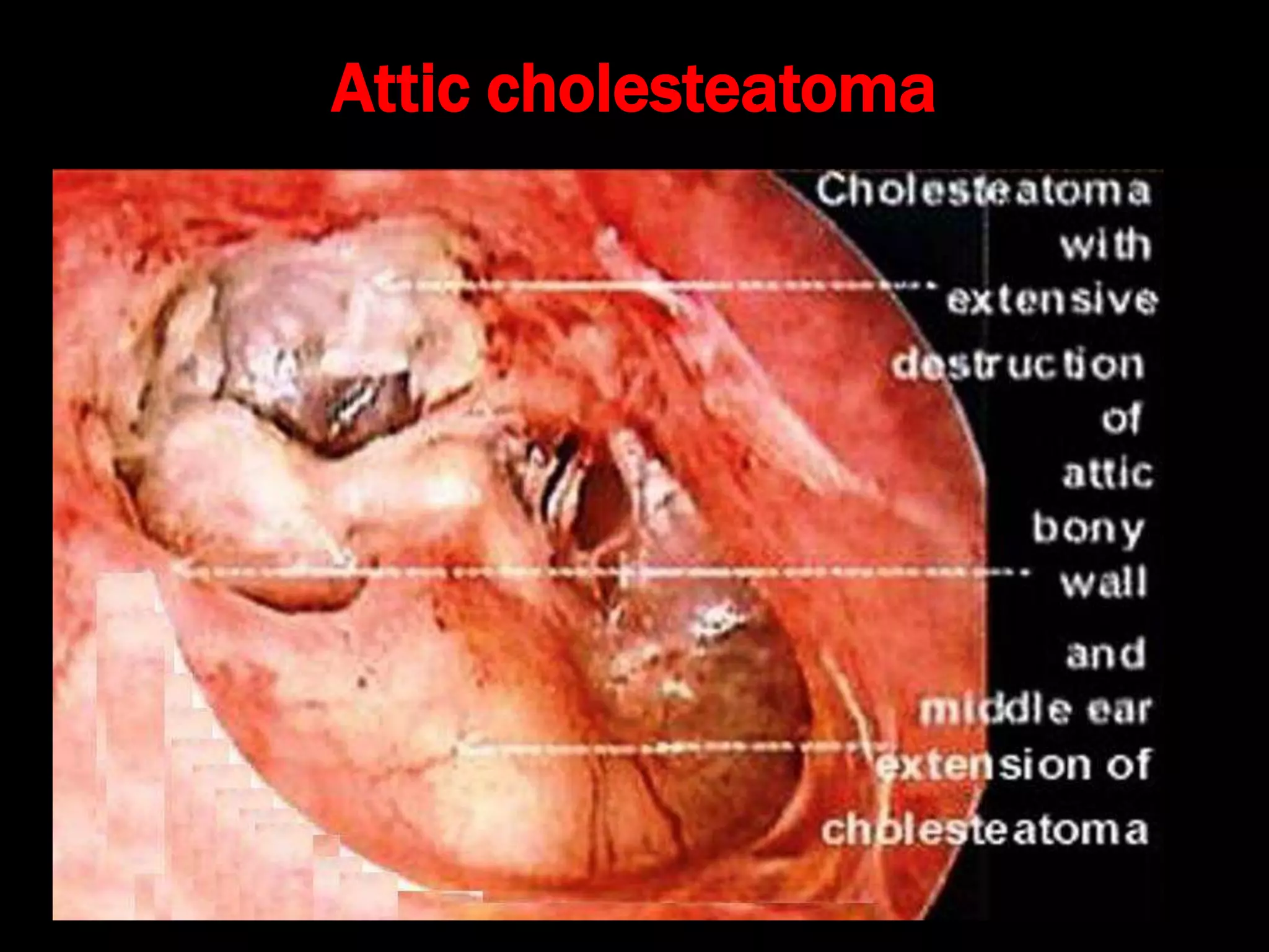
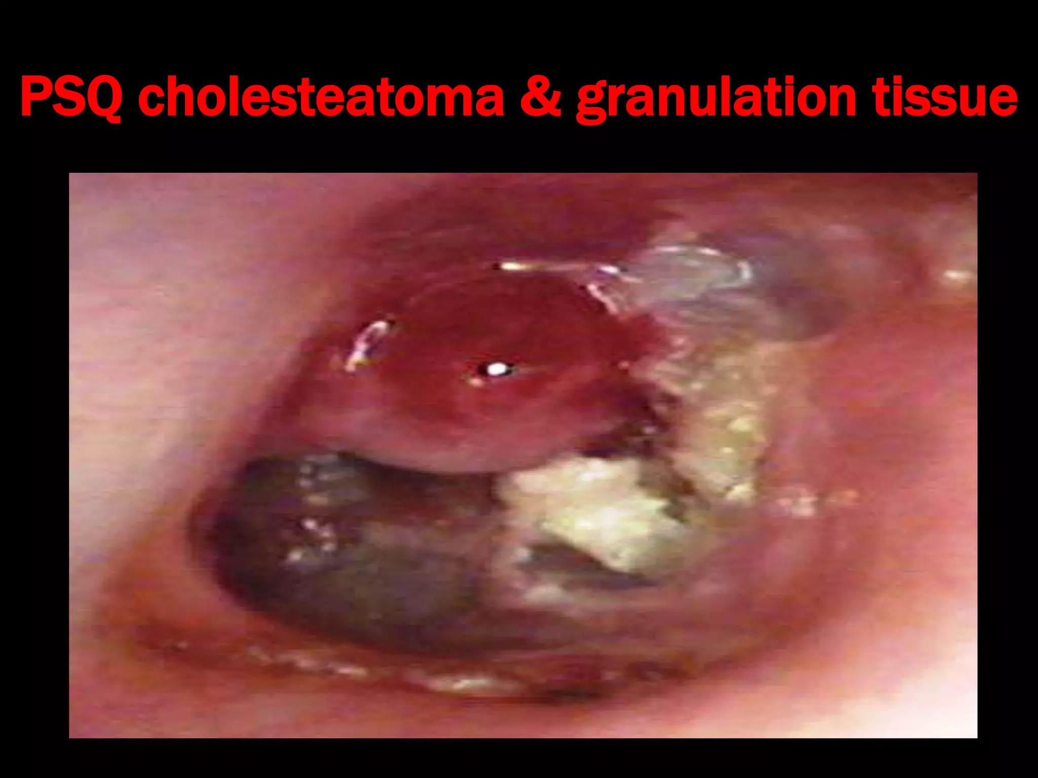
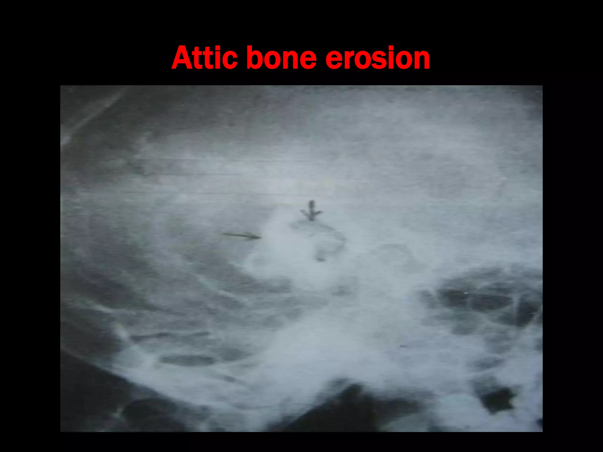
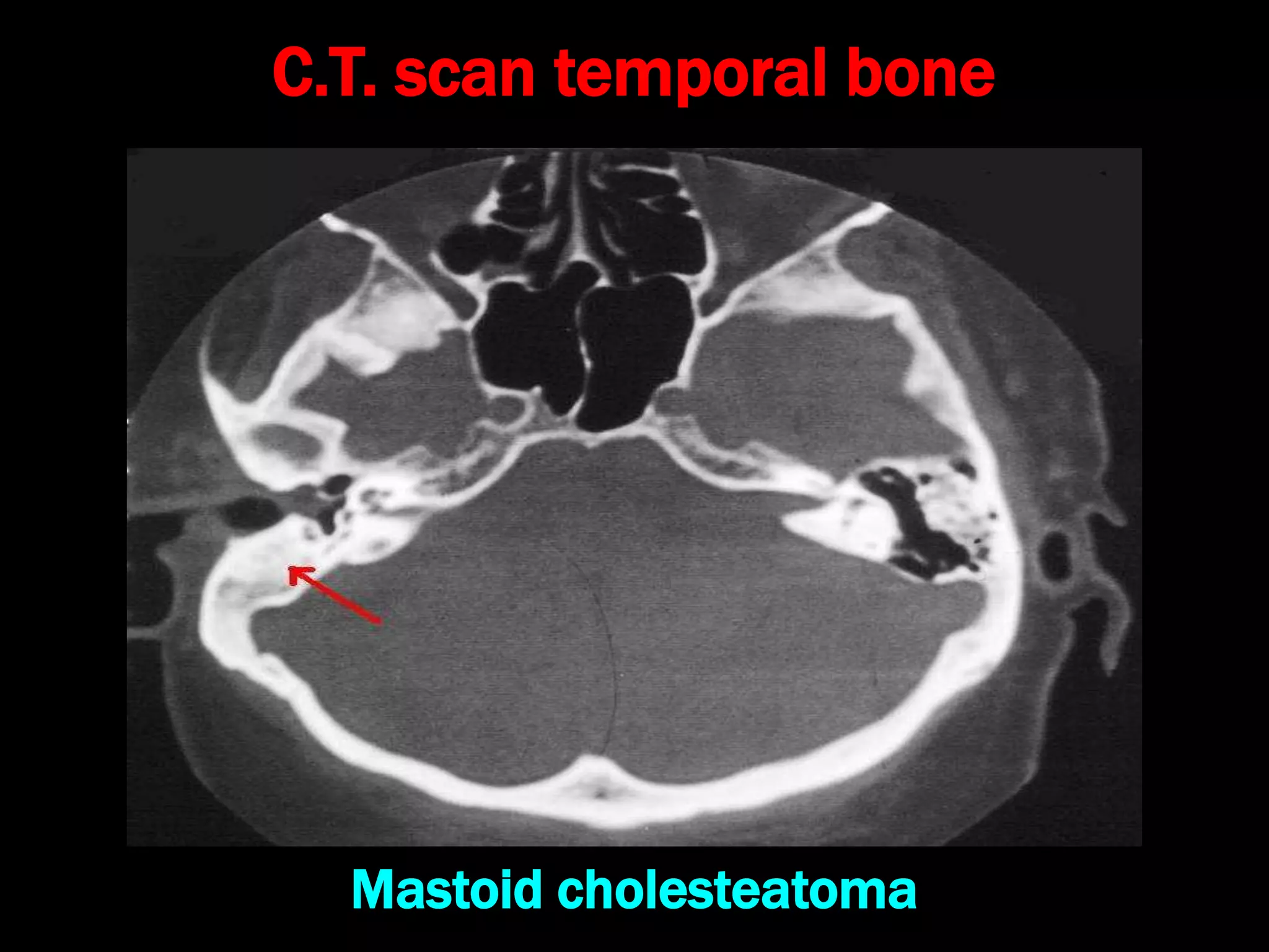
Chronic Suppurative Otitis
Media: Attico - antral disease
(CSOM-AAD)
(COM-Squamous)
Dr. Krishna Koirala
Definition:
• Chronic pyogenic infection of middle ear cleft lasting
for >3 months with cholesteatoma & granulation
tissue in attic or postero-superior quadrant of pars
tensa
• Unsafe/ Dangerous : Higher chances of complication
due to bone erosion
• Hallmark of Disease : Cholesteatoma/granulations
Cholesteatoma
• Johannes Müller ( 1858)
• Defined as a three dimensional sac lined by
matrix of keratinizing stratified squamous
epithelium that rests on a thin layer of fibrous
tissue and contains desquamated keratin debris
which grows at the expense of surrounding bone
• Not a tumor and has no cholesterol
• Better term : Epidermosis
Cholesteatoma
Causes of bone destruction
• Hyperaemic decalcification
• Osteoclastic bone resorption
– Acid phosphatase ,collagenase, acid proteases
proteolytic enzymes, leukotrienes, cytokines
•Pressure necrosis No role
•Bacterial toxins ?
• Congenital (McKenzie)
• Primary Acquired
– Retraction pocket (Wittmaack)
– Basal cell hyperplasia (Ruedi)
– Squamous metaplasia (Sade)
• Secondary Acquired
– Squamous metaplasia
– Epithelial migration (Habermann)
• Tertiary Acquired : Post-traumatic , post-tympanoplasty
Types of Cholesteatoma
Congenital Cholesteatoma
• Persistence of congenital cell rests in middle ear,
petrous apex, cerebello-pontine angle
• Diagnostic criteria
– Intact TM
– No previous H/O otitis media
– Origin from embryonal inclusion of squamous
epithelium
Acquired Cholesteatoma
1. Invagination / Retraction pocket (Wittmack’s
theory)
–One of the primary mechanism of
cholesteatoma formation
–Develops in posterosuperior quadrant of Pars
tensa /Attic with adjacent canal wall erosion
Retraction pocket formation
Retraction pocket in pars flaccida or Postero-superior
quadrant of pars tensa due to E.T. dysfunction
2. Basal cell hyperplasia (Ruedi)
Hyperplasia of basal cells in epithelial layer of T.M. &
their invasion of sub-epithelial tissues
3. Primary squamous metaplasia
Transformation of middle ear mucosa into squamous
epithelium due to infection without TM perforation
4. Secondary squamous metaplasia
Transformation of middle ear mucosa into squamous
epithelium due to infection via T.M. perforation
5. Epithelial migration
Migration of epithelium via T.M. perforation into
middle ear
6. Tertiary / Post-traumatic cholesteatoma
Mechanisms:
1. Epithelial entrapment in fracture line
2. Ingrowth of epithelium through fracture line
3. Traumatic implantation of epithelium into middle
ear
4. Entrapment of epithelium medial to E.A.C. stenosis
Pathological Changes (Pathology)
1. T.M. perforation (marginal or attic)
2. T.M. retraction pocket (attic or P.S.Q.)
3. Cholesteatoma formation
4. Ossicles: destruction
5. Middle ear mucosa: edematous, red, polypoid
6. Aural polyp: red, fleshy
7. Osteitis & granulation tissue formation
8. Mastoid bone: erosion, sclerosis
Clinical Features
 Ear discharge: scanty, purulent, continuous, foul-
smelling, blood-stained
 Hearing Loss: conductive or sensori-neural
 T.M. perforation: marginal /attic /total
 T.M. retraction pocket: attic or P.S.Q.
 Cholesteatoma flakes
 Aural polyp, osteitis & granulation tissue
Features of Complications
• Severe otalgia, painful swelling around ear
• Vertigo, nausea, vomiting
• Headache + blurred vision + projectile vomiting
• Fever + neck rigidity + irritability / drowsiness
• Facial asymmetry
• Headache/retro-orbital pain (apex petrositis)
• Ataxia
Attic cholesteatoma
PSQ cholesteatoma & granulation tissue
Attico-antral Tubo -Tympanic
Otorrhea Scanty Profuse
Continuous Intermittent
Purulent Mucoid
Blood-stained No
Foul smelling No
Perforation Attic / marginal
retraction pocket
Central perforation
Cholesteatoma,
granulation
Yes No
Investigations
• Examination under microscope
• Ear discharge swab: for culture and sensitivity
• Pure tone audiometry
• X-ray mastoid : B/L 300 lateral oblique
(Schuller)
• CT scan: revision surgery, complications,
children
Advantages of E.U.M.
• Confirmation of otoscopic findings
• Epithelial migration from margin of perforation
• Cholesteatoma & granulations
• Adhesions & tympanosclerosis
• Assessment of ossicular chain integrity
• Collection of discharge for culture sensitivity
Uses of X-ray mastoid
1. Position of dural & sinus plates
2. Type of pneumatization : Cellular (80%), Diploic
(<1%), Sclerotic (20%)small antrum, air cells absent
3. Cholesteatoma (cotton wool appearance)
4. Bone destruction: presence & extent
5. Mastoid cavity
Dural & sinus plates
Cellular mastoid
Sclerotic mastoid
Diploic mastoid
Attic bone erosion
Causes of big mastoid cavity
• Cholesteatoma erosion
• Mastoidectomy cavity
• Tubercular mastoiditis
• Coalescent mastoiditis
• Malignancy
• Eosinophilic granuloma
• Mega-antrum
• Large emissary vein
C.T. scan of temporal bone
Posterior canal wall erosion
C.T. scan temporal bone
Mastoid cholesteatoma
Medical - Conservative
• Topical ear drops + frequent suction clearance
• Indications:
– Early disease with shallow retraction pocket
– Only hearing ear with cholesteatoma
– Elderly patients
– Pts who are not fit for surgery under G.A.
– Pts who can regularly come for follow up
Treatment Options
 Canal Wall down
– Attico-antrostomy
– Modified Radical Mastoidectomy (MRM)
– Radical Mastoidectomy
 Canal Wall up
– Combined Approach Tympanoplasty (CAT)
Surgical Treatment - Mainstay

15-180318175059.pdf

  • 1.
    Chronic Suppurative Otitis Media:Attico - antral disease (CSOM-AAD) (COM-Squamous) Dr. Krishna Koirala
  • 2.
    Definition: • Chronic pyogenicinfection of middle ear cleft lasting for >3 months with cholesteatoma & granulation tissue in attic or postero-superior quadrant of pars tensa • Unsafe/ Dangerous : Higher chances of complication due to bone erosion • Hallmark of Disease : Cholesteatoma/granulations
  • 3.
    Cholesteatoma • Johannes Müller( 1858) • Defined as a three dimensional sac lined by matrix of keratinizing stratified squamous epithelium that rests on a thin layer of fibrous tissue and contains desquamated keratin debris which grows at the expense of surrounding bone • Not a tumor and has no cholesterol • Better term : Epidermosis
  • 4.
  • 5.
    Causes of bonedestruction • Hyperaemic decalcification • Osteoclastic bone resorption – Acid phosphatase ,collagenase, acid proteases proteolytic enzymes, leukotrienes, cytokines •Pressure necrosis No role •Bacterial toxins ?
  • 6.
    • Congenital (McKenzie) •Primary Acquired – Retraction pocket (Wittmaack) – Basal cell hyperplasia (Ruedi) – Squamous metaplasia (Sade) • Secondary Acquired – Squamous metaplasia – Epithelial migration (Habermann) • Tertiary Acquired : Post-traumatic , post-tympanoplasty Types of Cholesteatoma
  • 7.
    Congenital Cholesteatoma • Persistenceof congenital cell rests in middle ear, petrous apex, cerebello-pontine angle • Diagnostic criteria – Intact TM – No previous H/O otitis media – Origin from embryonal inclusion of squamous epithelium
  • 9.
    Acquired Cholesteatoma 1. Invagination/ Retraction pocket (Wittmack’s theory) –One of the primary mechanism of cholesteatoma formation –Develops in posterosuperior quadrant of Pars tensa /Attic with adjacent canal wall erosion
  • 10.
    Retraction pocket formation Retractionpocket in pars flaccida or Postero-superior quadrant of pars tensa due to E.T. dysfunction
  • 11.
    2. Basal cellhyperplasia (Ruedi) Hyperplasia of basal cells in epithelial layer of T.M. & their invasion of sub-epithelial tissues
  • 12.
    3. Primary squamousmetaplasia Transformation of middle ear mucosa into squamous epithelium due to infection without TM perforation
  • 13.
    4. Secondary squamousmetaplasia Transformation of middle ear mucosa into squamous epithelium due to infection via T.M. perforation
  • 14.
    5. Epithelial migration Migrationof epithelium via T.M. perforation into middle ear
  • 15.
    6. Tertiary /Post-traumatic cholesteatoma Mechanisms: 1. Epithelial entrapment in fracture line 2. Ingrowth of epithelium through fracture line 3. Traumatic implantation of epithelium into middle ear 4. Entrapment of epithelium medial to E.A.C. stenosis
  • 16.
    Pathological Changes (Pathology) 1.T.M. perforation (marginal or attic) 2. T.M. retraction pocket (attic or P.S.Q.) 3. Cholesteatoma formation 4. Ossicles: destruction 5. Middle ear mucosa: edematous, red, polypoid 6. Aural polyp: red, fleshy 7. Osteitis & granulation tissue formation 8. Mastoid bone: erosion, sclerosis
  • 17.
    Clinical Features  Eardischarge: scanty, purulent, continuous, foul- smelling, blood-stained  Hearing Loss: conductive or sensori-neural  T.M. perforation: marginal /attic /total  T.M. retraction pocket: attic or P.S.Q.  Cholesteatoma flakes  Aural polyp, osteitis & granulation tissue
  • 18.
    Features of Complications •Severe otalgia, painful swelling around ear • Vertigo, nausea, vomiting • Headache + blurred vision + projectile vomiting • Fever + neck rigidity + irritability / drowsiness • Facial asymmetry • Headache/retro-orbital pain (apex petrositis) • Ataxia
  • 19.
  • 20.
    PSQ cholesteatoma &granulation tissue
  • 21.
    Attico-antral Tubo -Tympanic OtorrheaScanty Profuse Continuous Intermittent Purulent Mucoid Blood-stained No Foul smelling No Perforation Attic / marginal retraction pocket Central perforation Cholesteatoma, granulation Yes No
  • 22.
    Investigations • Examination undermicroscope • Ear discharge swab: for culture and sensitivity • Pure tone audiometry • X-ray mastoid : B/L 300 lateral oblique (Schuller) • CT scan: revision surgery, complications, children
  • 23.
    Advantages of E.U.M. •Confirmation of otoscopic findings • Epithelial migration from margin of perforation • Cholesteatoma & granulations • Adhesions & tympanosclerosis • Assessment of ossicular chain integrity • Collection of discharge for culture sensitivity
  • 24.
    Uses of X-raymastoid 1. Position of dural & sinus plates 2. Type of pneumatization : Cellular (80%), Diploic (<1%), Sclerotic (20%)small antrum, air cells absent 3. Cholesteatoma (cotton wool appearance) 4. Bone destruction: presence & extent 5. Mastoid cavity
  • 25.
  • 26.
  • 27.
  • 28.
  • 29.
  • 30.
    Causes of bigmastoid cavity • Cholesteatoma erosion • Mastoidectomy cavity • Tubercular mastoiditis • Coalescent mastoiditis • Malignancy • Eosinophilic granuloma • Mega-antrum • Large emissary vein
  • 31.
    C.T. scan oftemporal bone Posterior canal wall erosion
  • 32.
    C.T. scan temporalbone Mastoid cholesteatoma
  • 33.
    Medical - Conservative •Topical ear drops + frequent suction clearance • Indications: – Early disease with shallow retraction pocket – Only hearing ear with cholesteatoma – Elderly patients – Pts who are not fit for surgery under G.A. – Pts who can regularly come for follow up Treatment Options
  • 34.
     Canal Walldown – Attico-antrostomy – Modified Radical Mastoidectomy (MRM) – Radical Mastoidectomy  Canal Wall up – Combined Approach Tympanoplasty (CAT) Surgical Treatment - Mainstay