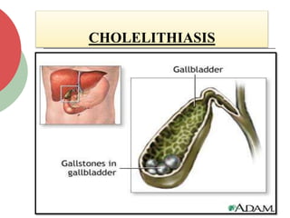
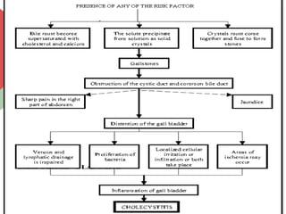
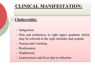
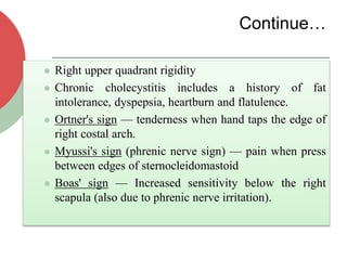
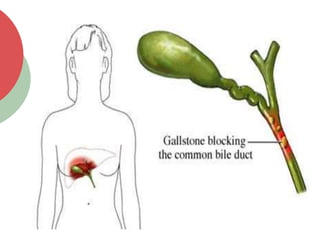
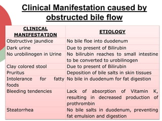
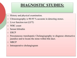
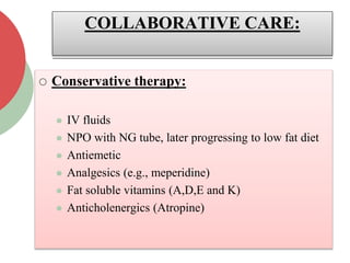
CHOLECYSTITIS AND
CHOLELITHIASIS
MS. VEDANTI PATEL
ASSI. PROFESSOR
INTRODUCTION:
 The most common disorder of biliary tract is
Cholelithiasis that is stones of the Gall Bladder.
 Cholecystitis is usually associated with
cholelithiasis.
 The stones may be lodged in the neck of the
Gall Bladder or in the cystic duct. Cholecystitis
may be acute or chronic. These conditions
usually occur together.
DEFINITION:
 Cholecystitis: It is defined as the
inflammation of the Gall Bladder.
 Cholelithiasis: Stones in the Gall Bladder
specially seen in the neck of the Gall
Bladder or the cystic duct.
CHOLECYSTITIS
COMMON RISK FACTORS:
 Incidence is higher in women, multiparous
women and persons over 40 years of age.
 Postmenopausal women on estrogen therapy
are at somewhat greater risk of having gall
bladder disease.
 Sedentary Life style
 Familial Tendency
 Obesity
CHOLECYSTITIS:
 Cholecystitis is mostly associated with
obstruction caused by gallstones or biliary
sludge.
 Acalculous cholecystitis is most often in older
adults, and in patients who have trauma,
extensive burns or recent surgery.
 Acalculous cholecystitis can also occur as a
result of prolonged immobility and fasting,
prolonged parenteral nutrition, and diabetes
mellitus.
Continue…
 Bacterial infection (Escherichia coli, streptococci
and salmonellae)
 Adhesions, neoplasm, anesthesia an narcotics
 In the majority of patients, gallstones are the cause
of acute cholecystitis.
 The process is one of physical obstruction of the
gallbladder by a gallstone, at the neck or in the
cystic duct. This obstruction results in increased
pressure in the gallbladder.
Continue…
 There are two factors which determine the
progression to acute cholecystitis — the degree of
obstruction and the duration of the obstruction.
 If the obstruction is partial and of short duration
the patient experiences biliary colic.
 If the obstruction is complete and of long duration
the patient develops acute cholecystitis.
CHOLELITHIASIS:
 The actual cause of gall stones is unknown.
 Cholelithiasis develops when the balance that keeps
cholesterol, bile salts, and calcium in solution is
altered due to that precipitation of these substances
occur. Conditions which upset these balance
includes:
 Infection
 Disturbances in the metabolism of cholesterol
CHOLELITHIASIS
Continue…
 Other components of bile that precipitate into stones
are bile salts, bilirubin, calcium and protein.
 In addition, increased levels of the
hormone estrogen, as a result of pregnancy or
hormonal therapy, or the use of combined (estrogen-
containing) forms of hormonal contraception, may
increase cholesterol levels in bile and also decrease
gallbladder movement, resulting in gallstone
formation.
Continue…
 The stones may remain in the gall bladder or
migrate to the cystic duct or to the common bile
duct. They cause pain as they pass to the duct or
else they may obstruct the duct.
 Additionally, prolonged use of proton pump
inhibitors has been shown to decrease gallbladder
function, potentially leading to gallstone
formation.
Pathophysiology of
Cholelithiasis and
Cholecystitis
CLINICAL MANIFESTATION:
 Cholecystitis:
 Indigestion
 Pain and tenderness in right upper quadrant which
may be referred to the right shoulder and scapula
 Nausea and vomiting
 Restlessness
 Diaphoresis
 Leucocytosis and fever due to infection
Continue…
 Right upper quadrant rigidity
 Chronic cholecystitis includes a history of fat
intolerance, dyspepsia, heartburn and flatulence.
 Ortner's sign — tenderness when hand taps the edge of
right costal arch.
 Myussi's sign (phrenic nerve sign) — pain when press
between edges of sternocleidomastoid
 Boas' sign — Increased sensitivity below the right
scapula (also due to phrenic nerve irritation).
CHOLELITHIASIS:
 Cholelithiasis may produce severe symptoms or
none at all.
 Many patients have silent cholelithiasis
 The severities of symptoms depend on whether
the stone are static or mobile and whether
obstruction is present or not.
 Spasm may be felt when a stone is moving to the
duct or passing through the duct.
Continue…
 Sometimes patient feels severe pain
referred to as Biliary Colic.
 The attacks of pain frequently occur after a
heavy meal or when the patient lies down.
 When total obstruction occurs, symptoms
related to bile blockage are manifested as
given in below table.
Clinical Manifestation caused by
obstructed bile flow
CLINICAL
MANIFESTATION
ETIOLOGY
Obstructive jaundice No bile floe into duodenum
Dark urine Due to present of Bilirubin
No urobilinogen in Urine No bilirubin reaches to small intestine
to be converted to urobilinogen
Clay colored stool Due to present of Bilirubin
Pruritus Deposition of bile salts in skin tissues
Intolerance for fatty
foods
No bile in duodenum for fat digestion
Bleeding tendencies Lack of absorption of Vitamin K,
resulting in decreased production of
prothrombin
Steatorrhea No bile salts in duodenum, preventing
fat emulsion and digestion
DIAGNOSTIC STUDIES:
 History and physical examination.
 Ultrasonography is 90-95 % accurate in detecting stones.
 Liver function test (LFT)
 WBC count
 Serum bilirubin
 ERCP
 Percutaneous transhepatic Cholangiography to diagnose obstructive
jaundice and to locate the stone within bile duct.
 MRCP
 Intraoperative cholangiogram
COLLABORATIVE CARE:
 Conservative therapy:
 IV fluids
 NPO with NG tube, later progressing to low fat diet
 Antiemetic
 Analgesics (e.g., meperidine)
 Fat soluble vitamins (A,D,E and K)
 Anticholenergics (Atropine)
Continue…
 Anticholenergics (Atropine)
 Antispasmodics
 Antibiotics for secondary infections
 For treatment of pruritus, Cholestyramine which
binds with the bile salts in intestine and increases
their excretion in feces. It may be administered in
powder form with milk or juice.
SURGICAL THERAPY:
Name of the surgery Description
Cholecystectomy Removal of gall bladder
Cholecystostomy Incision into gall bladder usually
for removal of stones
Choledocholithotomy Incision into common bile duct
for removal of stones
Cholecystogastrostomy Anastomosis between stomach
and gall bladder
Cholecystoduodenostomy Anastomosis between gall
bladder and duodenum to relieve
obstruction at distal end of
common bile duct
Laparoscopic Cholecystectomy Removal of gall bladder via
laparoscopy
Cholecystostomy
NURSING DIAGNOSIS:
 Acute pain secondary to biliary obstruction
 Ineffective coping related to nausea
 Deficient knowledge related to diagnosis
 Impaired gas exchange related to high abdominal
surgical incision.
 Impaired skin integrity related to altered biliary
drainage after surgical incision.
 Imbalanced nutrition related to inadequate bile
secretion.
NURSING MANAGEMENT:
 Provide nursing interventions during an acute
gallbladder attack.
 Intervene to relive pain; give prescribed
analgesics
 Promote adequate rest
 Administer IV fluids, monitor intake and output
 Monitor nasogastric tube and suctioning
 Administer antibiotics if prescribed.
Continue…
 Provide adequate nutrition.
 Assess nutritional status.
 Encourage a high-protein, high-carbohydrate,
low-fat diet.
 Give fat soluble vitamins
 Emphasis on more liquid diet when patient is
NPO and gradually progress towards low fat
diet.
Cholecystitis And Cholelithiasis slideshare

Cholecystitis And Cholelithiasis slideshare

  • 1.
  • 2.
    INTRODUCTION:  The mostcommon disorder of biliary tract is Cholelithiasis that is stones of the Gall Bladder.  Cholecystitis is usually associated with cholelithiasis.  The stones may be lodged in the neck of the Gall Bladder or in the cystic duct. Cholecystitis may be acute or chronic. These conditions usually occur together.
  • 3.
    DEFINITION:  Cholecystitis: Itis defined as the inflammation of the Gall Bladder.  Cholelithiasis: Stones in the Gall Bladder specially seen in the neck of the Gall Bladder or the cystic duct.
  • 4.
  • 5.
    COMMON RISK FACTORS: Incidence is higher in women, multiparous women and persons over 40 years of age.  Postmenopausal women on estrogen therapy are at somewhat greater risk of having gall bladder disease.  Sedentary Life style  Familial Tendency  Obesity
  • 6.
    CHOLECYSTITIS:  Cholecystitis ismostly associated with obstruction caused by gallstones or biliary sludge.  Acalculous cholecystitis is most often in older adults, and in patients who have trauma, extensive burns or recent surgery.  Acalculous cholecystitis can also occur as a result of prolonged immobility and fasting, prolonged parenteral nutrition, and diabetes mellitus.
  • 7.
    Continue…  Bacterial infection(Escherichia coli, streptococci and salmonellae)  Adhesions, neoplasm, anesthesia an narcotics  In the majority of patients, gallstones are the cause of acute cholecystitis.  The process is one of physical obstruction of the gallbladder by a gallstone, at the neck or in the cystic duct. This obstruction results in increased pressure in the gallbladder.
  • 8.
    Continue…  There aretwo factors which determine the progression to acute cholecystitis — the degree of obstruction and the duration of the obstruction.  If the obstruction is partial and of short duration the patient experiences biliary colic.  If the obstruction is complete and of long duration the patient develops acute cholecystitis.
  • 9.
    CHOLELITHIASIS:  The actualcause of gall stones is unknown.  Cholelithiasis develops when the balance that keeps cholesterol, bile salts, and calcium in solution is altered due to that precipitation of these substances occur. Conditions which upset these balance includes:  Infection  Disturbances in the metabolism of cholesterol
  • 10.
  • 11.
    Continue…  Other componentsof bile that precipitate into stones are bile salts, bilirubin, calcium and protein.  In addition, increased levels of the hormone estrogen, as a result of pregnancy or hormonal therapy, or the use of combined (estrogen- containing) forms of hormonal contraception, may increase cholesterol levels in bile and also decrease gallbladder movement, resulting in gallstone formation.
  • 12.
    Continue…  The stonesmay remain in the gall bladder or migrate to the cystic duct or to the common bile duct. They cause pain as they pass to the duct or else they may obstruct the duct.  Additionally, prolonged use of proton pump inhibitors has been shown to decrease gallbladder function, potentially leading to gallstone formation.
  • 13.
  • 15.
    CLINICAL MANIFESTATION:  Cholecystitis: Indigestion  Pain and tenderness in right upper quadrant which may be referred to the right shoulder and scapula  Nausea and vomiting  Restlessness  Diaphoresis  Leucocytosis and fever due to infection
  • 16.
    Continue…  Right upperquadrant rigidity  Chronic cholecystitis includes a history of fat intolerance, dyspepsia, heartburn and flatulence.  Ortner's sign — tenderness when hand taps the edge of right costal arch.  Myussi's sign (phrenic nerve sign) — pain when press between edges of sternocleidomastoid  Boas' sign — Increased sensitivity below the right scapula (also due to phrenic nerve irritation).
  • 17.
    CHOLELITHIASIS:  Cholelithiasis mayproduce severe symptoms or none at all.  Many patients have silent cholelithiasis  The severities of symptoms depend on whether the stone are static or mobile and whether obstruction is present or not.  Spasm may be felt when a stone is moving to the duct or passing through the duct.
  • 18.
    Continue…  Sometimes patientfeels severe pain referred to as Biliary Colic.  The attacks of pain frequently occur after a heavy meal or when the patient lies down.  When total obstruction occurs, symptoms related to bile blockage are manifested as given in below table.
  • 20.
    Clinical Manifestation causedby obstructed bile flow CLINICAL MANIFESTATION ETIOLOGY Obstructive jaundice No bile floe into duodenum Dark urine Due to present of Bilirubin No urobilinogen in Urine No bilirubin reaches to small intestine to be converted to urobilinogen Clay colored stool Due to present of Bilirubin Pruritus Deposition of bile salts in skin tissues Intolerance for fatty foods No bile in duodenum for fat digestion Bleeding tendencies Lack of absorption of Vitamin K, resulting in decreased production of prothrombin Steatorrhea No bile salts in duodenum, preventing fat emulsion and digestion
  • 21.
    DIAGNOSTIC STUDIES:  Historyand physical examination.  Ultrasonography is 90-95 % accurate in detecting stones.  Liver function test (LFT)  WBC count  Serum bilirubin  ERCP  Percutaneous transhepatic Cholangiography to diagnose obstructive jaundice and to locate the stone within bile duct.  MRCP  Intraoperative cholangiogram
  • 22.
    COLLABORATIVE CARE:  Conservativetherapy:  IV fluids  NPO with NG tube, later progressing to low fat diet  Antiemetic  Analgesics (e.g., meperidine)  Fat soluble vitamins (A,D,E and K)  Anticholenergics (Atropine)
  • 23.
    Continue…  Anticholenergics (Atropine) Antispasmodics  Antibiotics for secondary infections  For treatment of pruritus, Cholestyramine which binds with the bile salts in intestine and increases their excretion in feces. It may be administered in powder form with milk or juice.
  • 25.
    SURGICAL THERAPY: Name ofthe surgery Description Cholecystectomy Removal of gall bladder Cholecystostomy Incision into gall bladder usually for removal of stones Choledocholithotomy Incision into common bile duct for removal of stones Cholecystogastrostomy Anastomosis between stomach and gall bladder Cholecystoduodenostomy Anastomosis between gall bladder and duodenum to relieve obstruction at distal end of common bile duct Laparoscopic Cholecystectomy Removal of gall bladder via laparoscopy
  • 26.
  • 27.
    NURSING DIAGNOSIS:  Acutepain secondary to biliary obstruction  Ineffective coping related to nausea  Deficient knowledge related to diagnosis  Impaired gas exchange related to high abdominal surgical incision.  Impaired skin integrity related to altered biliary drainage after surgical incision.  Imbalanced nutrition related to inadequate bile secretion.
  • 28.
    NURSING MANAGEMENT:  Providenursing interventions during an acute gallbladder attack.  Intervene to relive pain; give prescribed analgesics  Promote adequate rest  Administer IV fluids, monitor intake and output  Monitor nasogastric tube and suctioning  Administer antibiotics if prescribed.
  • 29.
    Continue…  Provide adequatenutrition.  Assess nutritional status.  Encourage a high-protein, high-carbohydrate, low-fat diet.  Give fat soluble vitamins  Emphasis on more liquid diet when patient is NPO and gradually progress towards low fat diet.