Download to read offline

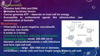
![Frei’s Test :-
• Also called as “skin test”
• 0.1ml antigen is injected intradermally into forearm
• The antigen is prepared either by
a] heating diluted bubo pus to 60°c for 3 hours to destroy infectivity
b] growing organisms in yolk sac and using purified and phenolised elementary bodies
• After 48 hours patient is examined for induration
• Positive reaction – development of induration of 7mm or more is seen, after 2-6 weeks of
infection
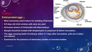
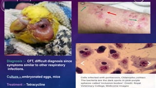
Culture :- embryonated eggs, mice
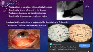
Treatment :- varies with stage of disease, Sulphadiazine is the drug of choice, Tetracycline
K R MICRO NOTES 11](https://image.slidesharecdn.com/chlamydiaekr-240404151237-e184d959/85/Chlamydiae-The-silent-killer-K-R-pptx-11-320.jpg)
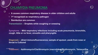


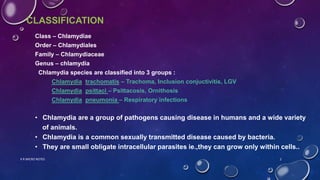
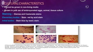
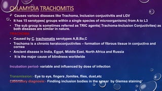
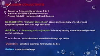
Chlamydiae are obligate intracellular parasites that cause disease in humans and animals. There are three main species: Chlamydia trachomatis, C. psittaci, and C. pneumoniae. C. trachomatis causes trachoma, inclusion conjunctivitis, and lymphogranuloma venereum. It has 15 serotypes and is transmitted sexually or from eye to eye. C. psittaci causes psittacosis in birds and ornithosis in humans via inhalation of bird droppings. C. pneumoniae is a common cause of respiratory infections in humans transmitted through coughing or sneezing. Chlamydiae have both RNA and DNA


![Polymer [ बहुलक ] Chemistry Notes PDF - Irfanullah Mehar - JJ Sir Chemistry.pdf](https://cdn.slidesharecdn.com/ss_thumbnails/polymerchemistrynotespdf-irfanullahmehar-jjsirchemistry-260210172118-3f9b37f7-thumbnail.jpg?width=640&height=640&fit=bounds)


