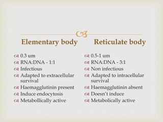
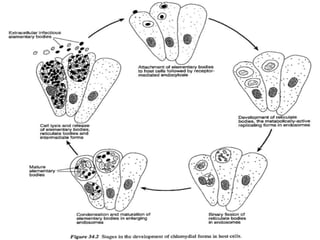
Submitted by: Dr. Deepika Sheoran
Submitted to: Dr. Parveen Kumar
Dr. Archana Sharma

Kingdom- Bacteria
Phylum- Chlamydiae
Class- Chlamydiae
Order- Chlamydiales
Family- Chlamydiaceae
Genus- Chlamydia
Taxonomy

Spherical intracellular bacteria with unique
developmental cycle
Staining procedures - modified Ziehl-Neelsen and
Giemsa
Unable to synthesize ATP and replicate only in living
cells
Cell walls lack peptidoglycan but contain genus-
specific lipopolysaccharide
Produce respiratory, enteric, plural and reproductive
tract diseases in animals and humans (Zoonotic)
Key Points

They replicate within cytoplasmic vacuoles
in host cells.
On account of their apparent inability to
generate ATP, with resultant dependence on
host cell metabolism, they have been termed
'energy parasites'.


 Formerly a single genus and 4 species, Chlamydia
trachomatis, C. psittaci, C. pneumoniae and C.
pecorum, were recognised.
 This classification was based on phenotypic
characteristics such as: host preference, inclusion
morphology, iodine staining for the presence of
glycogen, and sulphonamide susceptibility.
 However, recent nucleic acid sequencing studies of
the 16s and 23s rRNA genes confirm two distinct
lineages

 In the developmental cycle, infectious and
reproductive forms are morphologically distinct:
 1. Infectious extracellular form: Elementary bodies
 2. Reproductive form: Reticulate bodies

Small (200 to 300 nm)
Metabolically inert and osmotically stable
The periplasmic space does not contain a detectable
peptidoglycan layer and the EB relies on disulphide
cross-linked envelope proteins for osmotic stability
Elementary bodies enter host cells by receptor-
mediated endocytosis.
Elementary bodies

 Acidification of endosome and fusion with
lysosomes are prevented.
 Structural reorganization within the pathogen, of
several hours duration, results in the conversion of
an EB into a reticulate body (RB).

 The RB, about I um in diameter
 Metabolically active, osmotically fragile
 Replication : binary fission within the endosome.
 The endosome and its contents, when stained, is called an
inclusion.
 When a number of inclusions containing RBs of C.
trachomatis are formed in an infected cell, fusion of these
structures may occur.
 After 20 hours cycle becomes asynchronous with some
RBs continuing to divide while others condense and
mature to form EBs.
 In general, replication continues for up to 72 hours after
infection when the host cell lyses releasing EBs, RBs
and intermediate forms.
Reticulate body

Elementary body
 0.3 um
 RNA:DNA - 1:1
 Infectious
 Adapted to extracellular
survival
 Haemagglutinin present
 Induce endocytosis
 Metabollically active
Reticulate body
 0.5-1 um
 RNA:DNA - 3:1
 Non infectious
 Adapted to intracellular
survival
 Haemagglutinin absent
 Doesn’t induce
 Metabolically active



 In presence of gamma interferon or penicillin
 Or when the availability of tryptophan or cysteine is
limited
 Important in development of immunopathological
changes in humans associated with trachoma &
pelvic inflammatory reaction
Delay in replication

 Only RBs are susceptible to Penicillin G
 Penicillin G treated bacteria loss their control over
host cell apoptotic pathways & no longer express
pre-16S rRNA
 Chlamydia within inclusion fuse with lysosomal
compartments in pG treated cells. That lead to
recruitment of cathepsin D as early as pG treatment
is given ( an event preceding bacterial death by
several hours)
 Results in degradation of bacteria
Penicillin Mechanism

 The gastrointestinal tract - usual site
 Infections are often subclinical and persistent.
 Faecal shedding of the organisms
 EBs can survive in environment for several days.
Habitat

1. LPS- Lipoplysaccharide is a major surface
component- Responsible for infectivity of
host epithelial cells
2. MOMP (Major outer membrane protein)-
. Potential chlamydial cytoadhesin
3. OMC proteins- Bacterial adhesin that
mediates binding to heparan sulphate like
GAGs on host epithelial & endothelial cells
Major antigens

4. POMP (Polymorphic outer membrane
proteins)- Important role in avoidance of
host immune defences
5. Type 3 secretory system- Mechanism
capable of promoting chlamydial virulence
6. Heat shock proteins

 When bacteria is in stress certain system get
activated & one of these is Heat shock protein ( it is
like SOS response of bacteria when DNA damage
occur)
 These secreted proteins will cause high fever &
inflammatory conditions
 Chlamydial heat shock protein Hsp60 is a homolog
of E.coli GroEL, has been identified as one protein
capable of eliciting intense mononuclear
inflammation
Heat shock protein

 Many chlamydial infections are asymptomatic,
particularly when they are localized in superficial
epithelia
 Chlamydiae possess a number of heat shock proteins-
partially homologous to heat shock proteins in other
bacteria and to a number of human mitochondrial
proteins.
 Repeated stimulation of the immune system with these
proteins contributes to the delayed-type hypersensitivity
associated with trachoma and inflammatory pelvic
disease in man.
Pathogenesis

 Chlamydiae infect over 130 species of birds and a
large number of mammalian species including man.
 In recent years isolations have also been made from
invertebrate species

Intracellular survival of chlamydia is
dependent on the ability of organism to
prevent phagolysosome fusion
Survival in phagosome

 On basis of History, clinical signs, pathological
changes
 Isolation- Transport media- Sucrose phosphate
glutamate medium supplemented with Foetal calf
serum, aminoglycoside antibiotics & an antifungal
agent
 Samples should be kept at 4°C (thermolabile)
 For long term storage – frozen at -70°C
Diagnosis

 Direct microscopy-Smears or histological sections of
organs from aborted foetuses or from liver and
spleen in cases of avian chlamydiosis
 Placental smears from cases of chlamydial abortion
 Staining- modified Ziehl-Neelsen
 Giemsa
 Modified Machiavello
 Castaneda methods.
 Methylene blue-stained smears can be examined by
darkfield microscopy.

 Commercial kit sets- employing ELISA developed
for the detection of C. trachomatis.
 Many of these kit sets detect chlamydial
lipopolysaccharide (LPS) which is common to all
Chlamydia and Chlamydophila species.
 Chlamydiae can be isolated either in embryonated
eggs, inoculated into the yolk sac, or in a number of
continuous cell lines such as McCoy, L929, baby
hamster kidney and Vero.

 Attachment to cells- greatly enhanced by centrifugation of
the sample onto the monolayer.
 The sensitivity of the isolation procedure is also increased
by the use of non-replicating cells.
 This is achieved by the addition of cytotoxic chemicals
such as cycloheximide, 5-iodo-2-deoxyuridine,
cytochalasinB and emetine to the cell culture medium.
 After 2 to 3 days incubation at 37°C the monolayer is
fixed, stained and examined for the presence of
chlamydial inclusions.
 Antibiotics to which chlamydiae are sensitive -
oxytetracycline, erythromycin and penicillin, should not
be used in the cell culture medium.

 PCR techniques- Detection of Chlamydeal DNA in
samples
 Serological procedures-complement fixation, ELISA,
indirect immunofluorescence and micro-
immunofluorescence.
 Although the complement fixation test is the most
widely recognised serological test, time-consuming
and only moderately sensitive.

 Interpretation of results is complicated by the fact
that many of the available serological procedures
detect antibodies against chlamydial LPS and
therefore do not allow differentiation of the
chlamydial species involved in the infection.
 In addition, there is cross-reactivity between the LPS
of chlamydiae and that of some other Gram-negative
bacteria (like Neisseria gonorrhoeae)

 Ranging from inapparent infections to severe systemic
infections
 Conjunctivitis
 Arthritis
 Abortion
 Urethritis
 Enteritis
 Pneumonia
 Encephalomyelitis
 Human infections acquired from psittacine species -
psittacosis
 while those from other avian species are termed
ornithosis
Clinical infections

Chlamydial infections of veterinary &
medical importance


 Caused by- C. abortus
 Primarily a disease of intensively managed flocks
 Economically significant in most sheep producing
countries
Enzootic abortion of ewes(EAE)

 Large numbers of chlamydiae are shed in placentas
and uterine discharges from affected ewes.
 Organisms can remain viable in the environment for
several days at low temperatures.
 Infection occurs by ingestion
 Ewes infected late in pregnancy do not usually abort
but may do so in the next pregnancy.
 Infection early in pregnancy can result in abortion
during that pregnancy.
 Ewe lambs may acquire infection during the
neonatal period and abort during their first
pregnancy.
Epidemiology

 Abortion during late pregnancy or by the birth of
premature weak lambs.
 Aborted lambs are well developed and fresh.
 Necrosis of cotyledons and oedema of adjacent
intercotyledonary tissue in affected placentas along
with a dirty pink uterine exudate.
 Aborting ewes rarely show evidence of clinical
disease and their subsequent fertility is usually
unimpaired.
Clinical signs

 Well-preserved aborted lambs and evidence of
necrotic placentitis are suggestive of EAE.
 Large numbers of EBs can be demonstrated in
placental smears using suitable staining procedures.
Diagnosis


 Antibiotics- long-acting oxytetracycline.
 Transmission in an affected flock can be reduced by
isolating all aborted ewes for 2 to 3 weeks, removing
and destroying all placentas, thoroughly cleaning areas
where abortions occurred and administering long-acting
oxytetracycline to ewes which have not yet lambed.
 Eradicate the disease by vaccination & culling.
 A live attenuated vaccine – administered to ewes prior to
breeding.
 An inactivated vaccine is also available which can be
used in pregnant animals.
 ' Chlamydophila abortus infection is serious and potentially
life-threatening for pregnant women who should avoid
contact with ewes during the lambing season
Treatment & Control

 Chlamydophila felis (formerly known as feline strains
of C. psittaci) is associated with conjunctivitis and
less commonly rhinitis.
 Feline pneumonitis, the original name for feline
chlamydiosis, is now considered a misnomer
because of the rarity of lower respiratory tract
infection caused by C. felis in cats.
Feline chlamydiosis

 Serological surveys revealed that upto 10% cats
become infected with C. felis
 Transmitted by direct or indirect contact with
conjunctival or nasal secretions.
 Organisms may also be shed from the reproductive
tract
Epidemiology

 I.P- 5 days
 Unilateral or bilateral conjunctival congestion,
clear ocular discharge and blepharospasm
 If secondary infection with organisms such as
Mycoplasma felis and Staphylococcus species occurs,
the ocular discharge may become mucopurulent.
 Conjunctivitis may be accompanied by sneezing and
nasal discharge
 Usually resolves without treatment in a few weeks.
Clinical signs


 Stained Conjunctival smears may reveal
intracytoplasmic inclusions
Diagnosis

 Antibiotics- all in-contact cats should be treated at
the same time.
 Modified live vaccines- parenteral inoculation
 Inadvertent intraocular administration of the vaccine
can result in conjunctivitis
 A small number of cases of conjunctivitis in humans
involving C. felis have been reported.
Treatment & Control

 Neurological disease
 Caused by- C. pecorum
 Usually under 3 years of age develop-
 High fever, incoordination, depression, excessive
salivation, diarrhoea
 Terminally become recombinant & develop
opisthotonous
 Course of disease – about 2 weeks
 Mortality rate – 50%
Sporadic bovine
encephalomyelitis

 Clinical signs
 Presence of Serofibrinous peritonitis
 Histopathological changes in brain
 Isolation of organism from brain tissue
 High doses of antibiotics such as Tetracyclines &
Tyloin may be effective
 No vaccines are available
Diagnosis

 Caused by C.psittaci in psittacine birds
 EPIDEMIOLOGY-
 Wide range of wild & domestic avian species are
susceptible
 Organism is present in respiratory discharges &
faeces of infected birds
 Inhalation or Ingetion
 Subclinical infection_ common
Avian chlamydiosis

 I.P- 10 days
 Generalized infection, affecting particularly
digestive & respiratory tracts
 Loss of condition
 Nasal & occular discharges
 Diarrhoea
 Respiratory distress
 P.M findings- Hepatospleenomegaly, air sacculitis,
peritonitis
Clinical signs


 Tetracycline- Antibiotic of choice
 Imported birds – Held in quarantine & receive
tetracycline mediated feeds
 Proper husbandry & suitable transportation-
minimize occurance of clinical disease
 Isolates are Zoonotic-infection commonly follows
aerosol inhalation. Pulmonary involvement is
common. Meningitis or meningoencephalitis may
develop in severely affected individuals
 No vaccine available
Treatment & Control

THANKS

CHLAMYDIA.pptx veterinary microbiology .

  • 1.
    Submitted by: Dr.Deepika Sheoran Submitted to: Dr. Parveen Kumar Dr. Archana Sharma
  • 2.
     Kingdom- Bacteria Phylum- Chlamydiae Class-Chlamydiae Order- Chlamydiales Family- Chlamydiaceae Genus- Chlamydia Taxonomy
  • 3.
     Spherical intracellular bacteriawith unique developmental cycle Staining procedures - modified Ziehl-Neelsen and Giemsa Unable to synthesize ATP and replicate only in living cells Cell walls lack peptidoglycan but contain genus- specific lipopolysaccharide Produce respiratory, enteric, plural and reproductive tract diseases in animals and humans (Zoonotic) Key Points
  • 4.
     They replicate withincytoplasmic vacuoles in host cells. On account of their apparent inability to generate ATP, with resultant dependence on host cell metabolism, they have been termed 'energy parasites'.
  • 5.
  • 6.
      Formerly asingle genus and 4 species, Chlamydia trachomatis, C. psittaci, C. pneumoniae and C. pecorum, were recognised.  This classification was based on phenotypic characteristics such as: host preference, inclusion morphology, iodine staining for the presence of glycogen, and sulphonamide susceptibility.  However, recent nucleic acid sequencing studies of the 16s and 23s rRNA genes confirm two distinct lineages
  • 7.
      In thedevelopmental cycle, infectious and reproductive forms are morphologically distinct:  1. Infectious extracellular form: Elementary bodies  2. Reproductive form: Reticulate bodies
  • 8.
     Small (200 to300 nm) Metabolically inert and osmotically stable The periplasmic space does not contain a detectable peptidoglycan layer and the EB relies on disulphide cross-linked envelope proteins for osmotic stability Elementary bodies enter host cells by receptor- mediated endocytosis. Elementary bodies
  • 9.
      Acidification ofendosome and fusion with lysosomes are prevented.  Structural reorganization within the pathogen, of several hours duration, results in the conversion of an EB into a reticulate body (RB).
  • 10.
      The RB,about I um in diameter  Metabolically active, osmotically fragile  Replication : binary fission within the endosome.  The endosome and its contents, when stained, is called an inclusion.  When a number of inclusions containing RBs of C. trachomatis are formed in an infected cell, fusion of these structures may occur.  After 20 hours cycle becomes asynchronous with some RBs continuing to divide while others condense and mature to form EBs.  In general, replication continues for up to 72 hours after infection when the host cell lyses releasing EBs, RBs and intermediate forms. Reticulate body
  • 11.
     Elementary body  0.3um  RNA:DNA - 1:1  Infectious  Adapted to extracellular survival  Haemagglutinin present  Induce endocytosis  Metabollically active Reticulate body  0.5-1 um  RNA:DNA - 3:1  Non infectious  Adapted to intracellular survival  Haemagglutinin absent  Doesn’t induce  Metabolically active
  • 12.
  • 13.
  • 14.
      In presenceof gamma interferon or penicillin  Or when the availability of tryptophan or cysteine is limited  Important in development of immunopathological changes in humans associated with trachoma & pelvic inflammatory reaction Delay in replication
  • 15.
      Only RBsare susceptible to Penicillin G  Penicillin G treated bacteria loss their control over host cell apoptotic pathways & no longer express pre-16S rRNA  Chlamydia within inclusion fuse with lysosomal compartments in pG treated cells. That lead to recruitment of cathepsin D as early as pG treatment is given ( an event preceding bacterial death by several hours)  Results in degradation of bacteria Penicillin Mechanism
  • 16.
      The gastrointestinaltract - usual site  Infections are often subclinical and persistent.  Faecal shedding of the organisms  EBs can survive in environment for several days. Habitat
  • 17.
     1. LPS- Lipoplysaccharideis a major surface component- Responsible for infectivity of host epithelial cells 2. MOMP (Major outer membrane protein)- . Potential chlamydial cytoadhesin 3. OMC proteins- Bacterial adhesin that mediates binding to heparan sulphate like GAGs on host epithelial & endothelial cells Major antigens
  • 18.
     4. POMP (Polymorphicouter membrane proteins)- Important role in avoidance of host immune defences 5. Type 3 secretory system- Mechanism capable of promoting chlamydial virulence 6. Heat shock proteins
  • 19.
      When bacteriais in stress certain system get activated & one of these is Heat shock protein ( it is like SOS response of bacteria when DNA damage occur)  These secreted proteins will cause high fever & inflammatory conditions  Chlamydial heat shock protein Hsp60 is a homolog of E.coli GroEL, has been identified as one protein capable of eliciting intense mononuclear inflammation Heat shock protein
  • 20.
      Many chlamydialinfections are asymptomatic, particularly when they are localized in superficial epithelia  Chlamydiae possess a number of heat shock proteins- partially homologous to heat shock proteins in other bacteria and to a number of human mitochondrial proteins.  Repeated stimulation of the immune system with these proteins contributes to the delayed-type hypersensitivity associated with trachoma and inflammatory pelvic disease in man. Pathogenesis
  • 21.
      Chlamydiae infectover 130 species of birds and a large number of mammalian species including man.  In recent years isolations have also been made from invertebrate species
  • 22.
     Intracellular survival ofchlamydia is dependent on the ability of organism to prevent phagolysosome fusion Survival in phagosome
  • 23.
      On basisof History, clinical signs, pathological changes  Isolation- Transport media- Sucrose phosphate glutamate medium supplemented with Foetal calf serum, aminoglycoside antibiotics & an antifungal agent  Samples should be kept at 4°C (thermolabile)  For long term storage – frozen at -70°C Diagnosis
  • 24.
      Direct microscopy-Smearsor histological sections of organs from aborted foetuses or from liver and spleen in cases of avian chlamydiosis  Placental smears from cases of chlamydial abortion  Staining- modified Ziehl-Neelsen  Giemsa  Modified Machiavello  Castaneda methods.  Methylene blue-stained smears can be examined by darkfield microscopy.
  • 25.
      Commercial kitsets- employing ELISA developed for the detection of C. trachomatis.  Many of these kit sets detect chlamydial lipopolysaccharide (LPS) which is common to all Chlamydia and Chlamydophila species.  Chlamydiae can be isolated either in embryonated eggs, inoculated into the yolk sac, or in a number of continuous cell lines such as McCoy, L929, baby hamster kidney and Vero.
  • 26.
      Attachment tocells- greatly enhanced by centrifugation of the sample onto the monolayer.  The sensitivity of the isolation procedure is also increased by the use of non-replicating cells.  This is achieved by the addition of cytotoxic chemicals such as cycloheximide, 5-iodo-2-deoxyuridine, cytochalasinB and emetine to the cell culture medium.  After 2 to 3 days incubation at 37°C the monolayer is fixed, stained and examined for the presence of chlamydial inclusions.  Antibiotics to which chlamydiae are sensitive - oxytetracycline, erythromycin and penicillin, should not be used in the cell culture medium.
  • 27.
      PCR techniques-Detection of Chlamydeal DNA in samples  Serological procedures-complement fixation, ELISA, indirect immunofluorescence and micro- immunofluorescence.  Although the complement fixation test is the most widely recognised serological test, time-consuming and only moderately sensitive.
  • 28.
      Interpretation ofresults is complicated by the fact that many of the available serological procedures detect antibodies against chlamydial LPS and therefore do not allow differentiation of the chlamydial species involved in the infection.  In addition, there is cross-reactivity between the LPS of chlamydiae and that of some other Gram-negative bacteria (like Neisseria gonorrhoeae)
  • 29.
      Ranging frominapparent infections to severe systemic infections  Conjunctivitis  Arthritis  Abortion  Urethritis  Enteritis  Pneumonia  Encephalomyelitis  Human infections acquired from psittacine species - psittacosis  while those from other avian species are termed ornithosis Clinical infections
  • 30.
     Chlamydial infections ofveterinary & medical importance
  • 31.
  • 32.
      Caused by-C. abortus  Primarily a disease of intensively managed flocks  Economically significant in most sheep producing countries Enzootic abortion of ewes(EAE)
  • 33.
      Large numbersof chlamydiae are shed in placentas and uterine discharges from affected ewes.  Organisms can remain viable in the environment for several days at low temperatures.  Infection occurs by ingestion  Ewes infected late in pregnancy do not usually abort but may do so in the next pregnancy.  Infection early in pregnancy can result in abortion during that pregnancy.  Ewe lambs may acquire infection during the neonatal period and abort during their first pregnancy. Epidemiology
  • 34.
      Abortion duringlate pregnancy or by the birth of premature weak lambs.  Aborted lambs are well developed and fresh.  Necrosis of cotyledons and oedema of adjacent intercotyledonary tissue in affected placentas along with a dirty pink uterine exudate.  Aborting ewes rarely show evidence of clinical disease and their subsequent fertility is usually unimpaired. Clinical signs
  • 35.
      Well-preserved abortedlambs and evidence of necrotic placentitis are suggestive of EAE.  Large numbers of EBs can be demonstrated in placental smears using suitable staining procedures. Diagnosis
  • 36.
  • 37.
      Antibiotics- long-actingoxytetracycline.  Transmission in an affected flock can be reduced by isolating all aborted ewes for 2 to 3 weeks, removing and destroying all placentas, thoroughly cleaning areas where abortions occurred and administering long-acting oxytetracycline to ewes which have not yet lambed.  Eradicate the disease by vaccination & culling.  A live attenuated vaccine – administered to ewes prior to breeding.  An inactivated vaccine is also available which can be used in pregnant animals.  ' Chlamydophila abortus infection is serious and potentially life-threatening for pregnant women who should avoid contact with ewes during the lambing season Treatment & Control
  • 38.
      Chlamydophila felis(formerly known as feline strains of C. psittaci) is associated with conjunctivitis and less commonly rhinitis.  Feline pneumonitis, the original name for feline chlamydiosis, is now considered a misnomer because of the rarity of lower respiratory tract infection caused by C. felis in cats. Feline chlamydiosis
  • 39.
      Serological surveysrevealed that upto 10% cats become infected with C. felis  Transmitted by direct or indirect contact with conjunctival or nasal secretions.  Organisms may also be shed from the reproductive tract Epidemiology
  • 40.
      I.P- 5days  Unilateral or bilateral conjunctival congestion, clear ocular discharge and blepharospasm  If secondary infection with organisms such as Mycoplasma felis and Staphylococcus species occurs, the ocular discharge may become mucopurulent.  Conjunctivitis may be accompanied by sneezing and nasal discharge  Usually resolves without treatment in a few weeks. Clinical signs
  • 41.
  • 42.
      Stained Conjunctivalsmears may reveal intracytoplasmic inclusions Diagnosis
  • 43.
      Antibiotics- allin-contact cats should be treated at the same time.  Modified live vaccines- parenteral inoculation  Inadvertent intraocular administration of the vaccine can result in conjunctivitis  A small number of cases of conjunctivitis in humans involving C. felis have been reported. Treatment & Control
  • 44.
      Neurological disease Caused by- C. pecorum  Usually under 3 years of age develop-  High fever, incoordination, depression, excessive salivation, diarrhoea  Terminally become recombinant & develop opisthotonous  Course of disease – about 2 weeks  Mortality rate – 50% Sporadic bovine encephalomyelitis
  • 45.
      Clinical signs Presence of Serofibrinous peritonitis  Histopathological changes in brain  Isolation of organism from brain tissue  High doses of antibiotics such as Tetracyclines & Tyloin may be effective  No vaccines are available Diagnosis
  • 46.
      Caused byC.psittaci in psittacine birds  EPIDEMIOLOGY-  Wide range of wild & domestic avian species are susceptible  Organism is present in respiratory discharges & faeces of infected birds  Inhalation or Ingetion  Subclinical infection_ common Avian chlamydiosis
  • 47.
      I.P- 10days  Generalized infection, affecting particularly digestive & respiratory tracts  Loss of condition  Nasal & occular discharges  Diarrhoea  Respiratory distress  P.M findings- Hepatospleenomegaly, air sacculitis, peritonitis Clinical signs
  • 48.
  • 49.
      Tetracycline- Antibioticof choice  Imported birds – Held in quarantine & receive tetracycline mediated feeds  Proper husbandry & suitable transportation- minimize occurance of clinical disease  Isolates are Zoonotic-infection commonly follows aerosol inhalation. Pulmonary involvement is common. Meningitis or meningoencephalitis may develop in severely affected individuals  No vaccine available Treatment & Control
  • 50.