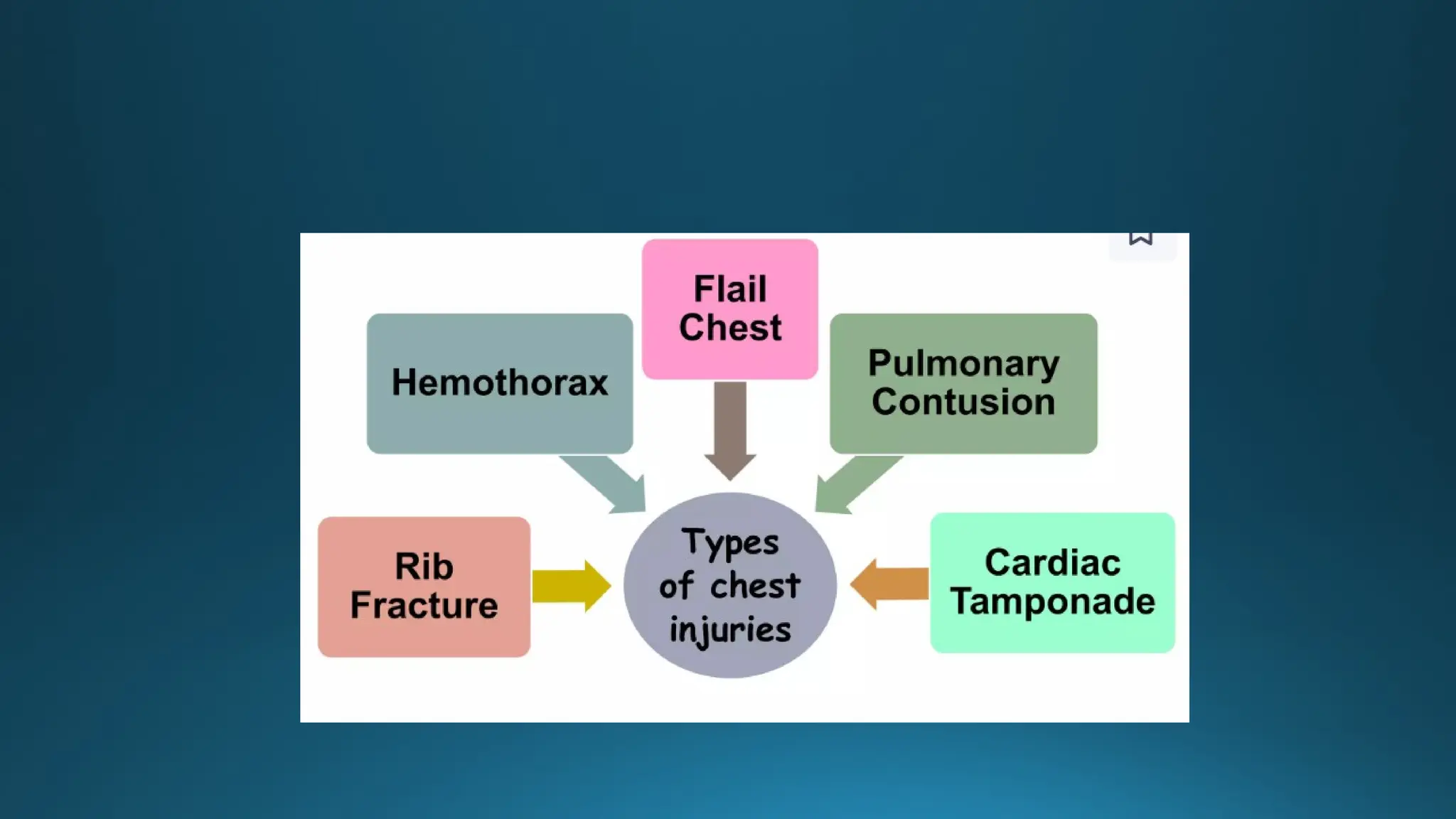
• Thoracic injuryaccounts for 25% of all severe injuries
• In a further 25%, it may be a significant contributor to the
subsequent death of the patient
• In most of these patients, the cause of death is
haemorrhage
• About 80% of patients with chest injury can be managed
nonoperatively
• The key to a good outcome is early physiological
resuscitation followed by a correct diagnosis
5.
Investigation
• Clinical examination,supplemented by chest radiography
• Ultrasound – extended focused assessment with sonar for
trauma Ultrasound can be used to differentiate between
contusion and the actual presence of blood
• The technique uses sonar assessment in the chest, looking
for a cardiac tamponade or free blood and air in the
hemithorax on each side, and assessment for blood in the
abdominal cavity, in the paracolic gutters,
subdiaphragmatic spaces and pelvis
6.
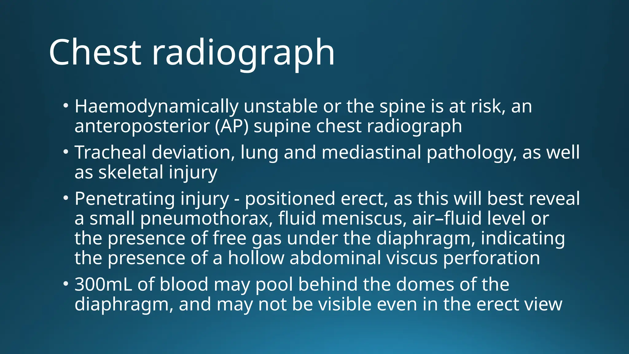
Chest radiograph
• Haemodynamicallyunstable or the spine is at risk, an
anteroposterior (AP) supine chest radiograph
• Tracheal deviation, lung and mediastinal pathology, as well
as skeletal injury
• Penetrating injury - positioned erect, as this will best reveal
a small pneumothorax, fluid meniscus, air–fluid level or
the presence of free gas under the diaphragm, indicating
the presence of a hollow abdominal viscus perforation
• 300mL of blood may pool behind the domes of the
diaphragm, and may not be visible even in the erect view
7.
• Should alertthe clinician to the possibility of adjacent
thoracic or abdominal visceral injury
• Rupture of the thoracic aorta can be related to fractures of
the first and second rib, bilateral clavicular fracture and
fracture of the sternum, thoracic spine or scapula
• Fracture of the lower ribs can be related to injury of liver or
spleen
• Fracture of ribs, irrespective of site, can be related to injury
to the lung parenchyma or thoracic wall vasculature,
causing pneumothorax, haemothorax or lung contusion.
8.
Computed tomography scan
•CT scan with contrast allows for three dimensional
reconstruction of the chest and abdomen, as well as of the
bony skeleton
• Blunt -definition of fractures, as well as showing
haematomas, pneumothoraces and pulmonary contusion
• Penetrating - show the track or presence of the missile and
allow the proper planning of definitive surgery
Tension pneumothorax
• Atension pneumothorax develops
when a ‘oneway valve’ air leak occurs
either from the lung or through the
chest wall.
• Air is sucked into the thoracic cavity
without any means of escape,
completely collapsing then
compressing the affected lung. The
mediastinum is displaced to the
opposite side, decreasing venous
return and compressing the opposite
lung
13.
• The mostcommon causes are penetrating chest trauma,
blunt chest trauma with a parenchymal lung injury and air
leak that did not spontaneously close, iatrogenic lung
injury (e.g. due to central venepuncture) and mechanical
positive pressure ventilation.
• The clinical presentation is dramatic. The patient is
increasingly restless with tachypnoea, dyspnoea and
distended neck veins (similar to pericardial tamponade).
• Clinical examination may reveal tracheal deviation; this is
a late finding and is not necessary to clinically confirm
diagnosis
14.
• There willalso be hyperresonance and decreased or absent
breath sounds over the affected hemithorax
• Tension pneumothorax is a clinical diagnosis and treatment
should never be delayed by waiting for radiological
confirmation
• Treatment consists of immediate decompression, initially by
rapid insertion of a large bore cannula into the second
intercostal space in the midclavicular line of the affected side,
then followed by insertion of a chest tube through the fifth
intercostal space in the anterior axillary line
17.
Open pneumothorax (‘suckingchest
wound’)
• This is due to a large open defect in the
chest (>3cm), leading to immediate
equilibration between intrathoracic and
atmospheric pressure
• If the opening in the chest wall exceeds
about two thirds of the diameter of the
trachea, then with each inspiratory cycle,
air will be preferentially drawn through
the defect, rather than through the
trachea
18.
• Air accumulatesin the
hemithorax (rather than in
the lung) with each
inspiration, leading to
profound hypoventilation on
the affected side and hypoxia.
If there is a valvular effect,
increasing amounts of air in
the pleura will result in a
tension pneumothorax
19.
Massive haemothorax
• Themost common cause of massive haemothorax in blunt
injury is continuing bleeding from torn intercostal vessels
or occasionally from the internal mammary artery
secondary to fractures of the ribs
• In penetrating injury, a variety of viscera, both thoracic
and abdominal (with blood leaking through a hole in the
diaphragm from the positive pressure abdomen into the
negative pressure thorax) may be involved
20.
• Accumulation ofblood in a haemothorax can significantly
compromise respiratory efforts, compressing the lung and
preventing adequate ventilation
• Presentation is with haemorrhagic shock, flat neck veins,
unilateral absence of breath sounds and dullness to
percussion
• The initial treatment consists of correcting the
hypovolaemic shock, insertion of an intercostal drain and,
in some cases, intubation
21.
• Initial drainageof more than 1500 mL of blood or ongoing
haemorrhage of more than 200 mL/h over 3–4 hours is
generally considered an indication for urgent thoracotomy
• Blood in the pleural space should be removed as
completely and rapidly as possible to prevent ongoing
bleeding, an empyema or fibrothorax later
22.
The following pointsare important in the management of an
open pneumothorax/haemothorax:
• A common problem is using too small a tube – a 28FG or
larger tube should be used in an adult;
• Clot occlusion of a chest drainage tube may result in ‘no’
drainage, even in the presence of ongoing bleeding;
• A second drain is sometimes necessary (but see
Tracheobronchial injuries);
• A chest radiograph can help identify the presence of blood,
physiotherapy and active mobilisation should begin as soon
as possible
23.
• Underwater chestdrain In the physiologically grossly
unstable patient, where physical examination is
inconclusive and there is no time for radiological
investigations, insertion of an underwater chest drainage
tube can be a diagnostic procedure as well as a
therapeutic one, and the benefits of insertion often
outweigh the risks
24.
Pericardial tamponade
• Pericardialtamponade needs to be differentiated from a
tension pneumothorax in the shocked patient with
distended neck veins
• It is most commonly the result of penetrating trauma
• Accumulation of a relatively small amount of blood into
the non distensible pericardial sac can produce
compression of the heart and obstruction of the venous
return, leading to decreased filling of the cardiac
chambers during diastole
• Classically, the presentation consists of central venous
pressure elevation, decline in arterial pressure with
tachycardia and muffled heart sounds
26.
• However, incases in which major bleeding from other sites has
taken place, the neck veins may be flat
• A central line should be inserted, checking for a rising central
venous pressure
• eFAST showing fluid in the pericardial sac. This is the most
expeditious and reliable diagnostic tool, or chest radiography
looking for an enlarged heart shadow
• Pericardiocentesis is a temporising measure only, with a high
complication rate and is not a substitute for immediate
operative intervention
27.
Flail chest
• Thiscondition usually results from blunt trauma associated with
multiple rib fractures, and is defined as three or more ribs
fractured in two or more places
• The blunt force typically also produces an underlying pulmonary
contusion
• The diagnosis is made clinically in patients who are not ventilated,
not by radiography
• To confirm the diagnosis the chest wall can be observed for
paradoxical motion of a chest wall segment
28.
• On inspiration,the loose segment
of the chest wall is displaced
inwards and therefore less air
moves into the lungs
• On expiration, the segment moves
outwards (paradoxical respiration)
• Voluntary splinting of the chest
wall occurs as a result of pain, so
mechanically impaired chest wall
movement and the associated
lung contusion all contribute to
the hypoxia
• There is a high risk of developing
a pneumothorax or haemothorax
29.
• The CTscan, with contrast to display the vascular
structures and a 3D reconstruction of the chest wall, is the
gold standard for diagnosis of this condition
30.
• Currently, treatmentconsists of oxygen administration,
adequate analgesia (including opiates) and physiotherapy
• If a chest tube is in situ, topical intrapleural local analgesia
introduced via the tube, can also be used
• Ventilation is reserved for cases developing respiratory
failure despite adequate analgesia and oxygen
• Surgery to stabilise the flail segment using internal
fixation of the ribs may be useful in a selected group of
patients with isolated or severe chest injury and
pulmonary contusion
31.
Potentially life-threatening injuries:
Thoracic aortic disruption
• Traumatic aortic rupture is a
common cause of sudden
death after an automobile
collision or fall from a great
height
• The vessel is relatively fixed
distal to the ligamentum
arteriosum, just distal to the
origin of the left subclavian
artery
32.
• Aortic disruptionshould be clinically suspected in patients with
gross asymmetry in systolic blood pressure (between the two
upper limbs, or between upper and lower limbs), widened pulse
pressure and chest wall contusion
• Erect chest radiography can also suggest thoracic aortic disruption,
the most common radiological finding being a widened
mediastinum
• The diagnosis is confirmed by a CT scan of the mediastinum, or
possibly by echocardiography, in unstable patients who cannot be
moved to the scanner
33.
• Initially, managementconsists of
control of the systolic arterial blood
pressure (to less than 120 mmHg)
• Thereafter, an endovascular intra-
aortic stent can be placed, or the
tear can be operatively repaired by
direct repair or excision and
grafting using a Dacron graft
34.
Tracheobronchial
injuries
• Severe subcutaneousemphysema
with respiratory compromise can
suggest tracheobronchial disruption
• A chest drain placed on the affected
side will reveal a large air leak and
the collapsed lung may fail to re-
expand
• Bronchoscopy is diagnostic
• Treatment involves intubation of the
unaffected bronchus followed by
operative repair
35.
Blunt myocardial injury
•Significant blunt cardiac injury that causes haemodynamic
instability is rare. Blunt myocardial injury should be suspected in
any patient sustaining blunt trauma who develops early ECG
abnormalities
• Two-dimensional echocardiography may show wall motion
abnormalities. A trans-oesophageal echocardiogram may also be
helpful
• All patients with myocardial contusion diagnosed with conduction
abnormalities are at risk of developing sudden dysrhythmias and
should be closely monitored
36.
Diaphragmatic injuries
• Anypenetrating injury below the fifth intercostal space should
raise suspicion of diaphragmatic penetration and, therefore,
injury to abdominal contents
• Blunt injury to the diaphragm is usually caused by a
compressive force applied to the pelvis and abdomen
• The diaphragmatic rupture is usually large, with herniation of
the abdominal contents into the chest
• Diagnosis of diaphragmatic rupture can easily be missed in
the acute phase, and may only be discovered at operation, or
through the presentation of complications
38.
• Chest radiographyafter placement of a nasogastric tube may be
helpful (as this may show the stomach herniated into the chest)
• The most accurate evaluation is by video-assisted thoracoscopy
(VATS) or laparoscopy, the latter offering the advantage of
allowing the surgeon to proceed to a repair and additional
evaluation of the abdominal organs
• Operative repair is recommended in all cases. All penetrating
diaphragmatic injury must be repaired via the abdomen and not
the chest, to rule out penetrating hollow viscus injury
39.
• The thoraxis at negative pressure and the abdomen is at
positive pressure. A complication of a breach of the
diaphragm is herniation of abdominal contents into the
chest. This may present much later, and strangulation of
any of the contents can then occur, with a high mortality
rate
• Operative repair is recommended in all cases. All
penetrating diaphragmatic injury must be repaired via the
abdomen and not the chest, to rule out penetrating hollow
viscus injury
40.
Oesophageal injury
• Mostoesophageal injuries result from penetrating trauma;
blunt injury is rare. A high index of suspicion is required
• The patient can present with odynophagia (pain on
swallowing saliva, foods or fluids), subcutaneous or
mediastinal emphysema, pleural effusion, air in the peri-
oesophageal space and unexplained fever
• Mediastinal and deep cervical emphysema are evidence of
an aerodigestive injury until proven otherwise
42.
• The mortalityrate rises exponentially if treatment is
delayed
• A combination of oesophagogram in the decubitus
position and oesophagoscopy confirm the diagnosis in the
great majority of cases
• The treatment is operative repair of any defect and
drainage
43.
Pulmonary contusion
• Pulmonarycontusion occurs more frequently following
blunt trauma, usually associated with a flail segment or
fractured ribs
• This is a very common, potentially lethal injury and the
major cause of hypoxaemia after blunt trauma
• Following gunshot wounds, there is an area of contusion
from the shock wave of the bullet
44.
• The naturalprogression of pulmonary contusion is
worsening hypoxaemia for the first 24–48 hours. Chest
radiographic findings may be typically delayed. Contrast
CT scanning can be confirmatory. Haemoptysis or blood in
the endotracheal tube is a sign of pulmonary contusion
• In mild contusion, the treatment is oxygen administration,
pulmonary toilet and adequate analgesia. In more severe
cases mechanical ventilation is necessary
45.
EMERGENCY THORACIC
SURGERY
It isimportant to make a distinction between:
• Immediate thoracotomy in the ED for the control of
haemorrhage, cardiac tamponade or internal cardiac
massage
• Emergency sternotomy for anterior mediastinal structures
and heart
• Planned thoracotomy for definitive correction of the
problem – this usually takes place in the more controlled
environment of the operating theatre
46.
• EDT shouldbe reserved for those patients suffering
penetrating injury in whom signs of life are still present
• Patients who have received cardiopulmonary resuscitation
(CPR) in the prehospital phase of their care are unlikely to
survive, and electrical activity must be present
47.
• The aimof EDT is to perform:
● internal cardiac massage; control of haemorrhage from
●
injury to the heart or lung; control of intrathoracic
●
haemorrhage from other sources; control of massive air
●
leak; clamping of the thoracic aorta to preserve the blood
●
supply to the heart and brain, and cutting off the arterial
supply distally, in a moribund patient with a major distal
penetrating injury
49.
• Some organsare best approached through a median
sternotomy. Otherwise the thoracotomy may be right or
left sided, and these may be joined, producing the so
called ‘clamshell incision’. This gives excellent exposure for
any surgeon who is not routinely entering the chest