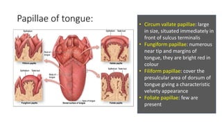
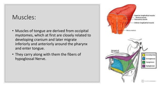
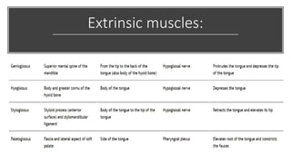
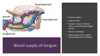
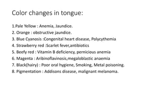
Anatomy of tongue
& its applied
aspects
L. VASAVI REDDY
IMDS
INTRODUCTION
• The tongue is a muscular structure that forms
part of the floor of the oral cavity and part of
the anterior wall of the oropharynx.
• Primary organ of taste (gustation)
• It has an oral part that lies in the mouth, and a
pharyngeal part that lies in the oropharynx.
• The oral and pharyngeal parts are separated by
a V-shaped sulcus, the sulcus terminalis.
Epithelium:
• Anterior 2/3rd – 1st branchial arch
• Posterior 1/3rd - 3rd branchial arch
• Posterior most part- 4th arch
Muscles:
• Occipital myotomes
Connective tissues:
From the local mesenchyme
External features:
• It has a fixed root, and a mobile
body & tip that can take on a variety
of shapes & positions.
• TIP- ◦ anterior end of the body.
◦ Rest on upper incisors.
• BODY- ◦ curved upper dorsal surface
◦ an inferior surface
• ROOT: It is attached to styloid
process , soft palate above and to
mandible ,hyoid bone below.
Dorsal surface Ventral surface
• Oral part
• Pharyngeal part
Papillae of tongue: • Circum vallate papillae: large
in size, situated immediately in
front of sulcus terminalis
• Fungiform papillae: numerous
near tip and margins of
tongue, they are bright red in
colour
• Filiform papillae: cover the
presulcular area of dorsum of
tongue giving a characteristic
velvety appearance
• Foliate papillae: few are
present
Muscles:
• Muscles of tongue are derived from occipital
myotomes, which at first are closely related to
developing cranium and later migrate
inferiorly and anteriorly around the pharynx
and enter tongue.
• They carry along with them the fibers of
hypoglossal Nerve.
Extrinsic muscles:
Blood supply of tongue:
• Arterial supply:
• Lingual artery
• Tonsillar- branch of facial
artery; ascending pharyngeal
branch
• Venouus drainage:
• Deep lingual vein is largest
and principal vein of tongue
Lymphatic
drainage:
Nerve supply:
Taste
pathway:
Taste sensation:
• Gustatory receptors detect following types of taste
sensation.
• Sweet: tip
• Bitter: base
• Sour: lateral margin
• Salty: anterolateral
Inspection:
• Wrap a piece of gauze around the tip of the
protruded tongue to hold Observe and note
1. The distribution of papillae
2. Margins of the tongue
3. Depapillated areas
4. Fissures, ulcers, and keratotic areas
5. Frenal attachment
6. Any deviations as the patient protrudes tongue
and attempts to move it to the right and left
7. Tongue thrust on swallowing
Applied aspects:
• Paralysis of the Genioglossus(safety muscle of tongue) ◦ tendency of
tongue to fall posteriorly, obstructing the airway.
• Injury to the Hypoglossal Nerve ◦ paralysis and eventual atrophy of affected
side of the tongue.
• Sublingual Absorption of Drugs ◦ quick absorption of a drug through deep
lingual veins
• Bilateral parasymphysis fracture- as genioglossus muscle is attached to the
superior genial tubercle, when bilateral parasymphysis fracture, tongue
might fall back
• Referred pain is felt in the ear in diseases of posterior tongue.
• Glossitis: Chemical irritants , drug reaction, sarcoidosis, sjögren syndrome
Systemic infections (e.g., syphilis)
Developmental disturbances:
• Aglossia & microglossia-Manifested as microglossia with glossoptosis.
• Macroglossia – tongue hypertrophy, pseudomacroglossia
• Ankyloglossia or tongue tie
• Cleft tongue/bifid tongue
• Fissured tongue-scortal tongue, lingual plicata -It is characterized by grooves that
vary in depth and are noted along the dorsal and lateral aspect of the tongue.
• Median rhomboid glossitis- the central papillary atrophy of the tongue,anterior to
circumvallate
• Geographic tongue- is serpiginous white lines surrounding areas of smooth,
depapillated mucosa.
• Hairy tongue-(lingua nigra,black hairy tongue,lingua villosa)- hypertrophy of
filliform papillae due to lack of mechanical debridement.
• Lingual varices- a dilated, tortuous vein, deep lingual vein.
• Lingual thyroid nodule- follicles of thyroid tissue are found in the substance of the
tongue
Malignant tumours:
• Squamous Cell Carcinoma
• Malignant lymphoma
• Malignant melanoma
• Metastatic tumors
• Sarcoma
Premalignant lesions:
Leukoplakia
erythroplakia
Bening tumours:
• Papilloma
• Rhabdomyoma
• Fibroma
• Glomus tumor
• Granular cell tumor
• Keratoacanthoma
• Leiomyoma
• Lipoma
Color changes in tongue:
1.Pale Yellow : Anemia, Jaundice.
2. Orange : obstructive jaundice.
3. Blue Cyanosis :Congenital heart disease, Polycythemia
4. Strawberry red :Scarlet fever,antibiotics
5. Beefy red : Vitamin B deficiency, pernicious anemia
6. Magenta : Aribinoflavinosis,megaloblastic anaemia
7. Black(hairy) : Poor oral hygiene, Smoking, Metal poisoning.
8. Pigmentation : Addisons disease, malignant melanoma.
Tongue coating evaluation in halitosis:
Miyazaki tongue coating
index

Tongue

  • 1.
    Anatomy of tongue &its applied aspects L. VASAVI REDDY IMDS
  • 2.
    INTRODUCTION • The tongueis a muscular structure that forms part of the floor of the oral cavity and part of the anterior wall of the oropharynx. • Primary organ of taste (gustation) • It has an oral part that lies in the mouth, and a pharyngeal part that lies in the oropharynx. • The oral and pharyngeal parts are separated by a V-shaped sulcus, the sulcus terminalis.
  • 3.
    Epithelium: • Anterior 2/3rd– 1st branchial arch • Posterior 1/3rd - 3rd branchial arch • Posterior most part- 4th arch Muscles: • Occipital myotomes Connective tissues: From the local mesenchyme
  • 4.
    External features: • Ithas a fixed root, and a mobile body & tip that can take on a variety of shapes & positions. • TIP- ◦ anterior end of the body. ◦ Rest on upper incisors. • BODY- ◦ curved upper dorsal surface ◦ an inferior surface • ROOT: It is attached to styloid process , soft palate above and to mandible ,hyoid bone below.
  • 5.
    Dorsal surface Ventralsurface • Oral part • Pharyngeal part
  • 6.
    Papillae of tongue:• Circum vallate papillae: large in size, situated immediately in front of sulcus terminalis • Fungiform papillae: numerous near tip and margins of tongue, they are bright red in colour • Filiform papillae: cover the presulcular area of dorsum of tongue giving a characteristic velvety appearance • Foliate papillae: few are present
  • 7.
    Muscles: • Muscles oftongue are derived from occipital myotomes, which at first are closely related to developing cranium and later migrate inferiorly and anteriorly around the pharynx and enter tongue. • They carry along with them the fibers of hypoglossal Nerve.
  • 8.
  • 9.
    Blood supply oftongue: • Arterial supply: • Lingual artery • Tonsillar- branch of facial artery; ascending pharyngeal branch • Venouus drainage: • Deep lingual vein is largest and principal vein of tongue
  • 10.
  • 11.
  • 12.
  • 13.
    Taste sensation: • Gustatoryreceptors detect following types of taste sensation. • Sweet: tip • Bitter: base • Sour: lateral margin • Salty: anterolateral
  • 14.
    Inspection: • Wrap apiece of gauze around the tip of the protruded tongue to hold Observe and note 1. The distribution of papillae 2. Margins of the tongue 3. Depapillated areas 4. Fissures, ulcers, and keratotic areas 5. Frenal attachment 6. Any deviations as the patient protrudes tongue and attempts to move it to the right and left 7. Tongue thrust on swallowing
  • 15.
    Applied aspects: • Paralysisof the Genioglossus(safety muscle of tongue) ◦ tendency of tongue to fall posteriorly, obstructing the airway. • Injury to the Hypoglossal Nerve ◦ paralysis and eventual atrophy of affected side of the tongue. • Sublingual Absorption of Drugs ◦ quick absorption of a drug through deep lingual veins • Bilateral parasymphysis fracture- as genioglossus muscle is attached to the superior genial tubercle, when bilateral parasymphysis fracture, tongue might fall back • Referred pain is felt in the ear in diseases of posterior tongue. • Glossitis: Chemical irritants , drug reaction, sarcoidosis, sjögren syndrome Systemic infections (e.g., syphilis)
  • 16.
    Developmental disturbances: • Aglossia& microglossia-Manifested as microglossia with glossoptosis. • Macroglossia – tongue hypertrophy, pseudomacroglossia • Ankyloglossia or tongue tie • Cleft tongue/bifid tongue • Fissured tongue-scortal tongue, lingual plicata -It is characterized by grooves that vary in depth and are noted along the dorsal and lateral aspect of the tongue. • Median rhomboid glossitis- the central papillary atrophy of the tongue,anterior to circumvallate • Geographic tongue- is serpiginous white lines surrounding areas of smooth, depapillated mucosa. • Hairy tongue-(lingua nigra,black hairy tongue,lingua villosa)- hypertrophy of filliform papillae due to lack of mechanical debridement. • Lingual varices- a dilated, tortuous vein, deep lingual vein. • Lingual thyroid nodule- follicles of thyroid tissue are found in the substance of the tongue
  • 17.
    Malignant tumours: • SquamousCell Carcinoma • Malignant lymphoma • Malignant melanoma • Metastatic tumors • Sarcoma Premalignant lesions: Leukoplakia erythroplakia Bening tumours: • Papilloma • Rhabdomyoma • Fibroma • Glomus tumor • Granular cell tumor • Keratoacanthoma • Leiomyoma • Lipoma
  • 18.
    Color changes intongue: 1.Pale Yellow : Anemia, Jaundice. 2. Orange : obstructive jaundice. 3. Blue Cyanosis :Congenital heart disease, Polycythemia 4. Strawberry red :Scarlet fever,antibiotics 5. Beefy red : Vitamin B deficiency, pernicious anemia 6. Magenta : Aribinoflavinosis,megaloblastic anaemia 7. Black(hairy) : Poor oral hygiene, Smoking, Metal poisoning. 8. Pigmentation : Addisons disease, malignant melanoma.
  • 19.
    Tongue coating evaluationin halitosis: Miyazaki tongue coating index

Editor's Notes

  • #7 Cv: cylindrical projection surrounded by circular sulcus Fungi: consists of narrow pedicle with large rounded head Fili: each is pointed and covered with keratin, the apex is often split into filamentous process
  • #8 Sl: makes dorsum concave: il : makes dorsum convex: transverse: makes tongue narrow and elongated; vertical makes tongue broad and flattened
  • #10 Unite to end at internal jugular vein