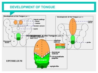
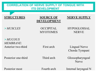
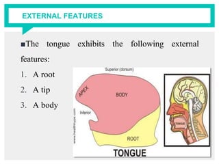
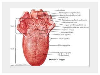
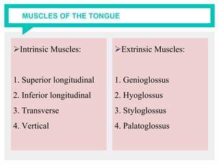
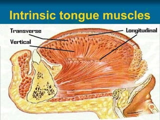
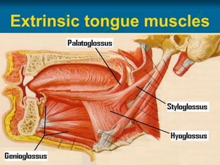
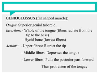
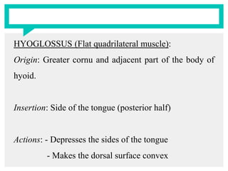
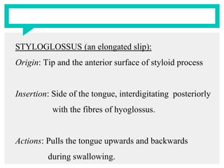
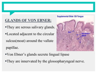
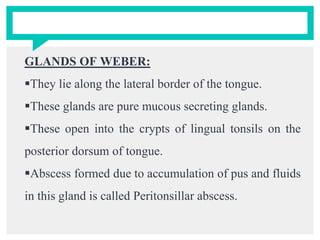
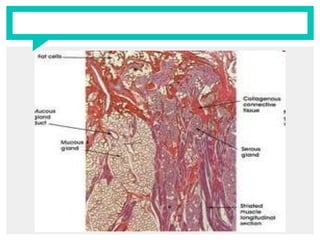
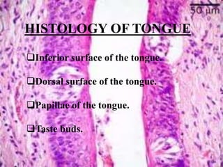
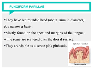
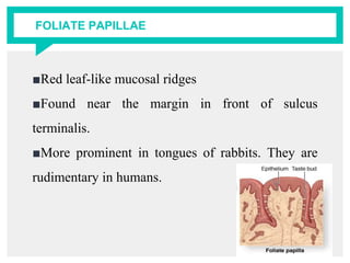
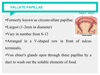
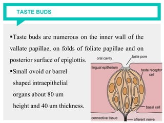
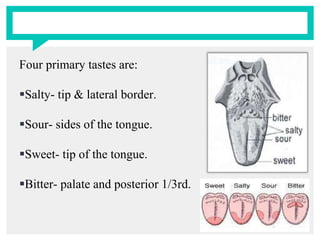
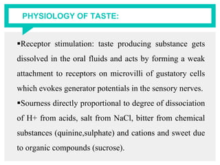
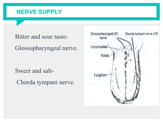
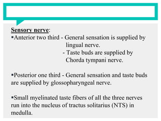
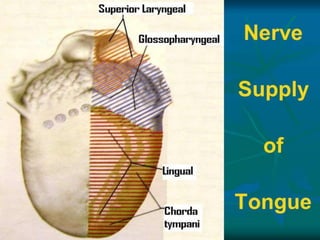
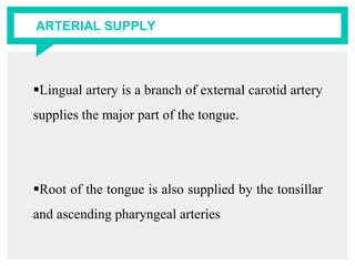
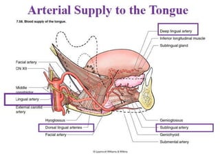
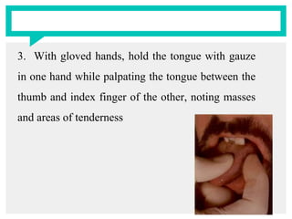
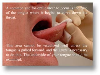
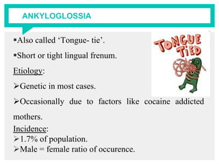
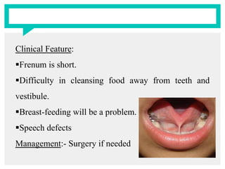
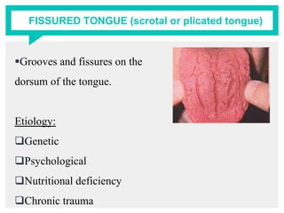
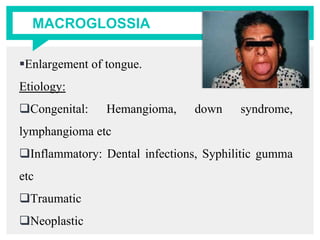
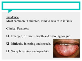
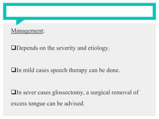
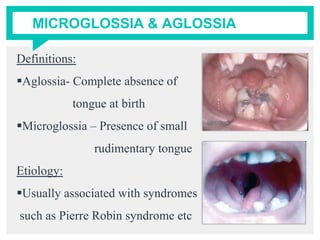
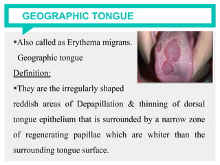
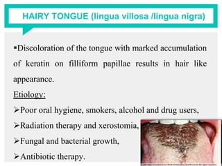
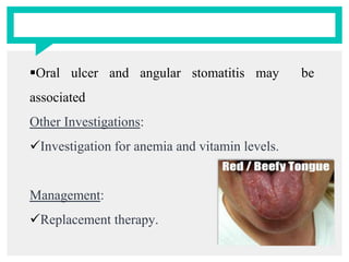
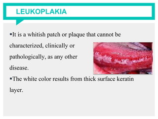
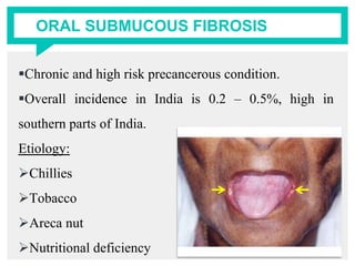
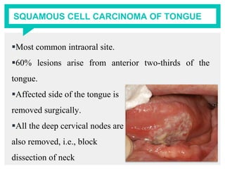
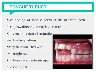
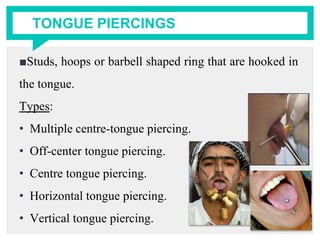
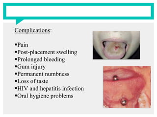
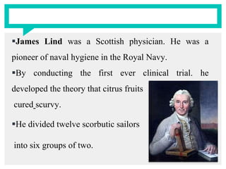
SEMINAR ON
TONGUE
PRESENTED BY:
DR. NABEELA BASHA
CONTENTS
■INTRODUCTION
■FUNCTIONS
■DEVELOPMENT OF TONGUE
■EXTERNAL FEATURES
■STRUCTURES OF TONGUE
■HISTOLOGY OF TONGUE
■NERVE, ARTERIAL, VENOUS SUPPLY AND
LYMPHATIC DRAINAGE OF TONGUE
■HOW TO EXAMINE TONGUE
■DISEASES OF TONGUE
■APPLIED ANATOMY
■CONCLUSION
■REFERENCES
■PREVIOUS YEAR QUESTIONS
INTRODUCTION
The word ‘tongue’ is derived from the Latin word
‘lingua’ and Greek word ‘glossa’.
The tongue is a mobile muscular organ in the oral
cavity which bulges upwards from the floor of the
mouth and its posterior part forms the anterior wall
of the oropharynx.
It is essentially a mass of skeletal muscle covered
by mucous membrane.
■Tongue is separated from teeth deep alveolo
lingual sulcus.
■The tongue may be affected as a part of oral
disease or as signs of a systemic disease.
FUNCTIONS
The tongue performs the following functions:
■Taste
■Speech
■Mastication
■Deglutition
■Barrier function
■Jaw development
■Thermal regulation
FUNCTIONS Contd
■Secretion
■Defence mechanism
■Maintenance of oral hygiene
■Sucking
■General sensitivity
DEVELOPMENT OF TONGUE
EPITHELIUM
■Muscles: develop from the occipital myotomes
which are supplied by Hypoglossal nerve
■Connective tissue: develops from the local
mesenchyme.
CORRELATION OF NERVE SUPPLY OF TONGUE WITH
ITS DEVELOPMENT
STRUCTURES
MUSCLES
MUCOUS
MEMBRANE:
Anterior two-third
Posterior one-third
Posterior most
SOURCE OF
DEVELOPMENT
OCCIPITAL
MYOTOMES
First arch
Third arch
Fourth arch
NERVE SUPPLY
HYPOGLOSSAL
NERVE
Lingual Nerve
Chorda Tympani
Glossopharyngeal
Nerve
Internal laryngeal N
EXTERNAL FEATURES
■The tongue exhibits the following external
features:
1. A root
2. A tip
3. A body
DORSAL SURFACE:
Convex on all the sides.
Divided by a V-Shaped sulcus into 2 parts:
Anterior two-third / Oral part
Posterior one-third / Pharyngeal part
Apex of sulcus terminalis is marked by Foramen
Caecum
Features of oral part:
■A median furrow, representing bilateral origin of
the tongue.
■Large number of papillae
■Embryological origin – 1st and 2nd pharyngeal
arches
Features of pharyngeal part:
■A large number of lymphoid follicles, which
together constitute the Lingual tonsil
■Large number of mucous and serous glands
■Embryological origin: 3rd and 4th pharyngeal
arches
VENTRAL (INFERIOR) SURFACE:
The mucous membrane lining this surface is smooth,
thin and purplish.
It presents the following features:
1. Frenulum Linguae – connecting the floor of the
mouth with tongue
2. Deep lingual veins – seen on either side of the
lingual frenum
3. Plica fimbriata – It is a fringed fold of mucous
membrane lateral to the lingual vein directed
forwards towards the tip of the tongue.
STRUCTURES OF THE TONGUE
■MUSCLES; tongue is made up of intrinsic and
extrinsic group of muscles.
■MUCOUS MEMBRANE; it is a layer of
connective tissue lined by stratified squamous
epithelium.
■GLANDS; numerous serous and mucous glands
lie deep to the mucous membrane
MUSCLES OF THE TONGUE
Intrinsic Muscles:
1. Superior longitudinal
2. Inferior longitudinal
3. Transverse
4. Vertical
Extrinsic Muscles:
1. Genioglossus
2. Hyoglossus
3. Styloglossus
4. Palatoglossus
SUPERIOR LONGITUDINAL:
Origin : Beneath the mucous membrane of the dorsal
surface of tongue
Insertion: Into the sides of the tongue
Actions: - Shortens the tongue
- Makes the dorsum concave
INFERIOR LONGITUDINAL:
Origin: Close to inferior surface between genioglossus
and hyoglossus
Insertion: Anterior part of median fibrous septum
Actions: - Shortens the tongue
- Makes the dorsum convex.
TRANSVERSUS LINGUAE:
Origin: Arise from the median fibrous septum
Insertion: Margins of the tongue
Actions: - Makes the tongue narrow and elongated.
VERTICALUS LINGUAE:
Origin: At the border of the anterior part of tongue
Insertion: Sides of the tongue
Action: - Makes the tongue broad and flattened.
GENIOGLOSSUS (fan shaped muscle):
Origin: Superior genial tubercle
Insertion: - Whole of the tongue (fibers radiate from the
tip to the base)
- Hyoid bone (lowest fibers)
Actions: - Upper fibres: Retract the tip
- Middle fibres: Depresses the tongue
- Lower fibres: Pulls the posterior part forward
Thus protrusion of the tongue
HYOGLOSSUS (Flat quadrilateral muscle):
Origin: Greater cornu and adjacent part of the body of
hyoid.
Insertion: Side of the tongue (posterior half)
Actions: - Depresses the sides of the tongue
- Makes the dorsal surface convex
STYLOGLOSSUS (an elongated slip):
Origin: Tip and the anterior surface of styloid process
Insertion: Side of the tongue, interdigitating posteriorly
with the fibres of hyoglossus.
Actions: Pulls the tongue upwards and backwards
during swallowing.
PALATOGLOSSUS (a slender slip):
Origin: Oral surface of palatine aponeurosis
Insertion: Side of the tongue (at the junction of its oral
& pharyngeal parts)
Actions: - Pulls up the root of the tongue
- Approximates the palatoglossal arches
MOVEMENTS OF THE TONGUE
Protrusion (most
important movement)
Retraction
Depression
Elevation (of posterior
one third)
Changes in shape
Genioglossus (of both
side acting together)
Styloglossus (of both
sides acting together)
Hypoglossus (of both
sides acting together)
Palatoglossus (of both
side acting together)
Intrinsic muscles
MUCOUS MEMBRANE
The mucosa on the oral dorsum part is moist and
pink and appears velvety due to presence of numerous
papillae. It is thicker than the ventral surface and is
adherent to muscular tissue covered by numerous
papillae.
Papillae are projections of lamina propria (corium)
of mucous membrane covered with epithelium.
■The mucous membrane over the dorsum of pharyngeal
part is devoid of papillae. It contain numerous lymphoid
follicles in the underlying submucosa.
■The mucous membrane in this part is continuous with
mucous membrane covering the palatine tonsil and the
pharynx.
■The lingual mucosa is thin, smooth and purplish on
the inferior surface of the tongue.
GLANDS
Mucous glands are numerous in the pharyngeal
part but are also present at the apex.
Serous glands of Von ebner are present near the
taste buds and their ducts open mostly into the sulci
of vallate papillae and their secretion is watery.
Mixed glands lie in the
ventral surface of the apex,
on either side of the
frenulum which are
covered by mucous
membrane.
GLANDS OF BLANDIN AND NUHN:
Anterior lingual glands (also called apical glands) are
deeply placed seromucous glands that are located near
the tip of the tongue on each side of lingual frenum.
They are between 12 to 25mm in length & approx.
8mm wide and each opens by 3 to 4 ducts on the
inferior surface of the tip.
GLANDS OF VON EBNER:
They are serous salivary glands.
Located adjacent to the circular
sulcus(moat) around the vallate
papillae.
Von Ebner’s glands secrete lingual lipase
They are innervated by the glossopharyngeal nerve.
GLANDS OF WEBER:
They lie along the lateral border of the tongue.
These glands are pure mucous secreting glands.
These open into the crypts of lingual tonsils on the
posterior dorsum of tongue.
Abscess formed due to accumulation of pus and fluids
in this gland is called Peritonsillar abscess.
HISTOLOGY OF TONGUE
Inferior surface of the tongue.
Dorsal surface of the tongue.
Papillae of the tongue.
Taste buds.
■Inferior surface:
a. The mucous membrane is thin and loosely
attached to the underlying surface for free mobility.
b. Made of non-keratinized epithelium.
c. Sub mucosa contains adipose tissue.
d. Sub lingual glands lie close to the sublingual
fold.
e. Mucous membrane is smooth and thin.
■Dorsal mucosa:
a. It is made up of specialized mucosa.
b. It is rough and irregular.
c. The dorsal surface of the tongue is a mixture of
thin, keratinized, filiform papillae interspersed with
pink mushroom-shaped fungiform papillae
PAPILLAE OF THE TONGUE
There are 4 types of papillae:
Filiform
Fungiform
Vallate
Foliate
Papillae simplex ( surface projections which can
only be seen under microscope
FILIFORM PAPILLAE
Narrowest and most numerous in number
Minute conical projections with sharply pointed tips.
Located abundantly on
the presulcal dorsal
area and are largely
responsible for its
velvety appearance.
FUNGIFORM PAPILLAE
They have red rounded head (about 1mm in diameter)
& a narrower base
Mostly found on the apex and margins of the tongue,
while some are scattered over the dorsal surface.
They are visible as discrete pink pinheads.
FOLIATE PAPILLAE
■Red leaf-like mucosal ridges
■Found near the margin in front of sulcus
terminalis.
■More prominent in tongues of rabbits. They are
rudimentary in humans.
VALLATE PAPILLAE
Formerly known as circumvallate papillae.
Largest (1-2mm in diameter)
Vary in number from 8-12
Arranged in a V-shaped row in front of sulcus
terminalis.
Von ebner's glands open through these papillae by a
duct to wash out the soluble elements of food.
TASTE BUDS
Taste buds are numerous on the inner wall of the
vallate papillae, on folds of foliate papillae and on
posterior surface of epiglottis.
Small ovoid or barrel
shaped intraepithelial
organs about 80 um
height and 40 um thickness.
■Outer surface has flat epithelial cells, surrounded
by a small opening called taste pore. Taste pore
leads to narrow space lined by supporting
cells(sustentacular cells).
■The other group of cells present in the taste buds
are the gustatory receptor cells which end with
microvilli.
■The afferent nerves from the gustatory receptor
cells begin as minute fibers which join to form 2 or
3 large fibers and each large fiber connects with 1
or more taste cells .
■Each taste bud has approx. 50 nerve fibers, and
each nerve fiber in turn receives input from about 5
taste buds.
TASTE SENSATION
■The taste receptor cells in the taste buds opens
through the pores to detect the various tastes.
■The taste receptors are chemoreceptors (located on
the edges, dorsum of the tongue,
epiglottis, soft palate and
pharynx) stimulated by
substances dissolved in the
oral fluids.
Four primary tastes are:
Salty- tip & lateral border.
Sour- sides of the tongue.
Sweet- tip of the tongue.
Bitter- palate and posterior 1/3rd.
PHYSIOLOGY OF TASTE:
Receptor stimulation: taste producing substance gets
dissolved in the oral fluids and acts by forming a weak
attachment to receptors on microvilli of gustatory cells
which evokes generator potentials in the sensory nerves.
Sourness directly proportional to degree of dissociation
of H+ from acids, salt from NaCl, bitter from chemical
substances (quinine,sulphate) and cations and sweet due
to organic compounds (sucrose).
NERVE SUPPLY
Bitter and sour taste-
Glossopharyngeal nerve.
Sweet and salt-
Chorda tympani nerve.
Motor nerve:
■Intrinsic and extrinsic muscles except
palatoglossus muscles are supplied by hypoglossal
nerve.
■Palatoglossus muscles are supplied by cranial part
of accessory nerve through the pharyngeal plexus.
Sensory nerve:
Anterior two third - General sensation is supplied by
lingual nerve.
- Taste buds are supplied by
Chorda tympani nerve.
Posterior one third - General sensation and taste buds
are supplied by glossopharyngeal nerve.
Small myelinated taste fibers of all the three nerves
run into the nucleus of tractus solitarius (NTS) in
medulla.
ARTERIAL SUPPLY
Lingual artery is a branch of external carotid artery
supplies the major part of the tongue.
Root of the tongue is also supplied by the tonsillar
and ascending pharyngeal arteries
LYMPHATIC DRAINAGE
Tip of the tongue drains into the submental lymph
nodes.
The right and the left halves of the anterior 2/3rd
tongue drains into the submandibular lymph nodes on
either side.
The posterior 1/3rd of the tongue drains into the
jugulo-omohyoid group of deep cervical lymph nodes.
VENOUS DRAINAGE
 Deep lingual vein is the largest and main vein, which
supplies the tongue.
 The vein is visible in the inferior surface of the
tongue.
 It runs backwards and crosses the genioglossus and
hyoglossus muscle.
HOW TO EXAMINE TONGUE…?
On physical examination, there are several
characteristics of the tongue that should be noted:
Color
Pink-red on dorsal and ventral surfaces. The ventral
surface may have some visible vasculature.
Texture
Rough dorsal surface owing to papillae. There
should be no hairs, furrows, or ulceration.
Size
Should fit comfortably in mouth, tip against lower
incisors. Sublingual glands should not be displaced.
The tongue can be checked for swelling or
abnormal color or texture as the tongue is extended
out.
If the tongue deviates to one side or the other, a
possible sign that something is affecting the nerves
which control its movement.
Then a small piece of gauze is used to gently pull
the tongue to one side, then the other to fully
visualize its edges (a common location for lesions
to occur).
The borders of the tongue will be felt for (for hard
spots) at the same time.
In general, the examination of the tongue should
occur in the following steps:
1. Have the patient touch the tip of the tongue to the
roof of their mouth and inspect the ventral surface.
2. Have the patient protrude the tongue straight out
and inspect for deviation, color, texture, and
masses
3. With gloved hands, hold the tongue with gauze
in one hand while palpating the tongue between the
thumb and index finger of the other, noting masses
and areas of tenderness
A common site for oral cancer to occur is the base
of the tongue where it begins to curve down the
throat.
This area cannot be visualized well unless the
tongue is pulled forward, and the gauze is necessary
to do this. The underside of your tongue should be
examined.
DISEASES OF THE TONGUE
Inherited, Congenital & Developmental anomalies
Disorders of the lingual mucosa
Diseases affecting the body of the tongue
Malignant tumors of the tongue
Inherited, Congenital & Developmental anomalies:
Variations in morphology:
oAnkyloglossia
oFissured Tongue
oMacroglossia
oMicroglossia
oLingual thyroid nodule
ANKYLOGLOSSIA
Also called ‘Tongue- tie’.
Short or tight lingual frenum.
Etiology:
Genetic in most cases.
Occasionally due to factors like cocaine addicted
mothers.
Incidence:
1.7% of population.
Male = female ratio of occurence.
Clinical Feature:
Frenum is short.
Difficulty in cleansing food away from teeth and
vestibule.
Breast-feeding will be a problem.
Speech defects
Management:- Surgery if needed
FISSURED TONGUE (scrotal or plicated tongue)
Grooves and fissures on the
dorsum of the tongue.
Etiology:
Genetic
Psychological
Nutritional deficiency
Chronic trauma
Rarely it may be associated with erythema migrans,
Melkersson-Rosenthal syndrome, Down syndrome and
psoriasis.
Incidence:
5% of the population.
Clinical Feature:
Multiple fissures on the dorsum of the tongue.
Mostly asymptomatic.
Management: Maintenance of tongue hygiene
MACROGLOSSIA
Enlargement of tongue.
Etiology:
Congenital: Hemangioma, down syndrome,
lymphangioma etc
Inflammatory: Dental infections, Syphilitic gumma
etc
Traumatic
Neoplastic
Incidence:
Most common in children, mild to severe in infants.
Clinical Features:
 Enlarged, diffuse, smooth and drooling tongue.
 Difficulty in eating and speech.
 Noisy breathing and open bite.
Management:
Depends on the severity and etiology.
In mild cases speech therapy can be done.
In sever cases glossectomy, a surgical removal of
excess tongue can be advised.
MICROGLOSSIA & AGLOSSIA
Definitions:
Aglossia- Complete absence of
tongue at birth
Microglossia – Presence of small
rudimentary tongue
Etiology:
Usually associated with syndromes
such as Pierre Robin syndrome etc
Also associated with cleft lip and palate.
Clinical Features:
Difficulty in eating and speaking.
High arched palate and narrow constricted mandible.
There may be airway obstruction, due to negative
pressure generated by deglutition and inspiration.
Management:
Non surgical techniques such as nasogastric
intubation, temporary endotracheal intubation may
be carried out to prevent airway obstruction.
Disorders of Lingual Mucosa:
Geographic Tongue
Hairy tongue
Non Keratotic & Keratotic white lesions:
Candidiasis
Leukoplakia
Nutritional deficiencies and hematological
abnormalities:
Vit B12 deficiency
Iron deficiency anemia
Infections:
Tertiary syphilis
GEOGRAPHIC TONGUE
Also called as Erythema migrans.
Geographic tongue
Definition:
They are the irregularly shaped
reddish areas of Depapillation & thinning of dorsal
tongue epithelium that is surrounded by a narrow zone
of regenerating papillae which are whiter than the
surrounding tongue surface.
Etiology:
Immunological reaction, allergic, emotional stress &
hereditary factors
Infections and nutritional deficiencies.
Clinical Features:
Common in young & middle aged, 5-84 years
Usually asymptomatic, can be sometimes associated
with burning sensation.
Management:
Bland diet, elimination of irritants
Zinc supplements – recent
Topical corticosteroids
HAIRY TONGUE (lingua villosa /lingua nigra)
Discoloration of the tongue with marked accumulation
of keratin on filliform papillae results in hair like
appearance.
Etiology:
Poor oral hygiene, smokers, alcohol and drug users,
Radiation therapy and xerostomia,
Fungal and bacterial growth,
Antibiotic therapy.
Clinical Features:
Appears usually in midline just anterior to
circumvallate papillae.
The papillae are elongated, usually yellow or black in
colour result of pigmentation.
asymptomatic.
Some time patient may complaints of bad taste and
breath.
Management:
Improve oral hygiene.
Treatment for the etiology.
Scrape or brush the tongue.
Sodium bicarbonate and hydrogen peroxide
mouthwash.
CANDIDAL GLOSSITIS
■Sore tongue due to candidal infection.
Etiology:
Opportunistic infection with
candida.albicans
Xerostomia,
Immune defects
Clinical Feature:
Diffuse erythema,
Soreness of the tongue,
White patches on the tongue.
Management:
Elimination of etiology
Antifungal drugs
DEFICIENCY GLOSSITIS
■Soreness of tongue due to deficiency of vitamins and
minerals.
■Etiology:
Deficiency of iron, folic acid and vitamin B12.
■Clinical Feature:
Linear patchy or red lesion,
Depapillated tongue,
Oral ulcer and angular stomatitis may be
associated
Other Investigations:
Investigation for anemia and vitamin levels.
Management:
Replacement therapy.
LEUKOPLAKIA
It is a whitish patch or plaque that cannot be
characterized, clinically or
pathologically, as any other
disease.
The white color results from thick surface keratin
layer.
Etiology:
 Classically known as the 6 ‘S’.
 Smoking, Sharp tooth, Syphilis, Sepsis, Spirit and
Spices.
Clinical Features:
White lesion on both sides of the tongue, vertically
corrugated.
Appears to be benign and self-limiting
Management:
Elimination of etiology
Conservative treatment
Surgical Management
ORAL SUBMUCOUS FIBROSIS
Chronic and high risk precancerous condition.
Overall incidence in India is 0.2 – 0.5%, high in
southern parts of India.
Etiology:
Chillies
Tobacco
Areca nut
Nutritional deficiency
Affects the tongue in 37% cases, most common site
being buccal mucosa.
Stage II – Stage of Fibrosis-:
Inability to open mouth completely.
Inability to protrude tongue.
Tongue movements become restricted.
Depapillation usually on the lateral margins, tongue
appears smooth.
Management:
Restriction of habit
Vitamin rich diet
Steroids
Lycopene
Surgery, if neoplastic changes seen or when there
is marked trismus and dysphagia.
SQUAMOUS CELL CARCINOMA OF TONGUE
Most common intraoral site.
60% lesions arise from anterior two-thirds of the
tongue.
Affected side of the tongue is
removed surgically.
All the deep cervical nodes are
also removed, i.e., block
dissection of neck
TONGUE THRUST
Positioning of tongue between the anterior teeth
during swallowing, speaking or at rest.
It is seen in retained infantile
swallowing pattern.
May be associated with
Macroglossia.
In these cases, anterior open
bite is present.
TONGUE PIERCINGS
■Studs, hoops or barbell shaped ring that are hooked in
the tongue.
Types:
• Multiple centre-tongue piercing.
• Off-center tongue piercing.
• Centre tongue piercing.
• Horizontal tongue piercing.
• Vertical tongue piercing.
Complications:
Pain
Post-placement swelling
Prolonged bleeding
Gum injury
Permanent numbness
Loss of taste
HIV and hepatitis infection
Oral hygiene problems
Management: Avoid piercing.
If pierced;
Use chlorhexidine mouthwash every half an hour
immediately after tongue piercing for 8 hours.
Tongue swelling will subside within 7 to 8 days, and
complete healing within 2 weeks.
 Advice not to take hot and spicy foods. Rinse mouth
before and after food.
Sterilize the jewellery before placing. Improve and
maintain oral hygiene.
 Regular visit to dentist at least once in 3 months.
APPLIED ANATOMY
Injury to the hypoglossal nerve leads to the paralysis of
the muscles of the tongue on the side of the lesion.
If the lesion is infranuclear, there is gradual atrophy of
the affected half of the tongue.
Supranuclear lesions produce paralysis without
wasting.
The tongue is stiff, small and moves very sluggishly
resulting in defective articulation.
The tongue becomes bald in anemias due to atrophy of
the filiform papillae.
The presence of rich network of lymphatics and loose
areolar tissue is responsible for enormous swelling of the
tongue in acute glossitis.
In unconcious patients the tongue may fall back to
obstruct the air passages.
Carcinoma of the posterior 1/3rd of the tongue is more
dangerous due to bilateral lymphatic spread.
In grandmal epilepsy, the tongue is commonly bitten
by the front incisors during attack.
Sublingual absorption of drugs: For quick
absorption, pill or spray is put under the tongue
where it dissolves and enters the lingual vein
E.g. Nitroglycerine in angina pectoris
CONCLUSION
Tongue is an important organ which contributes to
speech, mastication, deglutition and taste.
The examination of the tongue plays a major role
during the oral examination of the soft tissues.
The knowledge about the development, functions,
anatomy and diseases associated with tongue is
important to the dental professionals as it helps in
identifying or diagnosing many congenital and
systemic diseases leading to pathological changes of
the tongue at the earliest.
The early signs of cancer can be detected through
examination of the tongue routinely during
screening in masses which is of public health
importance.
REFERENCES
Anatomy of Head, Neck & Brain. Vishram singh
Essentials of Human Anatomy Head & Neck. 5th
Edn . A. K. DATTA
B. D Chaurasia’s Human Anatomy. 4th Edn
Textbook of Oral Medicine. 2nd Edn. Anil
Govindrao Ghom
http://stanfordmedicine25.stanford.edu/the25/tong
ue.html
Oral pathology- By shafer’s ;5thedition ;2003.
Human embryology- By Inderbir singh;6th
edition;1996.
http://www.livestrong.com/article/182822-how-to-
treat-a-fresh-tongue-piercing/
http://bodyjewelryblog.com/2011/08/04/whats-
that-called-dictionary-of-oral-piercings/
PREVIOUS YEAR QUESTIONS
 Short essays on (10marks each):
Muscles of the tongue. (MDS Degree Examination;
2002)
Tongue. ( MDS Degree Examination; 2005)
Taste buds. (MDS Degree Examination; 2001)
Musculature, Nerve supply & Lymphatic drainage of
tongue. (MDS Degree Examination; Apr/May 2007)
 Development of Tongue. (JSS University,
Mysore; April 2012) (7marks)
HAWTHORNE EFFECT
 It is the name of a place where the effect was first
encountered.
In 1958, the researcher, Henry A. Landsberger,
performed a study and analysis of data from experiments
performed between 1924 and 1932, by Elton Mayo, at
the Hawthorne Works near Chicago.
The company had commissioned studies to determine if
the level of light within their building affected the
productivity of the workers.
Ref: https://en.wikipedia.org/wiki/Hawthorne_effect
James Lind was a Scottish physician. He was a
pioneer of naval hygiene in the Royal Navy.
By conducting the first ever clinical trial. he
developed the theory that citrus fruits
cured scurvy.
He divided twelve scorbutic sailors
into six groups of two.
They all received the same diet but, in addition, group
one was given a quart of cider daily, group two twenty-
five drops of elixir of vitriol (sulfuric acid), group three
six spoonfuls of vinegar, group four half a pint of
seawater, group five received two oranges and
one lemon, and the last group a spicy paste plus a drink
of barley water.
The treatment of group five stopped after six days
when they ran out of fruit, but by that time one
sailor was fit for duty while the other had almost
recovered. Apart from that, only group one also
showed some effect of its treatment.
Ref:https://en.wikipedia.org/wiki/James_Lind
Tongue

Tongue

  • 2.
  • 3.
    CONTENTS ■INTRODUCTION ■FUNCTIONS ■DEVELOPMENT OF TONGUE ■EXTERNALFEATURES ■STRUCTURES OF TONGUE ■HISTOLOGY OF TONGUE ■NERVE, ARTERIAL, VENOUS SUPPLY AND LYMPHATIC DRAINAGE OF TONGUE ■HOW TO EXAMINE TONGUE ■DISEASES OF TONGUE ■APPLIED ANATOMY ■CONCLUSION ■REFERENCES ■PREVIOUS YEAR QUESTIONS
  • 4.
    INTRODUCTION The word ‘tongue’is derived from the Latin word ‘lingua’ and Greek word ‘glossa’. The tongue is a mobile muscular organ in the oral cavity which bulges upwards from the floor of the mouth and its posterior part forms the anterior wall of the oropharynx. It is essentially a mass of skeletal muscle covered by mucous membrane.
  • 5.
    ■Tongue is separatedfrom teeth deep alveolo lingual sulcus. ■The tongue may be affected as a part of oral disease or as signs of a systemic disease.
  • 6.
    FUNCTIONS The tongue performsthe following functions: ■Taste ■Speech ■Mastication ■Deglutition ■Barrier function ■Jaw development ■Thermal regulation
  • 7.
    FUNCTIONS Contd ■Secretion ■Defence mechanism ■Maintenanceof oral hygiene ■Sucking ■General sensitivity
  • 8.
  • 9.
    ■Muscles: develop fromthe occipital myotomes which are supplied by Hypoglossal nerve ■Connective tissue: develops from the local mesenchyme.
  • 10.
    CORRELATION OF NERVESUPPLY OF TONGUE WITH ITS DEVELOPMENT STRUCTURES MUSCLES MUCOUS MEMBRANE: Anterior two-third Posterior one-third Posterior most SOURCE OF DEVELOPMENT OCCIPITAL MYOTOMES First arch Third arch Fourth arch NERVE SUPPLY HYPOGLOSSAL NERVE Lingual Nerve Chorda Tympani Glossopharyngeal Nerve Internal laryngeal N
  • 11.
    EXTERNAL FEATURES ■The tongueexhibits the following external features: 1. A root 2. A tip 3. A body
  • 13.
    DORSAL SURFACE: Convex onall the sides. Divided by a V-Shaped sulcus into 2 parts: Anterior two-third / Oral part Posterior one-third / Pharyngeal part Apex of sulcus terminalis is marked by Foramen Caecum
  • 14.
    Features of oralpart: ■A median furrow, representing bilateral origin of the tongue. ■Large number of papillae ■Embryological origin – 1st and 2nd pharyngeal arches
  • 15.
    Features of pharyngealpart: ■A large number of lymphoid follicles, which together constitute the Lingual tonsil ■Large number of mucous and serous glands ■Embryological origin: 3rd and 4th pharyngeal arches
  • 17.
    VENTRAL (INFERIOR) SURFACE: Themucous membrane lining this surface is smooth, thin and purplish. It presents the following features: 1. Frenulum Linguae – connecting the floor of the mouth with tongue 2. Deep lingual veins – seen on either side of the lingual frenum
  • 18.
    3. Plica fimbriata– It is a fringed fold of mucous membrane lateral to the lingual vein directed forwards towards the tip of the tongue.
  • 19.
    STRUCTURES OF THETONGUE ■MUSCLES; tongue is made up of intrinsic and extrinsic group of muscles. ■MUCOUS MEMBRANE; it is a layer of connective tissue lined by stratified squamous epithelium. ■GLANDS; numerous serous and mucous glands lie deep to the mucous membrane
  • 20.
    MUSCLES OF THETONGUE Intrinsic Muscles: 1. Superior longitudinal 2. Inferior longitudinal 3. Transverse 4. Vertical Extrinsic Muscles: 1. Genioglossus 2. Hyoglossus 3. Styloglossus 4. Palatoglossus
  • 22.
    SUPERIOR LONGITUDINAL: Origin :Beneath the mucous membrane of the dorsal surface of tongue Insertion: Into the sides of the tongue Actions: - Shortens the tongue - Makes the dorsum concave
  • 23.
    INFERIOR LONGITUDINAL: Origin: Closeto inferior surface between genioglossus and hyoglossus Insertion: Anterior part of median fibrous septum Actions: - Shortens the tongue - Makes the dorsum convex.
  • 24.
    TRANSVERSUS LINGUAE: Origin: Arisefrom the median fibrous septum Insertion: Margins of the tongue Actions: - Makes the tongue narrow and elongated.
  • 25.
    VERTICALUS LINGUAE: Origin: Atthe border of the anterior part of tongue Insertion: Sides of the tongue Action: - Makes the tongue broad and flattened.
  • 27.
    GENIOGLOSSUS (fan shapedmuscle): Origin: Superior genial tubercle Insertion: - Whole of the tongue (fibers radiate from the tip to the base) - Hyoid bone (lowest fibers) Actions: - Upper fibres: Retract the tip - Middle fibres: Depresses the tongue - Lower fibres: Pulls the posterior part forward Thus protrusion of the tongue
  • 28.
    HYOGLOSSUS (Flat quadrilateralmuscle): Origin: Greater cornu and adjacent part of the body of hyoid. Insertion: Side of the tongue (posterior half) Actions: - Depresses the sides of the tongue - Makes the dorsal surface convex
  • 29.
    STYLOGLOSSUS (an elongatedslip): Origin: Tip and the anterior surface of styloid process Insertion: Side of the tongue, interdigitating posteriorly with the fibres of hyoglossus. Actions: Pulls the tongue upwards and backwards during swallowing.
  • 30.
    PALATOGLOSSUS (a slenderslip): Origin: Oral surface of palatine aponeurosis Insertion: Side of the tongue (at the junction of its oral & pharyngeal parts) Actions: - Pulls up the root of the tongue - Approximates the palatoglossal arches
  • 31.
    MOVEMENTS OF THETONGUE Protrusion (most important movement) Retraction Depression Elevation (of posterior one third) Changes in shape Genioglossus (of both side acting together) Styloglossus (of both sides acting together) Hypoglossus (of both sides acting together) Palatoglossus (of both side acting together) Intrinsic muscles
  • 32.
    MUCOUS MEMBRANE The mucosaon the oral dorsum part is moist and pink and appears velvety due to presence of numerous papillae. It is thicker than the ventral surface and is adherent to muscular tissue covered by numerous papillae. Papillae are projections of lamina propria (corium) of mucous membrane covered with epithelium.
  • 33.
    ■The mucous membraneover the dorsum of pharyngeal part is devoid of papillae. It contain numerous lymphoid follicles in the underlying submucosa. ■The mucous membrane in this part is continuous with mucous membrane covering the palatine tonsil and the pharynx. ■The lingual mucosa is thin, smooth and purplish on the inferior surface of the tongue.
  • 34.
    GLANDS Mucous glands arenumerous in the pharyngeal part but are also present at the apex. Serous glands of Von ebner are present near the taste buds and their ducts open mostly into the sulci of vallate papillae and their secretion is watery.
  • 35.
    Mixed glands liein the ventral surface of the apex, on either side of the frenulum which are covered by mucous membrane.
  • 36.
    GLANDS OF BLANDINAND NUHN: Anterior lingual glands (also called apical glands) are deeply placed seromucous glands that are located near the tip of the tongue on each side of lingual frenum. They are between 12 to 25mm in length & approx. 8mm wide and each opens by 3 to 4 ducts on the inferior surface of the tip.
  • 38.
    GLANDS OF VONEBNER: They are serous salivary glands. Located adjacent to the circular sulcus(moat) around the vallate papillae. Von Ebner’s glands secrete lingual lipase They are innervated by the glossopharyngeal nerve.
  • 39.
    GLANDS OF WEBER: Theylie along the lateral border of the tongue. These glands are pure mucous secreting glands. These open into the crypts of lingual tonsils on the posterior dorsum of tongue. Abscess formed due to accumulation of pus and fluids in this gland is called Peritonsillar abscess.
  • 41.
    HISTOLOGY OF TONGUE Inferiorsurface of the tongue. Dorsal surface of the tongue. Papillae of the tongue. Taste buds.
  • 42.
    ■Inferior surface: a. Themucous membrane is thin and loosely attached to the underlying surface for free mobility. b. Made of non-keratinized epithelium. c. Sub mucosa contains adipose tissue. d. Sub lingual glands lie close to the sublingual fold. e. Mucous membrane is smooth and thin.
  • 43.
    ■Dorsal mucosa: a. Itis made up of specialized mucosa. b. It is rough and irregular. c. The dorsal surface of the tongue is a mixture of thin, keratinized, filiform papillae interspersed with pink mushroom-shaped fungiform papillae
  • 44.
    PAPILLAE OF THETONGUE There are 4 types of papillae: Filiform Fungiform Vallate Foliate Papillae simplex ( surface projections which can only be seen under microscope
  • 45.
    FILIFORM PAPILLAE Narrowest andmost numerous in number Minute conical projections with sharply pointed tips. Located abundantly on the presulcal dorsal area and are largely responsible for its velvety appearance.
  • 46.
    FUNGIFORM PAPILLAE They havered rounded head (about 1mm in diameter) & a narrower base Mostly found on the apex and margins of the tongue, while some are scattered over the dorsal surface. They are visible as discrete pink pinheads.
  • 47.
    FOLIATE PAPILLAE ■Red leaf-likemucosal ridges ■Found near the margin in front of sulcus terminalis. ■More prominent in tongues of rabbits. They are rudimentary in humans.
  • 48.
    VALLATE PAPILLAE Formerly knownas circumvallate papillae. Largest (1-2mm in diameter) Vary in number from 8-12 Arranged in a V-shaped row in front of sulcus terminalis. Von ebner's glands open through these papillae by a duct to wash out the soluble elements of food.
  • 49.
    TASTE BUDS Taste budsare numerous on the inner wall of the vallate papillae, on folds of foliate papillae and on posterior surface of epiglottis. Small ovoid or barrel shaped intraepithelial organs about 80 um height and 40 um thickness.
  • 50.
    ■Outer surface hasflat epithelial cells, surrounded by a small opening called taste pore. Taste pore leads to narrow space lined by supporting cells(sustentacular cells). ■The other group of cells present in the taste buds are the gustatory receptor cells which end with microvilli.
  • 52.
    ■The afferent nervesfrom the gustatory receptor cells begin as minute fibers which join to form 2 or 3 large fibers and each large fiber connects with 1 or more taste cells . ■Each taste bud has approx. 50 nerve fibers, and each nerve fiber in turn receives input from about 5 taste buds.
  • 53.
    TASTE SENSATION ■The tastereceptor cells in the taste buds opens through the pores to detect the various tastes. ■The taste receptors are chemoreceptors (located on the edges, dorsum of the tongue, epiglottis, soft palate and pharynx) stimulated by substances dissolved in the oral fluids.
  • 54.
    Four primary tastesare: Salty- tip & lateral border. Sour- sides of the tongue. Sweet- tip of the tongue. Bitter- palate and posterior 1/3rd.
  • 55.
    PHYSIOLOGY OF TASTE: Receptorstimulation: taste producing substance gets dissolved in the oral fluids and acts by forming a weak attachment to receptors on microvilli of gustatory cells which evokes generator potentials in the sensory nerves. Sourness directly proportional to degree of dissociation of H+ from acids, salt from NaCl, bitter from chemical substances (quinine,sulphate) and cations and sweet due to organic compounds (sucrose).
  • 56.
    NERVE SUPPLY Bitter andsour taste- Glossopharyngeal nerve. Sweet and salt- Chorda tympani nerve.
  • 57.
    Motor nerve: ■Intrinsic andextrinsic muscles except palatoglossus muscles are supplied by hypoglossal nerve. ■Palatoglossus muscles are supplied by cranial part of accessory nerve through the pharyngeal plexus.
  • 58.
    Sensory nerve: Anterior twothird - General sensation is supplied by lingual nerve. - Taste buds are supplied by Chorda tympani nerve. Posterior one third - General sensation and taste buds are supplied by glossopharyngeal nerve. Small myelinated taste fibers of all the three nerves run into the nucleus of tractus solitarius (NTS) in medulla.
  • 60.
    ARTERIAL SUPPLY Lingual arteryis a branch of external carotid artery supplies the major part of the tongue. Root of the tongue is also supplied by the tonsillar and ascending pharyngeal arteries
  • 62.
    LYMPHATIC DRAINAGE Tip ofthe tongue drains into the submental lymph nodes. The right and the left halves of the anterior 2/3rd tongue drains into the submandibular lymph nodes on either side. The posterior 1/3rd of the tongue drains into the jugulo-omohyoid group of deep cervical lymph nodes.
  • 63.
  • 64.
     Deep lingualvein is the largest and main vein, which supplies the tongue.  The vein is visible in the inferior surface of the tongue.  It runs backwards and crosses the genioglossus and hyoglossus muscle.
  • 65.
    HOW TO EXAMINETONGUE…? On physical examination, there are several characteristics of the tongue that should be noted: Color Pink-red on dorsal and ventral surfaces. The ventral surface may have some visible vasculature.
  • 66.
    Texture Rough dorsal surfaceowing to papillae. There should be no hairs, furrows, or ulceration. Size Should fit comfortably in mouth, tip against lower incisors. Sublingual glands should not be displaced.
  • 67.
    The tongue canbe checked for swelling or abnormal color or texture as the tongue is extended out. If the tongue deviates to one side or the other, a possible sign that something is affecting the nerves which control its movement.
  • 68.
    Then a smallpiece of gauze is used to gently pull the tongue to one side, then the other to fully visualize its edges (a common location for lesions to occur). The borders of the tongue will be felt for (for hard spots) at the same time.
  • 69.
    In general, theexamination of the tongue should occur in the following steps: 1. Have the patient touch the tip of the tongue to the roof of their mouth and inspect the ventral surface. 2. Have the patient protrude the tongue straight out and inspect for deviation, color, texture, and masses
  • 70.
    3. With glovedhands, hold the tongue with gauze in one hand while palpating the tongue between the thumb and index finger of the other, noting masses and areas of tenderness
  • 71.
    A common sitefor oral cancer to occur is the base of the tongue where it begins to curve down the throat. This area cannot be visualized well unless the tongue is pulled forward, and the gauze is necessary to do this. The underside of your tongue should be examined.
  • 72.
    DISEASES OF THETONGUE Inherited, Congenital & Developmental anomalies Disorders of the lingual mucosa Diseases affecting the body of the tongue Malignant tumors of the tongue
  • 73.
    Inherited, Congenital &Developmental anomalies: Variations in morphology: oAnkyloglossia oFissured Tongue oMacroglossia oMicroglossia oLingual thyroid nodule
  • 74.
    ANKYLOGLOSSIA Also called ‘Tongue-tie’. Short or tight lingual frenum. Etiology: Genetic in most cases. Occasionally due to factors like cocaine addicted mothers. Incidence: 1.7% of population. Male = female ratio of occurence.
  • 75.
    Clinical Feature: Frenum isshort. Difficulty in cleansing food away from teeth and vestibule. Breast-feeding will be a problem. Speech defects Management:- Surgery if needed
  • 76.
    FISSURED TONGUE (scrotalor plicated tongue) Grooves and fissures on the dorsum of the tongue. Etiology: Genetic Psychological Nutritional deficiency Chronic trauma
  • 77.
    Rarely it maybe associated with erythema migrans, Melkersson-Rosenthal syndrome, Down syndrome and psoriasis. Incidence: 5% of the population. Clinical Feature: Multiple fissures on the dorsum of the tongue. Mostly asymptomatic. Management: Maintenance of tongue hygiene
  • 78.
    MACROGLOSSIA Enlargement of tongue. Etiology: Congenital:Hemangioma, down syndrome, lymphangioma etc Inflammatory: Dental infections, Syphilitic gumma etc Traumatic Neoplastic
  • 79.
    Incidence: Most common inchildren, mild to severe in infants. Clinical Features:  Enlarged, diffuse, smooth and drooling tongue.  Difficulty in eating and speech.  Noisy breathing and open bite.
  • 80.
    Management: Depends on theseverity and etiology. In mild cases speech therapy can be done. In sever cases glossectomy, a surgical removal of excess tongue can be advised.
  • 81.
    MICROGLOSSIA & AGLOSSIA Definitions: Aglossia-Complete absence of tongue at birth Microglossia – Presence of small rudimentary tongue Etiology: Usually associated with syndromes such as Pierre Robin syndrome etc
  • 82.
    Also associated withcleft lip and palate. Clinical Features: Difficulty in eating and speaking. High arched palate and narrow constricted mandible. There may be airway obstruction, due to negative pressure generated by deglutition and inspiration.
  • 83.
    Management: Non surgical techniquessuch as nasogastric intubation, temporary endotracheal intubation may be carried out to prevent airway obstruction.
  • 84.
    Disorders of LingualMucosa: Geographic Tongue Hairy tongue Non Keratotic & Keratotic white lesions: Candidiasis Leukoplakia
  • 85.
    Nutritional deficiencies andhematological abnormalities: Vit B12 deficiency Iron deficiency anemia Infections: Tertiary syphilis
  • 86.
    GEOGRAPHIC TONGUE Also calledas Erythema migrans. Geographic tongue Definition: They are the irregularly shaped reddish areas of Depapillation & thinning of dorsal tongue epithelium that is surrounded by a narrow zone of regenerating papillae which are whiter than the surrounding tongue surface.
  • 87.
    Etiology: Immunological reaction, allergic,emotional stress & hereditary factors Infections and nutritional deficiencies. Clinical Features: Common in young & middle aged, 5-84 years Usually asymptomatic, can be sometimes associated with burning sensation.
  • 88.
    Management: Bland diet, eliminationof irritants Zinc supplements – recent Topical corticosteroids
  • 89.
    HAIRY TONGUE (linguavillosa /lingua nigra) Discoloration of the tongue with marked accumulation of keratin on filliform papillae results in hair like appearance. Etiology: Poor oral hygiene, smokers, alcohol and drug users, Radiation therapy and xerostomia, Fungal and bacterial growth, Antibiotic therapy.
  • 90.
    Clinical Features: Appears usuallyin midline just anterior to circumvallate papillae. The papillae are elongated, usually yellow or black in colour result of pigmentation. asymptomatic. Some time patient may complaints of bad taste and breath.
  • 91.
    Management: Improve oral hygiene. Treatmentfor the etiology. Scrape or brush the tongue. Sodium bicarbonate and hydrogen peroxide mouthwash.
  • 92.
    CANDIDAL GLOSSITIS ■Sore tonguedue to candidal infection. Etiology: Opportunistic infection with candida.albicans Xerostomia, Immune defects
  • 93.
    Clinical Feature: Diffuse erythema, Sorenessof the tongue, White patches on the tongue. Management: Elimination of etiology Antifungal drugs
  • 94.
    DEFICIENCY GLOSSITIS ■Soreness oftongue due to deficiency of vitamins and minerals. ■Etiology: Deficiency of iron, folic acid and vitamin B12. ■Clinical Feature: Linear patchy or red lesion, Depapillated tongue,
  • 95.
    Oral ulcer andangular stomatitis may be associated Other Investigations: Investigation for anemia and vitamin levels. Management: Replacement therapy.
  • 96.
    LEUKOPLAKIA It is awhitish patch or plaque that cannot be characterized, clinically or pathologically, as any other disease. The white color results from thick surface keratin layer.
  • 97.
    Etiology:  Classically knownas the 6 ‘S’.  Smoking, Sharp tooth, Syphilis, Sepsis, Spirit and Spices. Clinical Features: White lesion on both sides of the tongue, vertically corrugated. Appears to be benign and self-limiting
  • 98.
  • 99.
    ORAL SUBMUCOUS FIBROSIS Chronicand high risk precancerous condition. Overall incidence in India is 0.2 – 0.5%, high in southern parts of India. Etiology: Chillies Tobacco Areca nut Nutritional deficiency
  • 100.
    Affects the tonguein 37% cases, most common site being buccal mucosa. Stage II – Stage of Fibrosis-: Inability to open mouth completely. Inability to protrude tongue. Tongue movements become restricted. Depapillation usually on the lateral margins, tongue appears smooth.
  • 101.
    Management: Restriction of habit Vitaminrich diet Steroids Lycopene Surgery, if neoplastic changes seen or when there is marked trismus and dysphagia.
  • 102.
    SQUAMOUS CELL CARCINOMAOF TONGUE Most common intraoral site. 60% lesions arise from anterior two-thirds of the tongue. Affected side of the tongue is removed surgically. All the deep cervical nodes are also removed, i.e., block dissection of neck
  • 104.
    TONGUE THRUST Positioning oftongue between the anterior teeth during swallowing, speaking or at rest. It is seen in retained infantile swallowing pattern. May be associated with Macroglossia. In these cases, anterior open bite is present.
  • 105.
    TONGUE PIERCINGS ■Studs, hoopsor barbell shaped ring that are hooked in the tongue. Types: • Multiple centre-tongue piercing. • Off-center tongue piercing. • Centre tongue piercing. • Horizontal tongue piercing. • Vertical tongue piercing.
  • 106.
    Complications: Pain Post-placement swelling Prolonged bleeding Guminjury Permanent numbness Loss of taste HIV and hepatitis infection Oral hygiene problems
  • 107.
    Management: Avoid piercing. Ifpierced; Use chlorhexidine mouthwash every half an hour immediately after tongue piercing for 8 hours. Tongue swelling will subside within 7 to 8 days, and complete healing within 2 weeks.  Advice not to take hot and spicy foods. Rinse mouth before and after food. Sterilize the jewellery before placing. Improve and maintain oral hygiene.  Regular visit to dentist at least once in 3 months.
  • 108.
    APPLIED ANATOMY Injury tothe hypoglossal nerve leads to the paralysis of the muscles of the tongue on the side of the lesion. If the lesion is infranuclear, there is gradual atrophy of the affected half of the tongue. Supranuclear lesions produce paralysis without wasting.
  • 109.
    The tongue isstiff, small and moves very sluggishly resulting in defective articulation. The tongue becomes bald in anemias due to atrophy of the filiform papillae. The presence of rich network of lymphatics and loose areolar tissue is responsible for enormous swelling of the tongue in acute glossitis.
  • 110.
    In unconcious patientsthe tongue may fall back to obstruct the air passages. Carcinoma of the posterior 1/3rd of the tongue is more dangerous due to bilateral lymphatic spread. In grandmal epilepsy, the tongue is commonly bitten by the front incisors during attack.
  • 111.
    Sublingual absorption ofdrugs: For quick absorption, pill or spray is put under the tongue where it dissolves and enters the lingual vein E.g. Nitroglycerine in angina pectoris
  • 112.
    CONCLUSION Tongue is animportant organ which contributes to speech, mastication, deglutition and taste. The examination of the tongue plays a major role during the oral examination of the soft tissues.
  • 113.
    The knowledge aboutthe development, functions, anatomy and diseases associated with tongue is important to the dental professionals as it helps in identifying or diagnosing many congenital and systemic diseases leading to pathological changes of the tongue at the earliest. The early signs of cancer can be detected through examination of the tongue routinely during screening in masses which is of public health importance.
  • 114.
    REFERENCES Anatomy of Head,Neck & Brain. Vishram singh Essentials of Human Anatomy Head & Neck. 5th Edn . A. K. DATTA B. D Chaurasia’s Human Anatomy. 4th Edn Textbook of Oral Medicine. 2nd Edn. Anil Govindrao Ghom http://stanfordmedicine25.stanford.edu/the25/tong ue.html
  • 115.
    Oral pathology- Byshafer’s ;5thedition ;2003. Human embryology- By Inderbir singh;6th edition;1996. http://www.livestrong.com/article/182822-how-to- treat-a-fresh-tongue-piercing/ http://bodyjewelryblog.com/2011/08/04/whats- that-called-dictionary-of-oral-piercings/
  • 116.
    PREVIOUS YEAR QUESTIONS Short essays on (10marks each): Muscles of the tongue. (MDS Degree Examination; 2002) Tongue. ( MDS Degree Examination; 2005) Taste buds. (MDS Degree Examination; 2001) Musculature, Nerve supply & Lymphatic drainage of tongue. (MDS Degree Examination; Apr/May 2007)
  • 117.
     Development ofTongue. (JSS University, Mysore; April 2012) (7marks)
  • 118.
    HAWTHORNE EFFECT  Itis the name of a place where the effect was first encountered. In 1958, the researcher, Henry A. Landsberger, performed a study and analysis of data from experiments performed between 1924 and 1932, by Elton Mayo, at the Hawthorne Works near Chicago.
  • 119.
    The company hadcommissioned studies to determine if the level of light within their building affected the productivity of the workers. Ref: https://en.wikipedia.org/wiki/Hawthorne_effect
  • 120.
    James Lind wasa Scottish physician. He was a pioneer of naval hygiene in the Royal Navy. By conducting the first ever clinical trial. he developed the theory that citrus fruits cured scurvy. He divided twelve scorbutic sailors into six groups of two.
  • 121.
    They all receivedthe same diet but, in addition, group one was given a quart of cider daily, group two twenty- five drops of elixir of vitriol (sulfuric acid), group three six spoonfuls of vinegar, group four half a pint of seawater, group five received two oranges and one lemon, and the last group a spicy paste plus a drink of barley water.
  • 122.
    The treatment ofgroup five stopped after six days when they ran out of fruit, but by that time one sailor was fit for duty while the other had almost recovered. Apart from that, only group one also showed some effect of its treatment. Ref:https://en.wikipedia.org/wiki/James_Lind