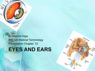
The document summarizes the anatomy and function of the human eye and ear. It describes the major parts of the eye, including the cornea, iris, pupil, lens, retina, and optic nerve. It explains how vision occurs as light enters the eye and is focused on the retina, where it is converted to electrical signals sent to the brain. It also outlines the three main parts of the ear - external, middle, and inner ear - and describes how sound waves are captured, vibrations are transmitted through the bones of the middle ear, and nerve signals are sent to the brain for interpretation of sounds.
anatomy of eye orbit, bones involved , boundaries of orbit, contents of orbit, orbital fat, openings of the orbit and contents passing through the openings
anatomy of eye orbit, bones involved , boundaries of orbit, contents of orbit, orbital fat, openings of the orbit and contents passing through the openings
structure of eye ball,eyeball is a specialized sense organ that helps us to understand our environment. It is a sensory unit composed of three parts: receptor, sensory pathway, and a brain center
The main parts of the human eye are The Conjunctiva,
Sclera,Choroid,
Cornea, Iris, Pupil,
Anterior Chamber,
Posterior Chamber, Aqueous humor, Lens, Vitreous humor, Retina,Macula and Optic nerve.
anatomy of an eye, internal structure of eye,layers of an eye ball, features of eye ball, cornea, sclera, retina, choroid, ciliary body lense, macula, blind spot, yellow spot, clinical aspect of eye , drainage of aqueous humor, cataract, glaucoma, causes of cataract and treatment.
This presentation provides a comprehensive review of major sulci of brain which help in defining the different lobes of brain.Very useful for first year residents.
Excelente presentación de Federico Pacheco, donde se dan a conocer aspecto referente a la Gestión de la Seguridad, estoy seguro sera de utilidad para muchos.
En estas laminas podrán observar como se puede realizar una búsquedas en Google con distintos resultados, al agregar ciertos comandos que Google tiene para realizar búsqueda mas eficiente.
Por medio de:
a) frases exactas
b) restricciones de palabras o frases
c) Por rangos
d) definiciones
e) tipos de archivos ya sea para uno o varios tipos de archivos
f) en sitios específicos, sitios oficiales o gubernamentales
Entre otros métodos
We looked at the data. Here’s a breakdown of some key statistics about the nation’s incoming presidents’ addresses, how long they spoke, how well, and more.
My books- Hacking Digital Learning Strategies http://hackingdls.com & Learning to Go https://gum.co/learn2go
Resources at http://shellyterrell.com/emoji
HEALTH ASSESSMENT AND DIAGNOSTIC TESTS OF EYE AND ENT DISORDERSDIAGNOSTIC TES...JishaSrivastava
HEALTH ASSESSMENT AND DIAGNOSTIC TESTS OF EYE AND ENT DISORDERS
At the end of the class students will be able to :
Describe the structures of the Eye and ENT.
Describe the functions of Eye and ENT.
Explain age affect on the Eye and ENT. .
Explain the techniques used in a physical examination of Eye and ENT.
List down the diagnostic tests for the disorders of the Eye and ENT.
Distinguish between normal and abnormal findings.
Explain the Nursing Interventions for diagnostic tests for the disorders of Eye and ENT.
ARTIFICIAL INTELLIGENCE IN HEALTHCARE.pdfAnujkumaranit
Artificial intelligence (AI) refers to the simulation of human intelligence processes by machines, especially computer systems. It encompasses tasks such as learning, reasoning, problem-solving, perception, and language understanding. AI technologies are revolutionizing various fields, from healthcare to finance, by enabling machines to perform tasks that typically require human intelligence.
Ethanol (CH3CH2OH), or beverage alcohol, is a two-carbon alcohol
that is rapidly distributed in the body and brain. Ethanol alters many
neurochemical systems and has rewarding and addictive properties. It
is the oldest recreational drug and likely contributes to more morbidity,
mortality, and public health costs than all illicit drugs combined. The
5th edition of the Diagnostic and Statistical Manual of Mental Disorders
(DSM-5) integrates alcohol abuse and alcohol dependence into a single
disorder called alcohol use disorder (AUD), with mild, moderate,
and severe subclassifications (American Psychiatric Association, 2013).
In the DSM-5, all types of substance abuse and dependence have been
combined into a single substance use disorder (SUD) on a continuum
from mild to severe. A diagnosis of AUD requires that at least two of
the 11 DSM-5 behaviors be present within a 12-month period (mild
AUD: 2–3 criteria; moderate AUD: 4–5 criteria; severe AUD: 6–11 criteria).
The four main behavioral effects of AUD are impaired control over
drinking, negative social consequences, risky use, and altered physiological
effects (tolerance, withdrawal). This chapter presents an overview
of the prevalence and harmful consequences of AUD in the U.S.,
the systemic nature of the disease, neurocircuitry and stages of AUD,
comorbidities, fetal alcohol spectrum disorders, genetic risk factors, and
pharmacotherapies for AUD.
Anti ulcer drugs and their Advance pharmacology ||
Anti-ulcer drugs are medications used to prevent and treat ulcers in the stomach and upper part of the small intestine (duodenal ulcers). These ulcers are often caused by an imbalance between stomach acid and the mucosal lining, which protects the stomach lining.
||Scope: Overview of various classes of anti-ulcer drugs, their mechanisms of action, indications, side effects, and clinical considerations.
Explore natural remedies for syphilis treatment in Singapore. Discover alternative therapies, herbal remedies, and lifestyle changes that may complement conventional treatments. Learn about holistic approaches to managing syphilis symptoms and supporting overall health.
Tom Selleck Health: A Comprehensive Look at the Iconic Actor’s Wellness Journeygreendigital
Tom Selleck, an enduring figure in Hollywood. has captivated audiences for decades with his rugged charm, iconic moustache. and memorable roles in television and film. From his breakout role as Thomas Magnum in Magnum P.I. to his current portrayal of Frank Reagan in Blue Bloods. Selleck's career has spanned over 50 years. But beyond his professional achievements. fans have often been curious about Tom Selleck Health. especially as he has aged in the public eye.
Follow us on: Pinterest
Introduction
Many have been interested in Tom Selleck health. not only because of his enduring presence on screen but also because of the challenges. and lifestyle choices he has faced and made over the years. This article delves into the various aspects of Tom Selleck health. exploring his fitness regimen, diet, mental health. and the challenges he has encountered as he ages. We'll look at how he maintains his well-being. the health issues he has faced, and his approach to ageing .
Early Life and Career
Childhood and Athletic Beginnings
Tom Selleck was born on January 29, 1945, in Detroit, Michigan, and grew up in Sherman Oaks, California. From an early age, he was involved in sports, particularly basketball. which played a significant role in his physical development. His athletic pursuits continued into college. where he attended the University of Southern California (USC) on a basketball scholarship. This early involvement in sports laid a strong foundation for his physical health and disciplined lifestyle.
Transition to Acting
Selleck's transition from an athlete to an actor came with its physical demands. His first significant role in "Magnum P.I." required him to perform various stunts and maintain a fit appearance. This role, which he played from 1980 to 1988. necessitated a rigorous fitness routine to meet the show's demands. setting the stage for his long-term commitment to health and wellness.
Fitness Regimen
Workout Routine
Tom Selleck health and fitness regimen has evolved. adapting to his changing roles and age. During his "Magnum, P.I." days. Selleck's workouts were intense and focused on building and maintaining muscle mass. His routine included weightlifting, cardiovascular exercises. and specific training for the stunts he performed on the show.
Selleck adjusted his fitness routine as he aged to suit his body's needs. Today, his workouts focus on maintaining flexibility, strength, and cardiovascular health. He incorporates low-impact exercises such as swimming, walking, and light weightlifting. This balanced approach helps him stay fit without putting undue strain on his joints and muscles.
Importance of Flexibility and Mobility
In recent years, Selleck has emphasized the importance of flexibility and mobility in his fitness regimen. Understanding the natural decline in muscle mass and joint flexibility with age. he includes stretching and yoga in his routine. These practices help prevent injuries, improve posture, and maintain mobilit
These lecture slides, by Dr Sidra Arshad, offer a quick overview of physiological basis of a normal electrocardiogram.
Learning objectives:
1. Define an electrocardiogram (ECG) and electrocardiography
2. Describe how dipoles generated by the heart produce the waveforms of the ECG
3. Describe the components of a normal electrocardiogram of a typical bipolar leads (limb II)
4. Differentiate between intervals and segments
5. Enlist some common indications for obtaining an ECG
Study Resources:
1. Chapter 11, Guyton and Hall Textbook of Medical Physiology, 14th edition
2. Chapter 9, Human Physiology - From Cells to Systems, Lauralee Sherwood, 9th edition
3. Chapter 29, Ganong’s Review of Medical Physiology, 26th edition
4. Electrocardiogram, StatPearls - https://www.ncbi.nlm.nih.gov/books/NBK549803/
5. ECG in Medical Practice by ABM Abdullah, 4th edition
6. ECG Basics, http://www.nataliescasebook.com/tag/e-c-g-basics
Title: Sense of Smell
Presenter: Dr. Faiza, Assistant Professor of Physiology
Qualifications:
MBBS (Best Graduate, AIMC Lahore)
FCPS Physiology
ICMT, CHPE, DHPE (STMU)
MPH (GC University, Faisalabad)
MBA (Virtual University of Pakistan)
Learning Objectives:
Describe the primary categories of smells and the concept of odor blindness.
Explain the structure and location of the olfactory membrane and mucosa, including the types and roles of cells involved in olfaction.
Describe the pathway and mechanisms of olfactory signal transmission from the olfactory receptors to the brain.
Illustrate the biochemical cascade triggered by odorant binding to olfactory receptors, including the role of G-proteins and second messengers in generating an action potential.
Identify different types of olfactory disorders such as anosmia, hyposmia, hyperosmia, and dysosmia, including their potential causes.
Key Topics:
Olfactory Genes:
3% of the human genome accounts for olfactory genes.
400 genes for odorant receptors.
Olfactory Membrane:
Located in the superior part of the nasal cavity.
Medially: Folds downward along the superior septum.
Laterally: Folds over the superior turbinate and upper surface of the middle turbinate.
Total surface area: 5-10 square centimeters.
Olfactory Mucosa:
Olfactory Cells: Bipolar nerve cells derived from the CNS (100 million), with 4-25 olfactory cilia per cell.
Sustentacular Cells: Produce mucus and maintain ionic and molecular environment.
Basal Cells: Replace worn-out olfactory cells with an average lifespan of 1-2 months.
Bowman’s Gland: Secretes mucus.
Stimulation of Olfactory Cells:
Odorant dissolves in mucus and attaches to receptors on olfactory cilia.
Involves a cascade effect through G-proteins and second messengers, leading to depolarization and action potential generation in the olfactory nerve.
Quality of a Good Odorant:
Small (3-20 Carbon atoms), volatile, water-soluble, and lipid-soluble.
Facilitated by odorant-binding proteins in mucus.
Membrane Potential and Action Potential:
Resting membrane potential: -55mV.
Action potential frequency in the olfactory nerve increases with odorant strength.
Adaptation Towards the Sense of Smell:
Rapid adaptation within the first second, with further slow adaptation.
Psychological adaptation greater than receptor adaptation, involving feedback inhibition from the central nervous system.
Primary Sensations of Smell:
Camphoraceous, Musky, Floral, Pepperminty, Ethereal, Pungent, Putrid.
Odor Detection Threshold:
Examples: Hydrogen sulfide (0.0005 ppm), Methyl-mercaptan (0.002 ppm).
Some toxic substances are odorless at lethal concentrations.
Characteristics of Smell:
Odor blindness for single substances due to lack of appropriate receptor protein.
Behavioral and emotional influences of smell.
Transmission of Olfactory Signals:
From olfactory cells to glomeruli in the olfactory bulb, involving lateral inhibition.
Primitive, less old, and new olfactory systems with different path
MANAGEMENT OF ATRIOVENTRICULAR CONDUCTION BLOCK.pdfJim Jacob Roy
Cardiac conduction defects can occur due to various causes.
Atrioventricular conduction blocks ( AV blocks ) are classified into 3 types.
This document describes the acute management of AV block.
micro teaching on communication m.sc nursing.pdfAnurag Sharma
Microteaching is a unique model of practice teaching. It is a viable instrument for the. desired change in the teaching behavior or the behavior potential which, in specified types of real. classroom situations, tends to facilitate the achievement of specified types of objectives.
3. The Eyeball
The Anatomy of the Eyeball
• Anterior Chamber: The space in the eye that is
behind the cornea and in front of the iris.
• Upper & lower lid: a thin fold of skin that covers and
protects the eye.
• Conjunctiva: a clear mucuse membrane consisting of
cells and underlying basement membrane
that covers the sclera (white part of the
eye) and lines the inside part of the eyelid.
• Pupil: a hole located in the center of the iris of the eye
that allows light to enter the eye
• Cornea: a transparent front part of the eye that covers the
iris and the pupil.
• Aqueous humor: The liquid between the lens and the
cornea.
• Posterior chamber: A narrow crack behind the
perpherial part of the iris
• Fovea centralis: The part of the eye located in the center
of the Macula region of the retina, this part is
responsible for the sharp central vision.
• Optic nerve: Transmits visual information from the
retina to the Brain.
• Central retinal artery: It pierces the optic nerve close to
the eyeball, sending branches over the internal
surface of the retina, and these terminal
branches are the only blood supply to the larger
part of the eye
• Sclera: fibrous, protective, outer layer of the eye
containing collagen and elastic fiber.
• Choroid: The vascular layer containing connective
tissue, of the eye lying between the retina and
the sclera. In humans its thickness is about 0.5 mm.
The choroid provides oxygen and nourishment to the
outer layers of the retina
• Retina: A light-sensitive tissue lining the inner
surface of the eye
4. Muscles of the eye
The extraocular
muscles are the six
muscles that control
the movements of the
(human) eye. The
actions of the
extraocular muscles
depend on the position
of the eye at the time
of muscle contraction.
5. Lacriminal apparatuse
The lacrimal apparatus is the
physiologic system containing the
orbital structures for tear production
and drainage[1]. It consists of
(a) the lacrimal gland, which
secretes the tears, and its
excretory ducts, which convey
the fluid to the surface of the
eye
(b) the lacrimal canaliculi, the
lacrimal sac, and the
nasolacrimal duct, by which the
fluid is conveyed into the cavity
of the nose, emptying
anterioinferiorly to the inferior
nasal conchae at the
nasolacrimal duct.
(c) the nerve supply of lacrimal
apparatus done by carotid
plexuse of nerves along artery
internal and external
sympathetically but
parasympathetic from lacrimal
nucleus of the facial nerve
6. • Vision begins when light rays are reflected off an
object and enter the eyes through the cornea, the
transparent outer covering of the eye. The cornea
bends or refracts the rays that pass through a round
hole called the pupil. The iris, or colored portion of the
eye that surrounds the pupil, opens and closes
(making the pupil bigger or smaller) to regulate the
amount of light passing through. The light rays then
pass through the lens, which actually changes shape
so it can further bend the rays and focus them on the
retina at the back of the eye. The retina is a thin layer
of tissue at the back of the eye that contains millions of
tiny light-sensing nerve cells called rods and cones,
which are named for their distinct shapes. Cones are
concentrated in the center of the retina, in an area
called the macula. In bright light conditions, cones
provide clear, sharp central vision and detect colors
and fine details. Rods are located outside the macula
and extend all the way to the outer edge of the retina.
They provide peripheral or side vision. Rods also allow
the eyes to detect motion and help us see in dim light
and at night. These cells in the retina convert the light
into electrical impulses. The optic nerve sends these
impulses to the brain where an image is produced.
How we see
8. External ear
The outer ear has no
bones. It is the
external portion of the
ear, which consists of
the pinna, concha, and
auditory meatus. It
gathers sound energy
and focuses it on the
eardrum (tympanic
membrane).
9. Middle ear
The middle ear is the portion
of the ear internal to the
eardrum, and external to the
oval window of the cochlea.
The mammalian middle ear
contains three ossicles, which
couple vibration of the
eardrum into waves in the
fluid and membranes of the
inner ear. The hollow space of
the middle ear has also been
called the tympanic cavity, or
cavum tympani. The
eustachian tube joins the
tympanic cavity with the nasal
cavity (nasopharynx),
allowing pressure to equalize
between the middle ear and
throat.
10. Inner ear
The inner ear is the
innermost part of the
vertebrate ear. It consists of
the bony labyrinth, a
system of passages
comprising two main
functional parts:The
cochlea is dedicated to
hearingThe vestibular
system is dedicated to
balanceThe inner ear is
found in all vertebrates,
with substantial variations
in form and function. The
inner ear is innervated by
the eight cranial nerve in all
vertebrates.
11. How we hear
When something makes a noise, it sends
vibrations, or sound waves, through the
air. The human eardrum is a stretched
membrane, like the skin of a drum.
When the sound waves hit your eardrum,
it vibrates and the brain interprets these
vibrations as. After the vibrations hit
your eardrum, a chain reaction is set off.
Your eardrum, which is smaller and
thinner than the nail on your pinky
finger, sends the vibrations to the three
smallest bones in your body. First the
hammer, then the anvil, and finally, the
stirrup. The stirrup passes those
vibrations along a coiled tub in the inner
ear called the cochlea. Inside the cochlea
there are thousands of hair-like nerve
endings, cilia. When the Cochlea
vibrates, the cilia move. Your brain is
sent these messages (translated from
vibrations by the cilia) through the
auditory nerve. Your brain then
translates all that and tells you what you
are hearing. Neurologists don't yet fully
understand how we process raw sound
data once it enters the cerebral cortex in
the brain.