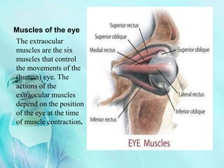
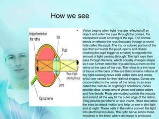
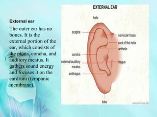
This ppt file belongs to Mr. Yonas Akalu one of my best instructors ...
Vision is by far the most used of the five senses and is one of the primary means that we use to gather information from our surroundings. More than 75% of the information we receive about the world around us consists of visual information.
The eye is often compared to a camera. Each gathers light and then transforms that light into a "picture." Both also have lenses to focus the incoming light. Just as a camera focuses light onto the film to create a picture, the eye focuses light onto a specialized layer of cells, called the retina.
The human eye is an organ which reacts to light and pressure. As a sense organ, the mammalian eye allows vision. Human eyes help to provide a three dimensional, moving image, normally coloured in daylight. Rod and cone cells in the retina allow conscious light perception and vision including color differentiation and the perception of depth. The human eye can differentiate between about 10 million colors[1] and is possibly capable of detecting a single photon.
Following points are explain in this article:
Introduction of eye
Eyelids & tears
Morphology of eye-ball
Components of eye
Functions of eye parts
Diagrams
HEALTH ASSESSMENT AND DIAGNOSTIC TESTS OF EYE AND ENT DISORDERSDIAGNOSTIC TES...JishaSrivastava
HEALTH ASSESSMENT AND DIAGNOSTIC TESTS OF EYE AND ENT DISORDERS
At the end of the class students will be able to :
Describe the structures of the Eye and ENT.
Describe the functions of Eye and ENT.
Explain age affect on the Eye and ENT. .
Explain the techniques used in a physical examination of Eye and ENT.
List down the diagnostic tests for the disorders of the Eye and ENT.
Distinguish between normal and abnormal findings.
Explain the Nursing Interventions for diagnostic tests for the disorders of Eye and ENT.
- Video recording of this lecture in English language: https://youtu.be/lK81BzxMqdo
- Video recording of this lecture in Arabic language: https://youtu.be/Ve4P0COk9OI
- Link to download the book free: https://nephrotube.blogspot.com/p/nephrotube-nephrology-books.html
- Link to NephroTube website: www.NephroTube.com
- Link to NephroTube social media accounts: https://nephrotube.blogspot.com/p/join-nephrotube-on-social-media.html
Flu Vaccine Alert in Bangalore Karnatakaaddon Scans
As flu season approaches, health officials in Bangalore, Karnataka, are urging residents to get their flu vaccinations. The seasonal flu, while common, can lead to severe health complications, particularly for vulnerable populations such as young children, the elderly, and those with underlying health conditions.
Dr. Vidisha Kumari, a leading epidemiologist in Bangalore, emphasizes the importance of getting vaccinated. "The flu vaccine is our best defense against the influenza virus. It not only protects individuals but also helps prevent the spread of the virus in our communities," he says.
This year, the flu season is expected to coincide with a potential increase in other respiratory illnesses. The Karnataka Health Department has launched an awareness campaign highlighting the significance of flu vaccinations. They have set up multiple vaccination centers across Bangalore, making it convenient for residents to receive their shots.
To encourage widespread vaccination, the government is also collaborating with local schools, workplaces, and community centers to facilitate vaccination drives. Special attention is being given to ensuring that the vaccine is accessible to all, including marginalized communities who may have limited access to healthcare.
Residents are reminded that the flu vaccine is safe and effective. Common side effects are mild and may include soreness at the injection site, mild fever, or muscle aches. These side effects are generally short-lived and far less severe than the flu itself.
Healthcare providers are also stressing the importance of continuing COVID-19 precautions. Wearing masks, practicing good hand hygiene, and maintaining social distancing are still crucial, especially in crowded places.
Protect yourself and your loved ones by getting vaccinated. Together, we can help keep Bangalore healthy and safe this flu season. For more information on vaccination centers and schedules, residents can visit the Karnataka Health Department’s official website or follow their social media pages.
Stay informed, stay safe, and get your flu shot today!
micro teaching on communication m.sc nursing.pdfAnurag Sharma
Microteaching is a unique model of practice teaching. It is a viable instrument for the. desired change in the teaching behavior or the behavior potential which, in specified types of real. classroom situations, tends to facilitate the achievement of specified types of objectives.
Title: Sense of Taste
Presenter: Dr. Faiza, Assistant Professor of Physiology
Qualifications:
MBBS (Best Graduate, AIMC Lahore)
FCPS Physiology
ICMT, CHPE, DHPE (STMU)
MPH (GC University, Faisalabad)
MBA (Virtual University of Pakistan)
Learning Objectives:
Describe the structure and function of taste buds.
Describe the relationship between the taste threshold and taste index of common substances.
Explain the chemical basis and signal transduction of taste perception for each type of primary taste sensation.
Recognize different abnormalities of taste perception and their causes.
Key Topics:
Significance of Taste Sensation:
Differentiation between pleasant and harmful food
Influence on behavior
Selection of food based on metabolic needs
Receptors of Taste:
Taste buds on the tongue
Influence of sense of smell, texture of food, and pain stimulation (e.g., by pepper)
Primary and Secondary Taste Sensations:
Primary taste sensations: Sweet, Sour, Salty, Bitter, Umami
Chemical basis and signal transduction mechanisms for each taste
Taste Threshold and Index:
Taste threshold values for Sweet (sucrose), Salty (NaCl), Sour (HCl), and Bitter (Quinine)
Taste index relationship: Inversely proportional to taste threshold
Taste Blindness:
Inability to taste certain substances, particularly thiourea compounds
Example: Phenylthiocarbamide
Structure and Function of Taste Buds:
Composition: Epithelial cells, Sustentacular/Supporting cells, Taste cells, Basal cells
Features: Taste pores, Taste hairs/microvilli, and Taste nerve fibers
Location of Taste Buds:
Found in papillae of the tongue (Fungiform, Circumvallate, Foliate)
Also present on the palate, tonsillar pillars, epiglottis, and proximal esophagus
Mechanism of Taste Stimulation:
Interaction of taste substances with receptors on microvilli
Signal transduction pathways for Umami, Sweet, Bitter, Sour, and Salty tastes
Taste Sensitivity and Adaptation:
Decrease in sensitivity with age
Rapid adaptation of taste sensation
Role of Saliva in Taste:
Dissolution of tastants to reach receptors
Washing away the stimulus
Taste Preferences and Aversions:
Mechanisms behind taste preference and aversion
Influence of receptors and neural pathways
Impact of Sensory Nerve Damage:
Degeneration of taste buds if the sensory nerve fiber is cut
Abnormalities of Taste Detection:
Conditions: Ageusia, Hypogeusia, Dysgeusia (parageusia)
Causes: Nerve damage, neurological disorders, infections, poor oral hygiene, adverse drug effects, deficiencies, aging, tobacco use, altered neurotransmitter levels
Neurotransmitters and Taste Threshold:
Effects of serotonin (5-HT) and norepinephrine (NE) on taste sensitivity
Supertasters:
25% of the population with heightened sensitivity to taste, especially bitterness
Increased number of fungiform papillae
These lecture slides, by Dr Sidra Arshad, offer a quick overview of physiological basis of a normal electrocardiogram.
Learning objectives:
1. Define an electrocardiogram (ECG) and electrocardiography
2. Describe how dipoles generated by the heart produce the waveforms of the ECG
3. Describe the components of a normal electrocardiogram of a typical bipolar leads (limb II)
4. Differentiate between intervals and segments
5. Enlist some common indications for obtaining an ECG
Study Resources:
1. Chapter 11, Guyton and Hall Textbook of Medical Physiology, 14th edition
2. Chapter 9, Human Physiology - From Cells to Systems, Lauralee Sherwood, 9th edition
3. Chapter 29, Ganong’s Review of Medical Physiology, 26th edition
4. Electrocardiogram, StatPearls - https://www.ncbi.nlm.nih.gov/books/NBK549803/
5. ECG in Medical Practice by ABM Abdullah, 4th edition
6. ECG Basics, http://www.nataliescasebook.com/tag/e-c-g-basics
3. The Eyeball
The Anatomy of the Eyeball
• Anterior Chamber: The space in the eye that is
behind the cornea and in front of the iris.
• Upper & lower lid: a thin fold of skin that covers and
protects the eye.
• Conjunctiva: a clear mucuse membrane consisting of
cells and underlying basement membrane
that covers the sclera (white part of the
eye) and lines the inside part of the eyelid.
• Pupil: a hole located in the center of the iris of the eye
that allows light to enter the eye
• Cornea: a transparent front part of the eye that covers the
iris and the pupil.
• Aqueous humor: The liquid between the lens and the
cornea.
• Posterior chamber: A narrow crack behind the
perpherial part of the iris
• Fovea centralis: The part of the eye located in the center
of the Macula region of the retina, this part is
responsible for the sharp central vision.
• Optic nerve: Transmits visual information from the
retina to the Brain.
• Central retinal artery: It pierces the optic nerve close to
the eyeball, sending branches over the internal
surface of the retina, and these terminal
branches are the only blood supply to the larger
part of the eye
• Sclera: fibrous, protective, outer layer of the eye
containing collagen and elastic fiber.
• Choroid: The vascular layer containing connective
tissue, of the eye lying between the retina and
the sclera. In humans its thickness is about 0.5 mm.
The choroid provides oxygen and nourishment to the
outer layers of the retina
• Retina: A light-sensitive tissue lining the inner
surface of the eye
4. Muscles of the eye
The extraocular
muscles are the six
muscles that control
the movements of the
(human) eye. The
actions of the
extraocular muscles
depend on the position
of the eye at the time
of muscle contraction.
5. Lacriminal apparatuse
The lacrimal apparatus is the
physiologic system containing the
orbital structures for tear production
and drainage[1]. It consists of
(a) the lacrimal gland, which
secretes the tears, and its
excretory ducts, which convey
the fluid to the surface of the
eye
(b) the lacrimal canaliculi, the
lacrimal sac, and the
nasolacrimal duct, by which the
fluid is conveyed into the cavity
of the nose, emptying
anterioinferiorly to the inferior
nasal conchae at the
nasolacrimal duct.
(c) the nerve supply of lacrimal
apparatus done by carotid
plexuse of nerves along artery
internal and external
sympathetically but
parasympathetic from lacrimal
nucleus of the facial nerve
6. • Vision begins when light rays are reflected off an
object and enter the eyes through the cornea, the
transparent outer covering of the eye. The cornea
bends or refracts the rays that pass through a round
hole called the pupil. The iris, or colored portion of the
eye that surrounds the pupil, opens and closes
(making the pupil bigger or smaller) to regulate the
amount of light passing through. The light rays then
pass through the lens, which actually changes shape
so it can further bend the rays and focus them on the
retina at the back of the eye. The retina is a thin layer
of tissue at the back of the eye that contains millions of
tiny light-sensing nerve cells called rods and cones,
which are named for their distinct shapes. Cones are
concentrated in the center of the retina, in an area
called the macula. In bright light conditions, cones
provide clear, sharp central vision and detect colors
and fine details. Rods are located outside the macula
and extend all the way to the outer edge of the retina.
They provide peripheral or side vision. Rods also allow
the eyes to detect motion and help us see in dim light
and at night. These cells in the retina convert the light
into electrical impulses. The optic nerve sends these
impulses to the brain where an image is produced.
How we see
8. External ear
The outer ear has no
bones. It is the
external portion of the
ear, which consists of
the pinna, concha, and
auditory meatus. It
gathers sound energy
and focuses it on the
eardrum (tympanic
membrane).
9. Middle ear
The middle ear is the portion
of the ear internal to the
eardrum, and external to the
oval window of the cochlea.
The mammalian middle ear
contains three ossicles, which
couple vibration of the
eardrum into waves in the
fluid and membranes of the
inner ear. The hollow space of
the middle ear has also been
called the tympanic cavity, or
cavum tympani. The
eustachian tube joins the
tympanic cavity with the nasal
cavity (nasopharynx),
allowing pressure to equalize
between the middle ear and
throat.
10. Inner ear
The inner ear is the
innermost part of the
vertebrate ear. It consists of
the bony labyrinth, a
system of passages
comprising two main
functional parts:The
cochlea is dedicated to
hearingThe vestibular
system is dedicated to
balanceThe inner ear is
found in all vertebrates,
with substantial variations
in form and function. The
inner ear is innervated by
the eight cranial nerve in all
vertebrates.
11. How we hear
When something makes a noise, it sends
vibrations, or sound waves, through the
air. The human eardrum is a stretched
membrane, like the skin of a drum.
When the sound waves hit your eardrum,
it vibrates and the brain interprets these
vibrations as. After the vibrations hit
your eardrum, a chain reaction is set off.
Your eardrum, which is smaller and
thinner than the nail on your pinky
finger, sends the vibrations to the three
smallest bones in your body. First the
hammer, then the anvil, and finally, the
stirrup. The stirrup passes those
vibrations along a coiled tub in the inner
ear called the cochlea. Inside the cochlea
there are thousands of hair-like nerve
endings, cilia. When the Cochlea
vibrates, the cilia move. Your brain is
sent these messages (translated from
vibrations by the cilia) through the
auditory nerve. Your brain then
translates all that and tells you what you
are hearing. Neurologists don't yet fully
understand how we process raw sound
data once it enters the cerebral cortex in
the brain.