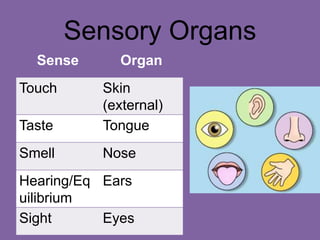
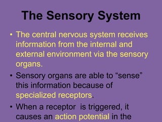
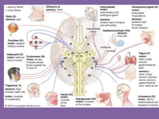
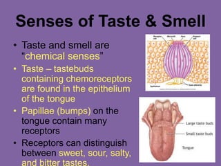
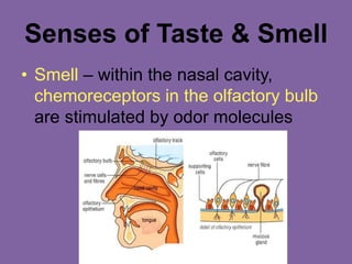
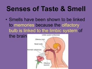
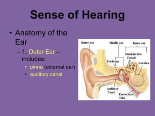
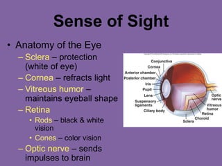
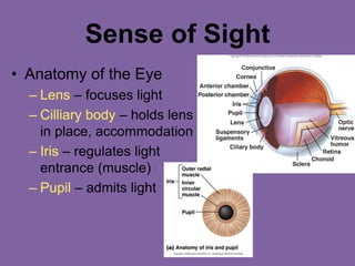
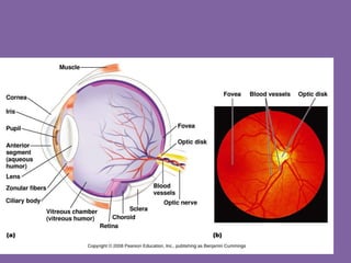
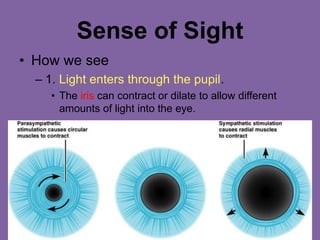
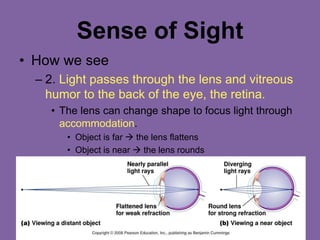
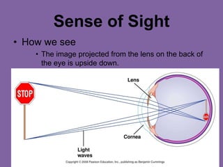
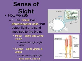
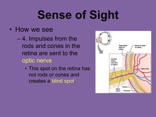
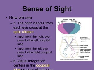
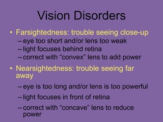
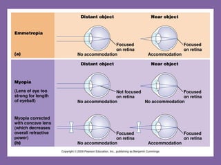
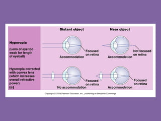
Sensory System
Chapter 9
Sensory Organs
Sense Organ
Touch Skin
(external)
Taste Tongue
Smell Nose
Hearing/Eq
uilibrium
Ears
Sight Eyes
The Sensory System
• The central nervous system receives
information from the internal and
external environment via the sensory
organs.
• Sensory organs are able to “sense”
this information because of
specialized receptors.
• When a receptor is triggered, it
causes an action potential in the
Types of Sensory
Receptors
• 1. Mechanoreceptors – stimulated by
changes in pressure or movement
– Found in skin and muscles
• 2. Thermoreceptors – stimulated by
changes in temperature
– Found in skin
• 3. Pain receptors – stimulated by
tissue damage
– Found in skin and viscera
Types of Sensory
Receptors (continued)
• 4. Chemoreceptors – stimulated by
changes in chemical concentration of
substances
– Used for taste and smell
• 5. Photoreceptors – stimulated by light
– Found only in the eye
Sense of Touch
• Mechanorecept
ors in the skin
and viscera
detect varying
degrees of
pressure.
• Free nerve
endings have
pain receptors
and
Sense of Touch – Pain
• Pain is caused by
chemicals released by
inflamed tissues.
– Aspirin and ibuprofen reduce
pain by blocking synthesis of
these chemicals
• Referred pain – inside the
body’s organs, pain is often
felt in another area.
– Ex: Pain from the heart is felt
Senses of Taste & Smell
• Taste and smell are
“chemical senses”
• Taste – tastebuds
containing chemoreceptors
are found in the epithelium
of the tongue
• Papillae (bumps) on the
tongue contain many
receptors
• Receptors can distinguish
between sweet, sour, salty,
and bitter tastes.
Senses of Taste & Smell
• Smell – within the nasal cavity,
chemoreceptors in the olfactory bulb
are stimulated by odor molecules
Senses of Taste & Smell
• Smells have been shown to be linked
to memories because the olfactory
bulb is linked to the limbic system of
the brain.
Sense of Hearing
• Anatomy of the
Ear
– 1. Outer Ear –
includes:
• pinna (external ear)
• auditory canal
Sense of Hearing
• Anatomy of the Ear
–2. Middle Ear- includes:
• Eardrum (tympanic
membrane)
• Ossicles – 3 small bones
– 1) Malleus (hammer)
– 2) Incus (anvil)
– 3) Stapes (stirrup)
• Eustachian tube –
equalization of air
pressure (“pops” ear)
Sense of Hearing
• Anatomy of the
Ear
– 3. Inner Ear –
includes:
• Semicircular
canals – involved
with equilibrium
• Cochlea – snail-
shaped structure
involved with
hearing
Sense of Hearing
• How we Hear
– 1. Sound waves travel through the auditory canal
to the eardrum.
– 2. The sound waves cause the eardrum to
vibrate.
– 3. The vibration causes the malleus (hammer) to
hit the incus (anvil) and then the stapes (stirrup).
– 4. The vibration passes to the fluid in the cochlea
of the inner ear.
– 5. Each part of the spiral cochlea is sensitive to
different frequencies of sound.
– 6. The auditory nerve takes impulses to the brain.
Sense of Hearing
• Equilibrium
– Mechanoreceptors in
the semicircular canals
detect rotation and
movement of the head
– Little hair cells send
information to the
brain to cause
appropriate motor
output so as to correct
position when it is
unbalanced.
– Vertigo (dizziness)
Sense of Sight
• Anatomy of the Eye
– Sclera – protection
(white of eye)
– Cornea – refracts light
– Vitreous humor –
maintains eyeball shape
– Retina
• Rods – black & white
vision
• Cones – color vision
– Optic nerve – sends
impulses to brain
Sense of Sight
• Anatomy of the Eye
– Lens – focuses light
– Cilliary body – holds lens
in place, accommodation
– Iris – regulates light
entrance (muscle)
– Pupil – admits light
Sense of Sight
• How we see
– 1. Light enters through the pupil.
• The iris can contract or dilate to allow different
amounts of light into the eye.
Sense of Sight
• How we see
– 2. Light passes through the lens and vitreous
humor to the back of the eye, the retina.
• The lens can change shape to focus light through
accommodation.
• Object is far  the lens flattens
• Object is near  the lens rounds
Sense of Sight
• How we see
• The image projected from the lens on the back of
the eye is upside down.
Sense of
Sight
• How we see
– 3. The retina has
photoreceptor cells that
detect light and send
impulses to the brain.
• Rods – black and white
vision
– sensitive to light; night
vision
• Cones – color vision &
detail
– Sensitive to bright light
– Blue, green, and red
Sense of Sight
• How we see
– 4. Impulses from the
rods and cones in the
retina are sent to the
optic nerve
• This spot on the retina has
not rods or cones and
creates a blind spot.
Sense of Sight
• How we see
– 5. The optic nerves from
each eye cross at the
optic chiasm.
• Input from the right eye
goes to the left occipital
lobe
• Input from the left eye
goes to the right occipital
lobe
– 6. Visual integration
centers in the occipital
Vision Disorders
• Farsightedness: trouble seeing close-up
– eye too short and/or lens too weak
– light focuses behind retina
– correct with “convex” lens to add power
• Nearsightedness: trouble seeing far
away
– eye is too long and/or lens is too powerful
– light focuses in front of retina
– correct with “concave” lens to reduce
power
• Presbyopia: Oldsightedness
• The crystalline lens tends to harden with
age
• The near point of distinct vision moves
further and further away from the eye
with age.
Astigmatism
• Abnormal curvature of the cornea
• Light from vertical and horizontal direction
do not focuses in the same point
• Correct with “cylindrical” lens to
compensate
Color Blindness
– Red-green color-blindness – occurs when
red or green cones or pigments are
missing
– Due to sex-linked gene (on X chromosomes)
so more common in men.
• Non-sex-linked condition
– Blue-color blindness- missing blue cones or
pigments
– Monochromats: people who are totally
colorblind, more severe
Disorders of the Eye
• Glaucoma
– Damage to the optic
nerve occurs due to
increased eye
pressure
– Can lead to blindness
• Cataracts
– Clouding of the lens
that affects vision
– Very common in older
people
Figure 10.27b

sensory_systems chapter 9 for medical physiology

  • 1.
  • 2.
    Sensory Organs Sense Organ TouchSkin (external) Taste Tongue Smell Nose Hearing/Eq uilibrium Ears Sight Eyes
  • 3.
    The Sensory System •The central nervous system receives information from the internal and external environment via the sensory organs. • Sensory organs are able to “sense” this information because of specialized receptors. • When a receptor is triggered, it causes an action potential in the
  • 5.
    Types of Sensory Receptors •1. Mechanoreceptors – stimulated by changes in pressure or movement – Found in skin and muscles • 2. Thermoreceptors – stimulated by changes in temperature – Found in skin • 3. Pain receptors – stimulated by tissue damage – Found in skin and viscera
  • 6.
    Types of Sensory Receptors(continued) • 4. Chemoreceptors – stimulated by changes in chemical concentration of substances – Used for taste and smell • 5. Photoreceptors – stimulated by light – Found only in the eye
  • 7.
    Sense of Touch •Mechanorecept ors in the skin and viscera detect varying degrees of pressure. • Free nerve endings have pain receptors and
  • 8.
    Sense of Touch– Pain • Pain is caused by chemicals released by inflamed tissues. – Aspirin and ibuprofen reduce pain by blocking synthesis of these chemicals • Referred pain – inside the body’s organs, pain is often felt in another area. – Ex: Pain from the heart is felt
  • 9.
    Senses of Taste& Smell • Taste and smell are “chemical senses” • Taste – tastebuds containing chemoreceptors are found in the epithelium of the tongue • Papillae (bumps) on the tongue contain many receptors • Receptors can distinguish between sweet, sour, salty, and bitter tastes.
  • 10.
    Senses of Taste& Smell • Smell – within the nasal cavity, chemoreceptors in the olfactory bulb are stimulated by odor molecules
  • 11.
    Senses of Taste& Smell • Smells have been shown to be linked to memories because the olfactory bulb is linked to the limbic system of the brain.
  • 12.
    Sense of Hearing •Anatomy of the Ear – 1. Outer Ear – includes: • pinna (external ear) • auditory canal
  • 13.
    Sense of Hearing •Anatomy of the Ear –2. Middle Ear- includes: • Eardrum (tympanic membrane) • Ossicles – 3 small bones – 1) Malleus (hammer) – 2) Incus (anvil) – 3) Stapes (stirrup) • Eustachian tube – equalization of air pressure (“pops” ear)
  • 14.
    Sense of Hearing •Anatomy of the Ear – 3. Inner Ear – includes: • Semicircular canals – involved with equilibrium • Cochlea – snail- shaped structure involved with hearing
  • 15.
    Sense of Hearing •How we Hear – 1. Sound waves travel through the auditory canal to the eardrum. – 2. The sound waves cause the eardrum to vibrate. – 3. The vibration causes the malleus (hammer) to hit the incus (anvil) and then the stapes (stirrup). – 4. The vibration passes to the fluid in the cochlea of the inner ear. – 5. Each part of the spiral cochlea is sensitive to different frequencies of sound. – 6. The auditory nerve takes impulses to the brain.
  • 16.
    Sense of Hearing •Equilibrium – Mechanoreceptors in the semicircular canals detect rotation and movement of the head – Little hair cells send information to the brain to cause appropriate motor output so as to correct position when it is unbalanced. – Vertigo (dizziness)
  • 17.
    Sense of Sight •Anatomy of the Eye – Sclera – protection (white of eye) – Cornea – refracts light – Vitreous humor – maintains eyeball shape – Retina • Rods – black & white vision • Cones – color vision – Optic nerve – sends impulses to brain
  • 18.
    Sense of Sight •Anatomy of the Eye – Lens – focuses light – Cilliary body – holds lens in place, accommodation – Iris – regulates light entrance (muscle) – Pupil – admits light
  • 20.
    Sense of Sight •How we see – 1. Light enters through the pupil. • The iris can contract or dilate to allow different amounts of light into the eye.
  • 21.
    Sense of Sight •How we see – 2. Light passes through the lens and vitreous humor to the back of the eye, the retina. • The lens can change shape to focus light through accommodation. • Object is far  the lens flattens • Object is near  the lens rounds
  • 22.
    Sense of Sight •How we see • The image projected from the lens on the back of the eye is upside down.
  • 23.
    Sense of Sight • Howwe see – 3. The retina has photoreceptor cells that detect light and send impulses to the brain. • Rods – black and white vision – sensitive to light; night vision • Cones – color vision & detail – Sensitive to bright light – Blue, green, and red
  • 24.
    Sense of Sight •How we see – 4. Impulses from the rods and cones in the retina are sent to the optic nerve • This spot on the retina has not rods or cones and creates a blind spot.
  • 25.
    Sense of Sight •How we see – 5. The optic nerves from each eye cross at the optic chiasm. • Input from the right eye goes to the left occipital lobe • Input from the left eye goes to the right occipital lobe – 6. Visual integration centers in the occipital
  • 26.
    Vision Disorders • Farsightedness:trouble seeing close-up – eye too short and/or lens too weak – light focuses behind retina – correct with “convex” lens to add power • Nearsightedness: trouble seeing far away – eye is too long and/or lens is too powerful – light focuses in front of retina – correct with “concave” lens to reduce power
  • 29.
    • Presbyopia: Oldsightedness •The crystalline lens tends to harden with age • The near point of distinct vision moves further and further away from the eye with age.
  • 30.
    Astigmatism • Abnormal curvatureof the cornea • Light from vertical and horizontal direction do not focuses in the same point • Correct with “cylindrical” lens to compensate
  • 31.
    Color Blindness – Red-greencolor-blindness – occurs when red or green cones or pigments are missing – Due to sex-linked gene (on X chromosomes) so more common in men. • Non-sex-linked condition – Blue-color blindness- missing blue cones or pigments – Monochromats: people who are totally colorblind, more severe
  • 32.
    Disorders of theEye • Glaucoma – Damage to the optic nerve occurs due to increased eye pressure – Can lead to blindness • Cataracts – Clouding of the lens that affects vision – Very common in older people Figure 10.27b