Downloaded 38 times
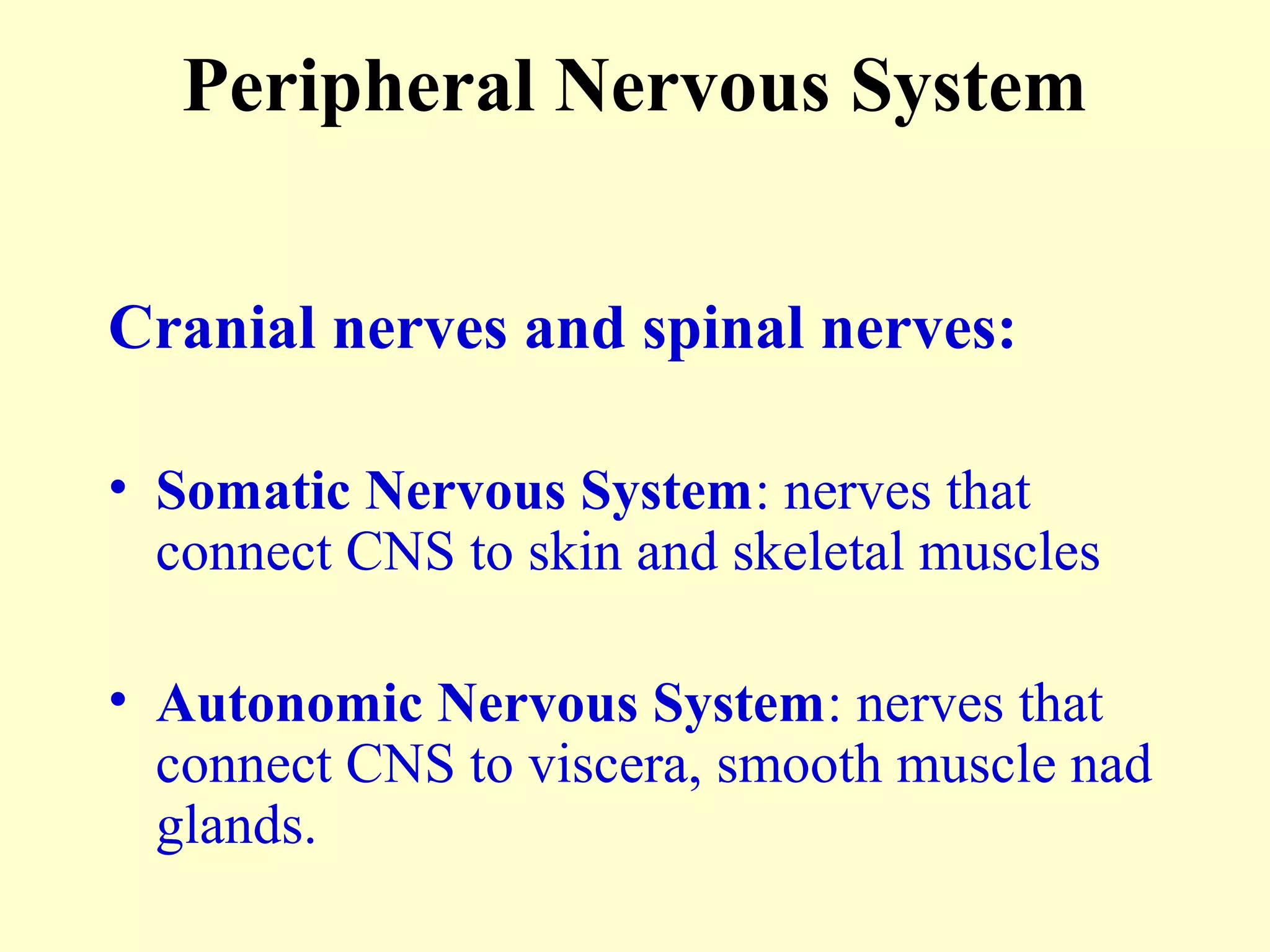

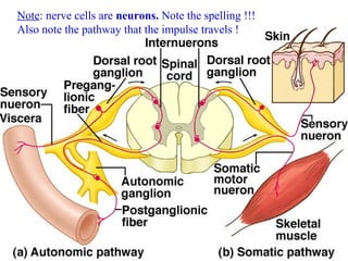
![Fig. 11.36 – note the meninges, dorsal and ventral roots.
Note that the spinal nerves are mixed function nerves [AM-PS].](https://image.slidesharecdn.com/ch11-ppt-nervoussystem11d-101128140006-phpapp02/85/Ch11-ppt-nervous-system-11-d-10-320.jpg)


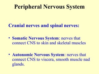
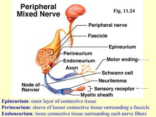
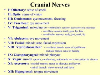
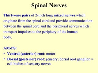
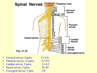
The peripheral nervous system consists of the cranial nerves and spinal nerves. The cranial nerves originate from the brain stem and cerebrum and include 12 pairs of nerves responsible for functions like smell, vision, eye movement, facial expressions, and hearing. The spinal nerves originate from the spinal cord and include 31 pairs that provide communication between the spinal cord and peripheral nerves. The peripheral nervous system also includes the autonomic nervous system which regulates involuntary functions like heart rate and digestion.


![Nov Dec 2009 Gb[1]](https://cdn.slidesharecdn.com/ss_thumbnails/nov-dec-2009gb1-100207104133-phpapp02-thumbnail.jpg?width=640&height=640&fit=bounds)


