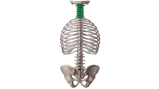
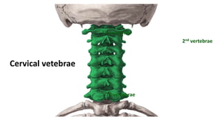
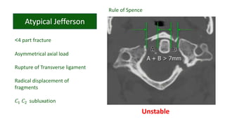
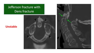
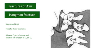
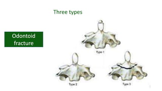
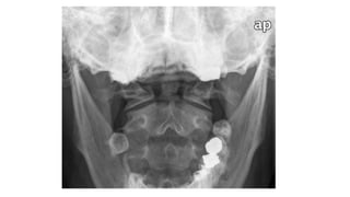
CERVICAL SPINE
PATHOLOGY
DR. PRAMOD NAGURE
Cervical vetebrae
1st vertebrae
2nd vertebrae
7th vertebrae
Atlanto occipital joint
Anterior tubercle
Dental fovea
Spinal nerve passes
along
Vertebrae prominance
Only cervical vertebrae
whose spinous process is
not Bifid
Anterior vertebral line
Posterior vertebral line
Spinal laminar line
Line across the spinous process
1. Disc spaces
2. Facet joints
3. Interspinous distances
1. Subluxation
2. Distraction
3. Dislocation
• Distraction with variable flexion and
extension
• Disruption of tentorial, apical, alar
ligaments
• Severe neurological deficits, often with
multi trauma
Axial load on straight C- spine
Force: Vertex → Occipital condyles → Lateral masses
of 𝐶1
• 4 part fracture
2 anterior arch fractures
2 posterior arch fractures
• Decompressive fracture
Stable
<4 part fracture
Asymmetrical axial load
Rupture of Transverse ligament
Radical displacement of
fragments
𝐶1 𝐶2 subluxation
Rule of Spence
Unstable
Unstable
Sub mental knot
Forceful Hyper extension
Bilateral 𝐶2 arch fracture and
anterior sub luxation of 𝐶2 on 𝐶3
• Bilateral Pars fracture
• No angulation
• No significant translation
• Disk and ligaments intact
• MOI: Hyper extension and axial loading
Stable
• MOI: Hyper extension and axial loading and Hyper
flexion
Unstable
• MOI: Flexion- distraction
Unstable
• MOI: Flexion- distraction
Unstable
PLC X
PLL OK
ALL OK
Hyperflexion
sprain
PLC X
PLL X
ALL OK
Anterior sub luxation
MOI : Hyperflexion and compression
INJURY
• Odontoid fracture
• Type 1
• Type 2
• Type 3
• Clay Shoveler’s fracture
• VB wedge fracture
• Anterior Subluxation
• Facet dislocation
• Atlanto occipital dislocation
• Tear drop fracture
Incidence: 46-79%
Three types
• Odontoid fractures should not
be confused with
a) Os Odontoideum
b) Ossiculum terminale
Cervical vertebrae anatomy and pathology

Cervical vertebrae anatomy and pathology

Editor's Notes

  • #5 No vertebral body No spinous process Atlanto occipital joint
  • #6 With occipital condyles Synovial joint
  • #7 Thickened portions of atlas that support the skull Superior facet and inf fac Sup- occipital cond Inf- axis
  • #8 Ant longus colli and all Dental fovea with dens of axis also called odontoid process
  • #9 Post tubercle Groove for ver art, c1 spinal nerve Provides att to rectus capitis post musucl, nuchal lig
  • #10 It is easily identifiable due to dens/odontoid process which extends sup from ant portion of vert Has trasverse process, large bifid spinous process
  • #11 Apex, ant art sur, post art surface Apical lig att to apex
  • #12 Art with trans lig
  • #13 It ensures that the odointoid process maintains contact with atlas Firmly attached on either side to the med surface of lateral masses
  • #14 They connect the sides of odointoid process of axis to the tubercles on med side of occipital condyles
  • #16 OPENING SEEN ON THE transverse process of all cer vert Ver art and vein
  • #17 Groove found on trans process of c3-c7 Spinal n passes laong the groove as it exits intervert foramina
  • #18 Longus colli muscle atach
  • #19 Rectus capiyis post minor attach
  • #21 Cox its long spinous process protrudes from underthe skin and is visible to the naked eye
  • #22 Flooe of post cranial fossa Angle of mand Ant post arch of atlas Dens of axis Harris ring is a ring like structure resulting from proj of lat masses of c2 on its body Laminae of c2 Retropharyngeal retrotracheal
  • #23 Ant vert body line Post vert body line Spinal laminar line Line across the spinous process
  • #24 Too wide or narrow or uniform Too wide or parallel Too wide too narrow
  • #27 dens occipital bone spinous process transverse band of t1 trachea first rib
  • #28 Height of cervical bodies Height of each joint space Spinous process in alignment
  • #30 Sub- head is not properly aligned with the spine. Occipital cond to latermass relation ruined Distra- elevated increased distance bte occ cond and latermal masses
  • #32 Occ cond floating And the whole cer spine is pushed backwardsin relation to the head
  • #33 Basion dense interval <9.5mm
  • #34 For true jeff Force hits the vertex of the head, go down through the occ condyles land on c1 and drive c1 to sides and disperse the fragments radially
  • #37 Any tilt on the neck when the force is being applied
  • #39 Motor vehicle crashes falls
  • #40 Typical hang amn With frac of pars interarticularis If vert body or transverse formen inv atypical
  • #41 Vert body comp of c2 Not the same mech as judicial hanging
  • #42 Bilateral Pars fracture Anterior angulation No significant translation Disk and ligaments intact
  • #43 Bilateral Pars fracture Significant angulation No significant translation Disk and ligaments intact
  • #44 Bilateral Pars fracture Angulation and Translation C2 c3 facet sub luxation or dislocation
  • #45 No mvm of c2 ver body
  • #46 Soft tissue swelling Spinolaminar line c2 does not fall on the line Harris ring is a ring like structure resulting from proj of lat masses of c2 on its body. Ring disrupted Fat Body of c2 Thses could be the subtle radiolographic signs in hangman
  • #47 these ligaments are present throughout the entire vertebral column. Anterior and posterior longitudinal ligaments – long ligaments that run the length of the vertebral column, covering the vertebral bodies and intervertebral discs. Ligamentum flavum – connects the laminae of adjacent vertebrae. Interspinous ligament – connects the spinous processes of adjacent vertebrae. Nuchal ligament – a continuation of the supraspinous ligament. It attaches to the tips of the spinous processes from C1-C7 and provides the proximal attachment for the rhomboids and trapezius.
  • #48 Hit on the back Hyoer flexion
  • #49 Problem arising when you rip post long lig Unstable
  • #50 Facets parallel, v shaped or slide across one another
  • #52 Little more force sub continue dislocation Gross dislocation
  • #54 Last one in hyper flexion is flexion tear drop Compression fractures the ant inf column of the vertebrae Post lig dis and ant comp frac of vert body
  • #55 Tear droop fragment Spinal cord compression
  • #57 Oblique frac through sup tip of dens Rare Stable when isolated With a o dis 2- common. Transverse frac through base of the dens Unstable prone to non union 3- through body of axis Best prognosis
  • #59 Normal variant developmental anomaly of the odontoid process in which an ossification center that gives rise to the tip of the dens fails to fuse properly with the body of the axis. Os- separation of odontoid process from body of axis The ossiculum terminale appears as a secondary ossification center of the dens between 3-6 years and normally fuses by 12 years. Failure of fusion results in a persistent ossiculum terminale (also called Bergmann's ossicle or ossiculum terminale of Bergmann) and is considered a normal anatomical variant of the axis.