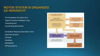
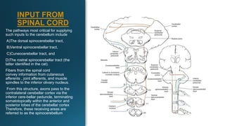
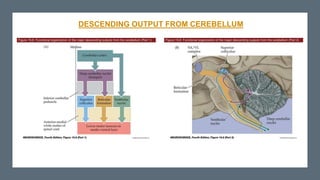
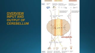
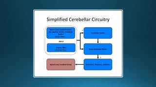
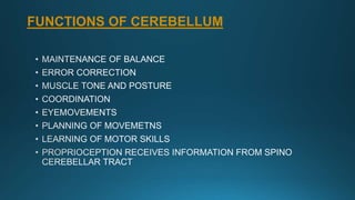
1) The cerebellum receives sensory information from the spinal cord, brainstem, and cerebral cortex, and coordinates movement through its output to motor areas. It acts as a feedback loop to smooth and refine motor commands.
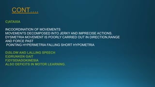
2) Lesions of the cerebellum cause ataxia, tremors, balance and posture problems, and difficulties with motor learning and coordination.
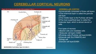
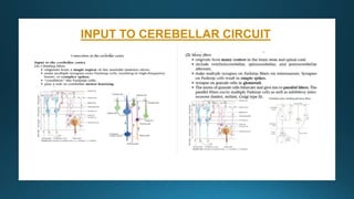
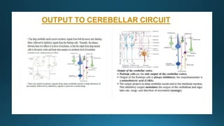
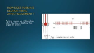
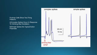
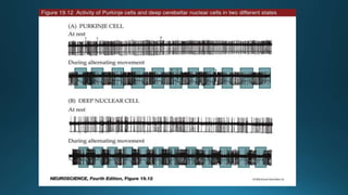
3) Purkinje neurons in the cerebellar cortex integrate inputs from mossy fibers and climbing fibers and inhibit targets in the deep cerebellar nuclei through GABA signaling.


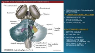
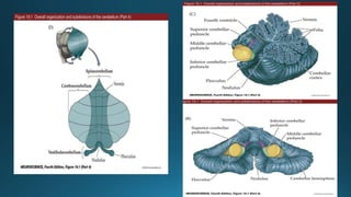
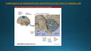
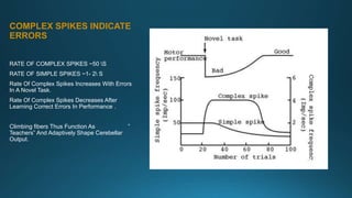
![CEREBELLAR PEDUNCLES
connect the cerebellum to the brain
stem.[1]
1) Superior cerebellar peduncle
(brachium conjunctiva)
Connects cerebellum to mid-brain.
2) Middle cerebellar peduncles
(Brachium pontis)
Connects cerebellum to the pons
3) Inferior cerebellar peduncle
(Restiform body )
Connects with medulla oblongata.](https://image.slidesharecdn.com/cerebellumphysiologyppt-200505045549/85/Cerebellum-physiology-ppt-7-320.jpg)