CEREBELLUM
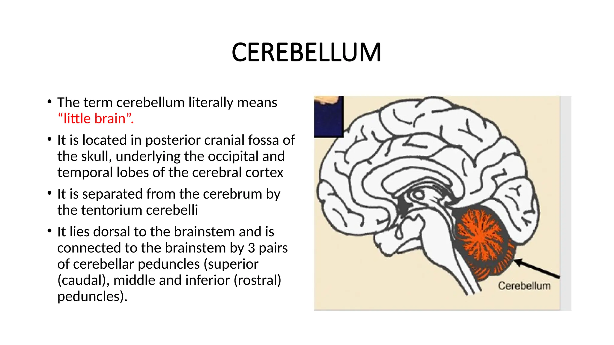
• The termcerebellum literally means
“little brain”.
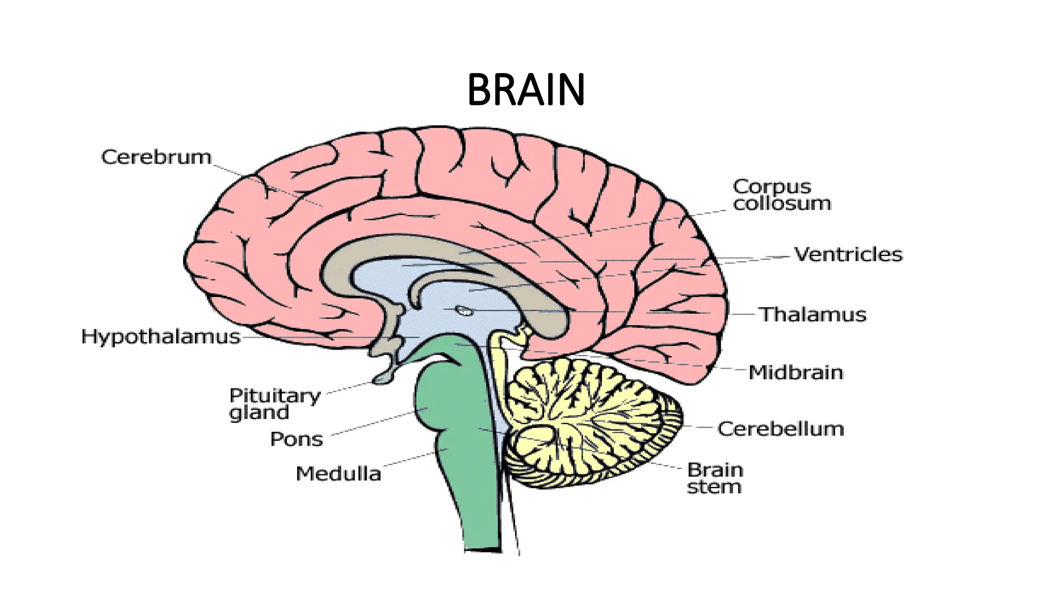
• It is located in posterior cranial fossa of
the skull, underlying the occipital and
temporal lobes of the cerebral cortex
• It is separated from the cerebrum by
the tentorium cerebelli
• It lies dorsal to the brainstem and is
connected to the brainstem by 3 pairs
of cerebellar peduncles (superior
(caudal), middle and inferior (rostral)
peduncles).
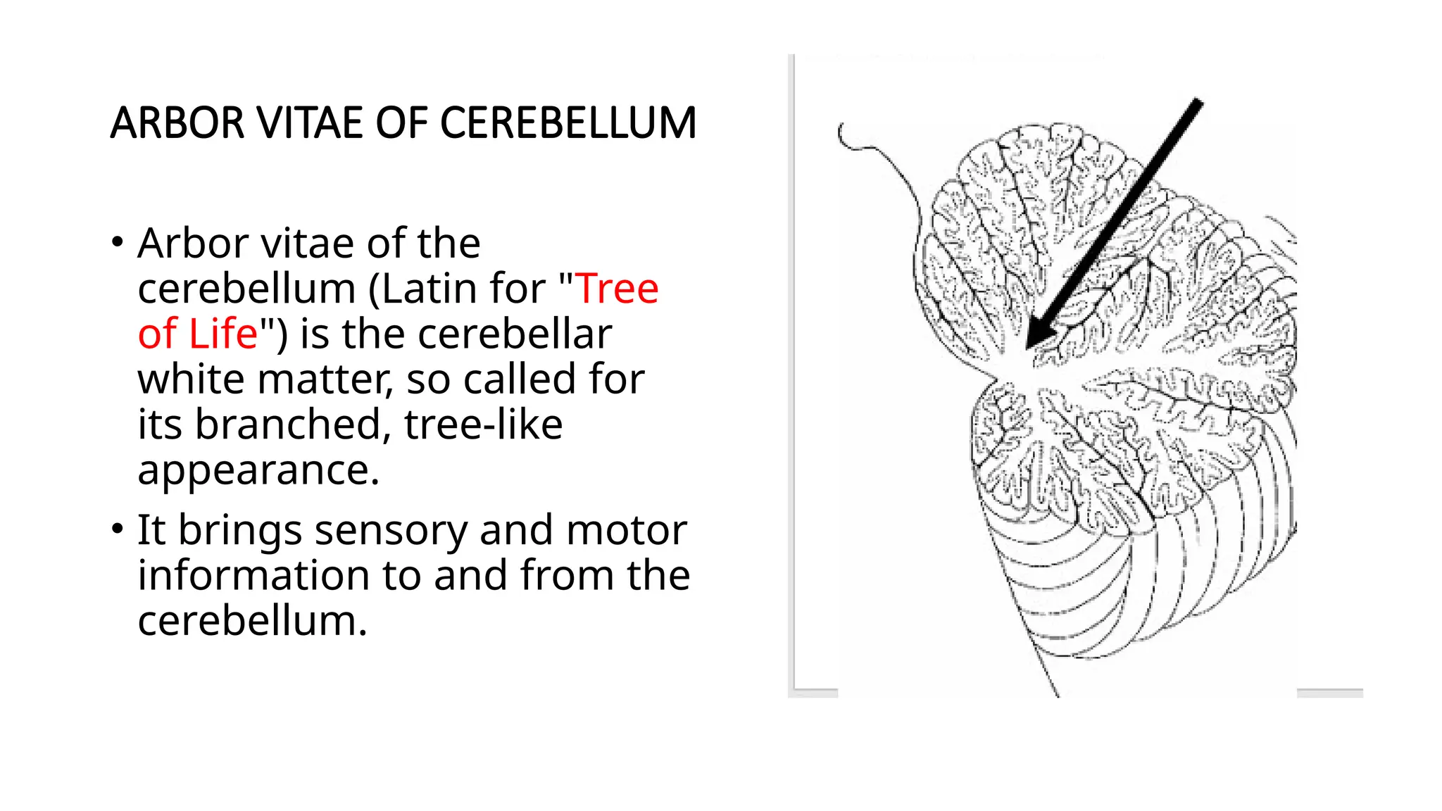
ARBOR VITAE OFCEREBELLUM
• Arbor vitae of the
cerebellum (Latin for "Tree
of Life") is the cerebellar
white matter, so called for
its branched, tree-like
appearance.
• It brings sensory and motor
information to and from the
cerebellum.
6.
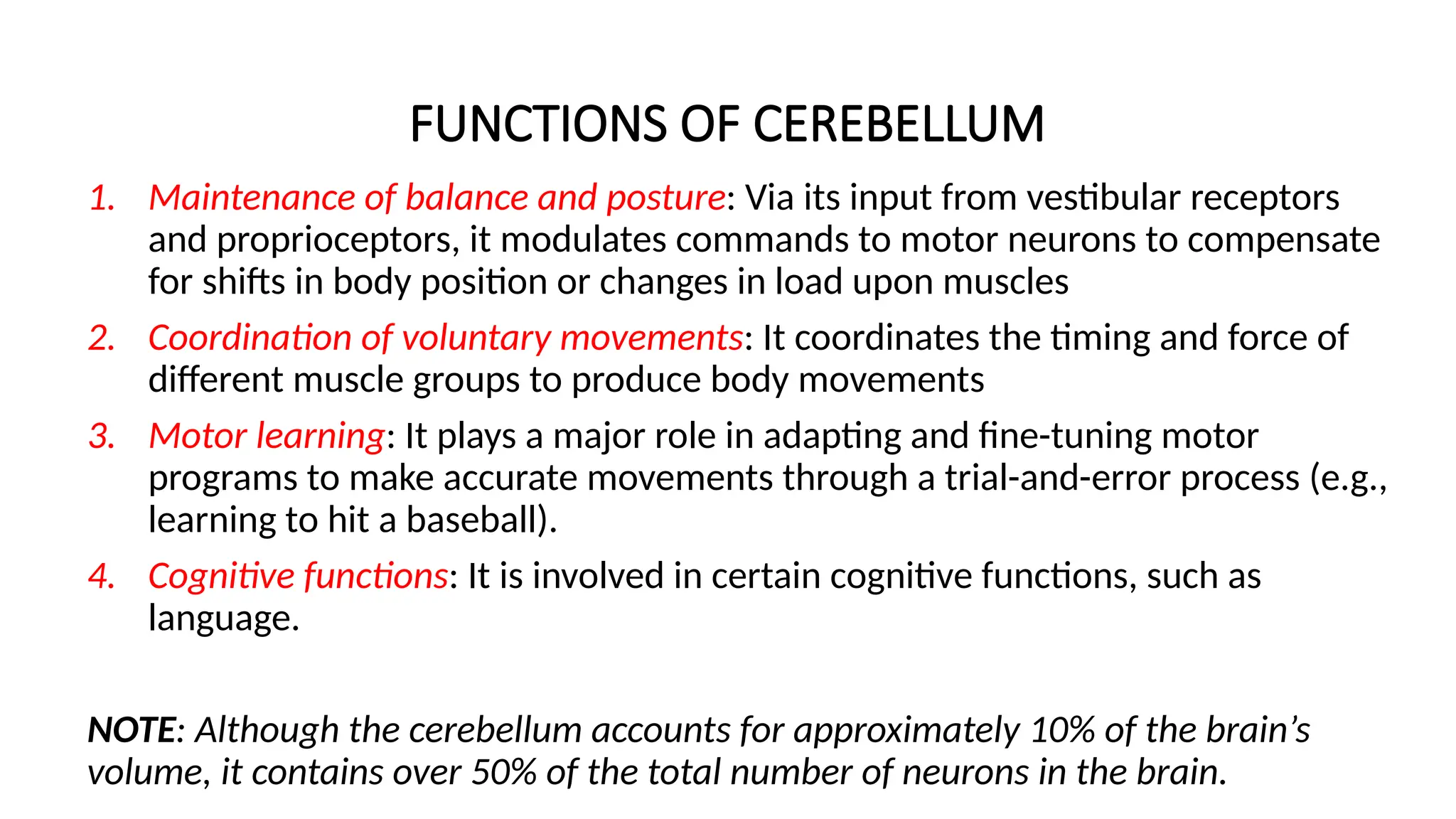
FUNCTIONS OF CEREBELLUM
1.Maintenance of balance and posture: Via its input from vestibular receptors
and proprioceptors, it modulates commands to motor neurons to compensate
for shifts in body position or changes in load upon muscles
2. Coordination of voluntary movements: It coordinates the timing and force of
different muscle groups to produce body movements
3. Motor learning: It plays a major role in adapting and fine-tuning motor
programs to make accurate movements through a trial-and-error process (e.g.,
learning to hit a baseball).
4. Cognitive functions: It is involved in certain cognitive functions, such as
language.
NOTE: Although the cerebellum accounts for approximately 10% of the brain’s
volume, it contains over 50% of the total number of neurons in the brain.
7.
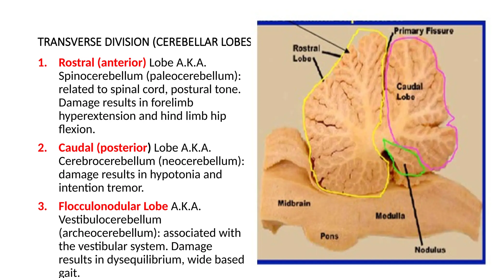
TRANSVERSE DIVISION (CEREBELLARLOBES)
1. Rostral (anterior) Lobe A.K.A.
Spinocerebellum (paleocerebellum):
related to spinal cord, postural tone.
Damage results in forelimb
hyperextension and hind limb hip
flexion.
2. Caudal (posterior) Lobe A.K.A.
Cerebrocerebellum (neocerebellum):
damage results in hypotonia and
intention tremor.
3. Flocculonodular Lobe A.K.A.
Vestibulocerebellum
(archeocerebellum): associated with
the vestibular system. Damage
results in dysequilibrium, wide based
gait.
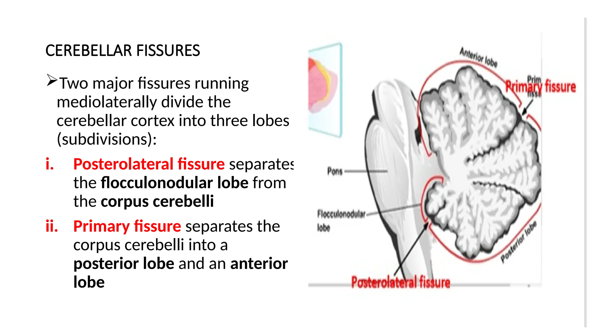
CEREBELLAR FISSURES
Two majorfissures running
mediolaterally divide the
cerebellar cortex into three lobes
(subdivisions):
i. Posterolateral fissure separates
the flocculonodular lobe from
the corpus cerebelli
ii. Primary fissure separates the
corpus cerebelli into a
posterior lobe and an anterior
lobe
10.
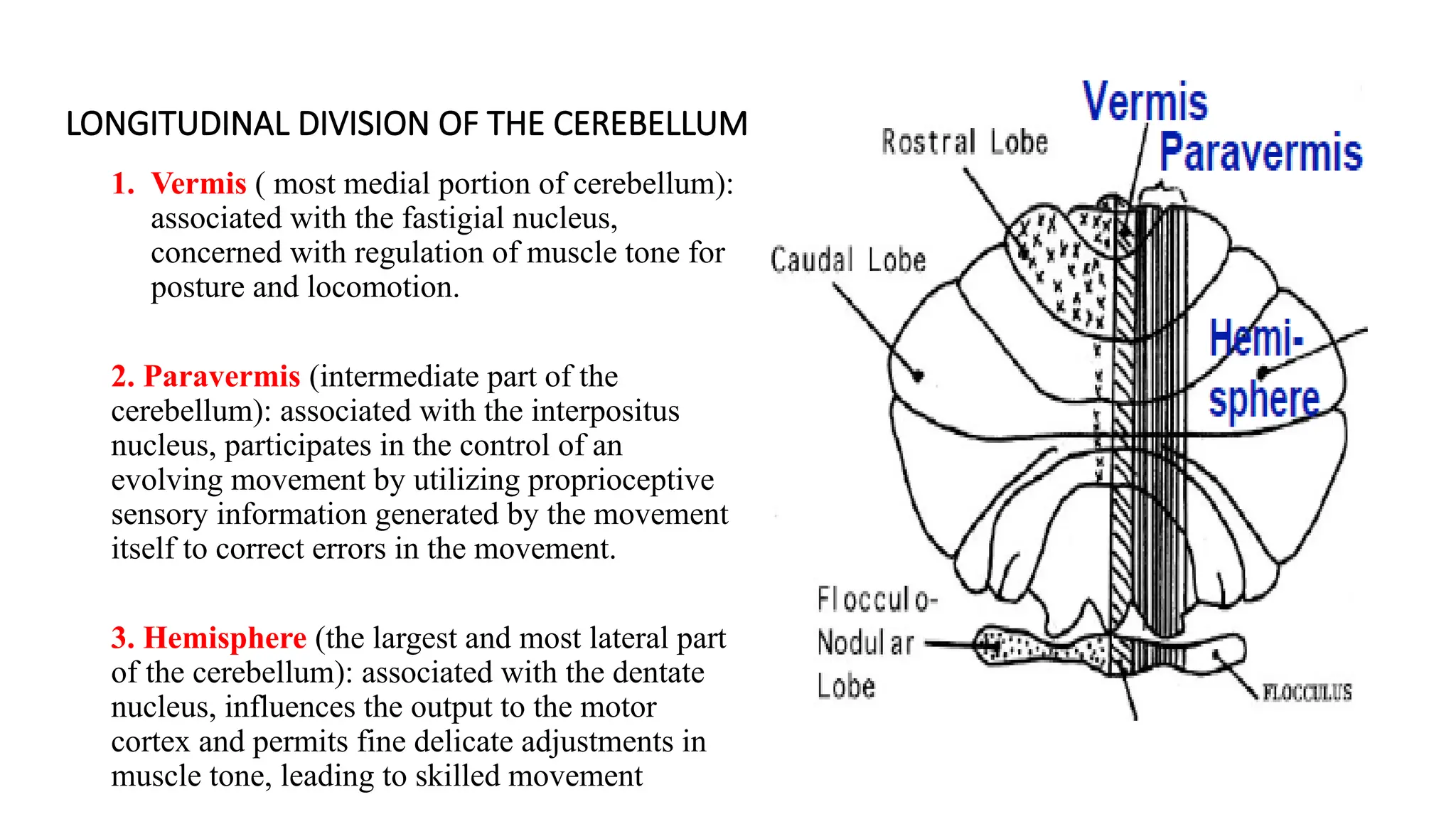
LONGITUDINAL DIVISION OFTHE CEREBELLUM
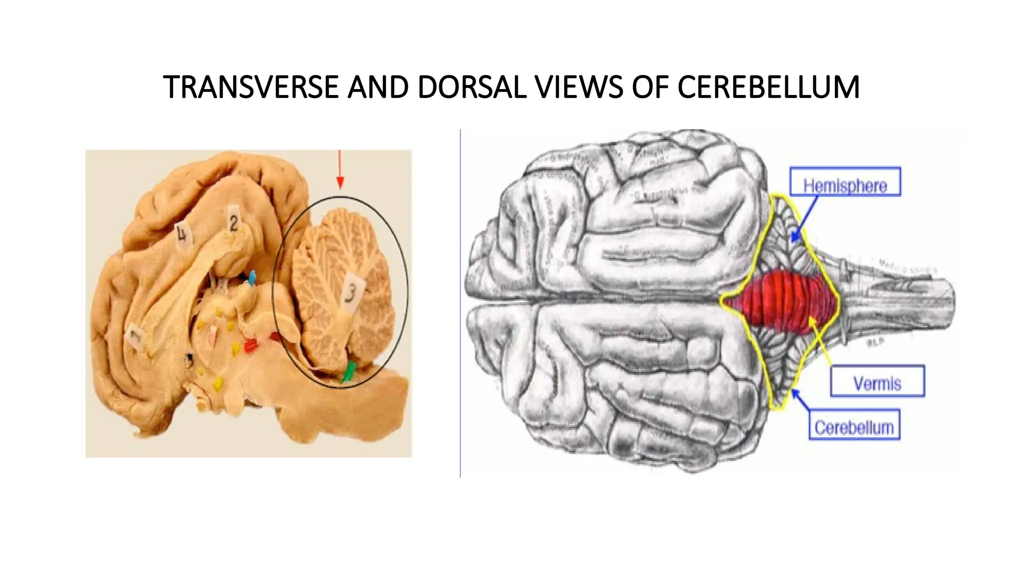
1. Vermis ( most medial portion of cerebellum):
associated with the fastigial nucleus,
concerned with regulation of muscle tone for
posture and locomotion.
2. Paravermis (intermediate part of the
cerebellum): associated with the interpositus
nucleus, participates in the control of an
evolving movement by utilizing proprioceptive
sensory information generated by the movement
itself to correct errors in the movement.
3. Hemisphere (the largest and most lateral part
of the cerebellum): associated with the dentate
nucleus, influences the output to the motor
cortex and permits fine delicate adjustments in
muscle tone, leading to skilled movement
11.
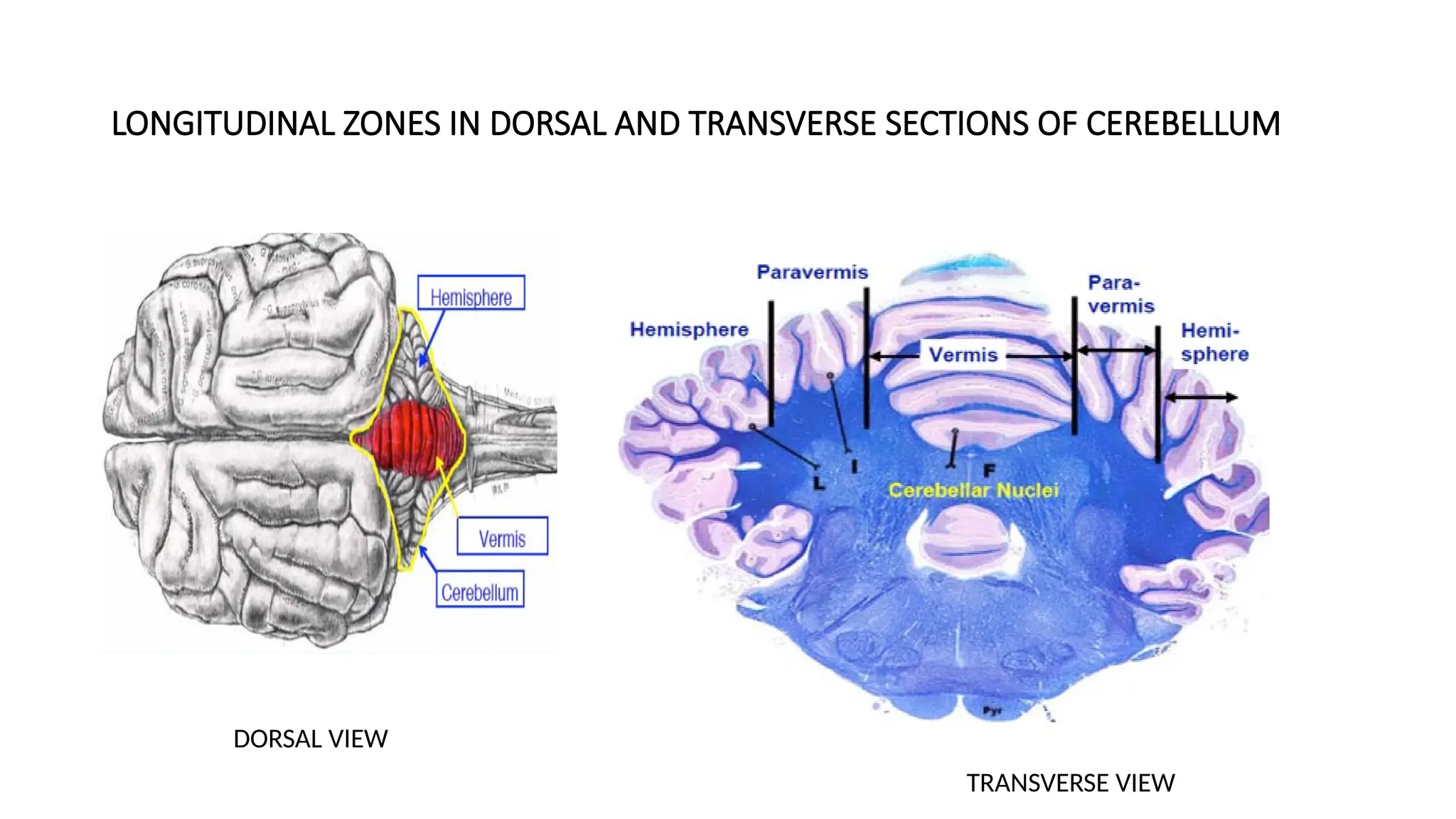
LONGITUDINAL ZONES INDORSAL AND TRANSVERSE SECTIONS OF CEREBELLUM
DORSAL VIEW
TRANSVERSE VIEW
12.
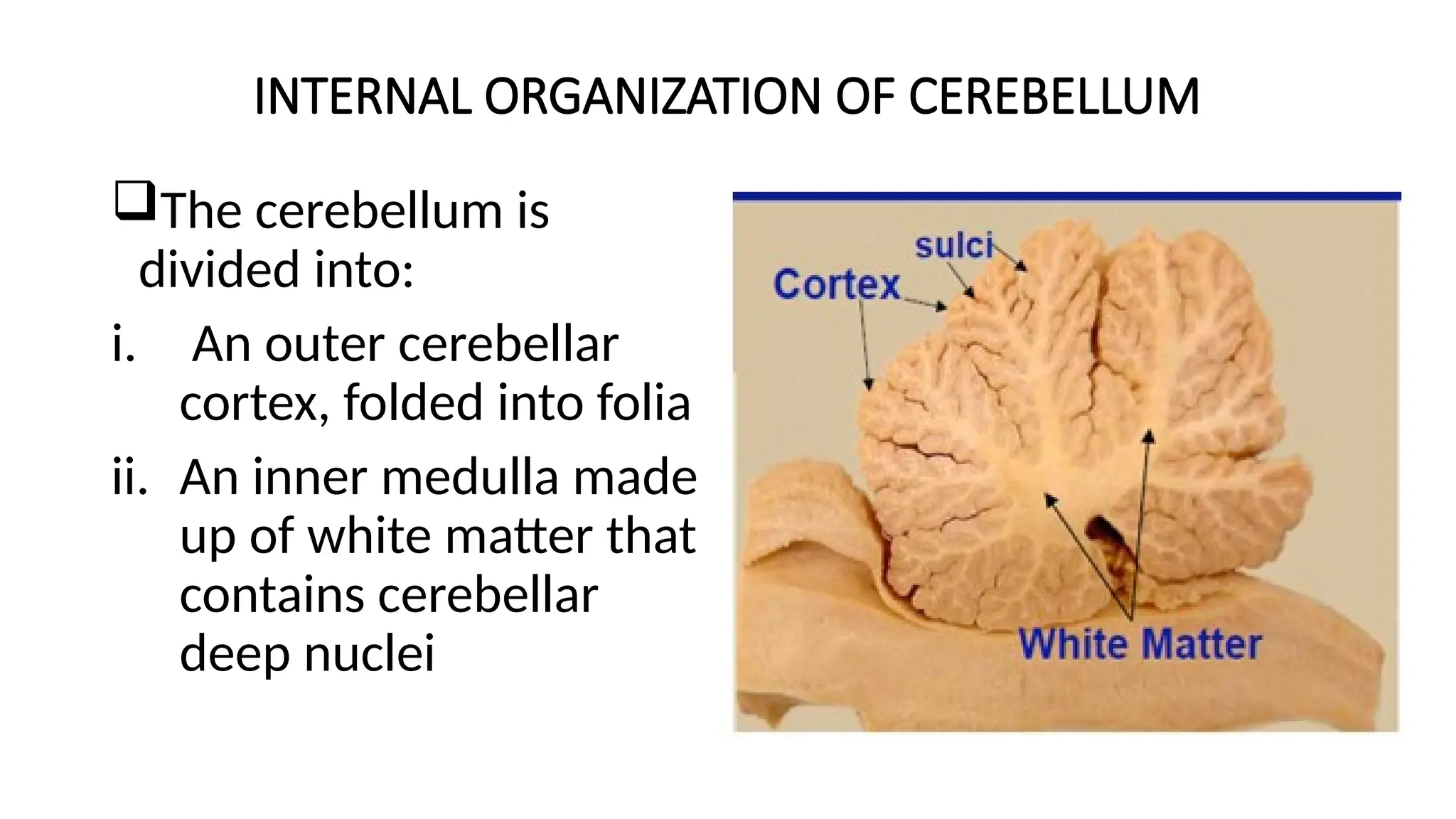
INTERNAL ORGANIZATION OFCEREBELLUM
The cerebellum is
divided into:
i. An outer cerebellar
cortex, folded into folia
ii. An inner medulla made
up of white matter that
contains cerebellar
deep nuclei
13.
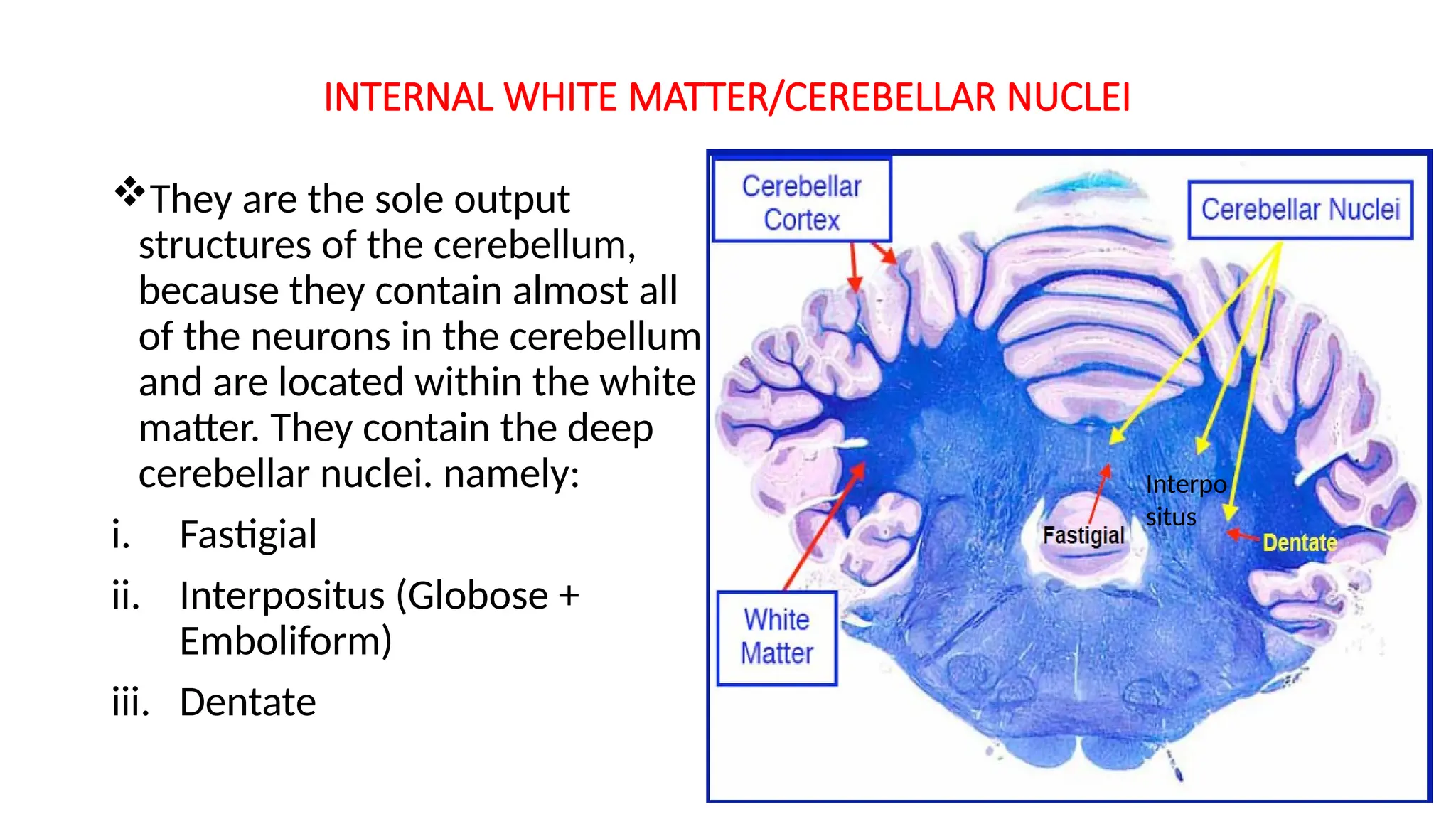
INTERNAL WHITE MATTER/CEREBELLARNUCLEI
They are the sole output
structures of the cerebellum,
because they contain almost all
of the neurons in the cerebellum
and are located within the white
matter. They contain the deep
cerebellar nuclei. namely:
i. Fastigial
ii. Interpositus (Globose +
Emboliform)
iii. Dentate
Interpo
situs
14.
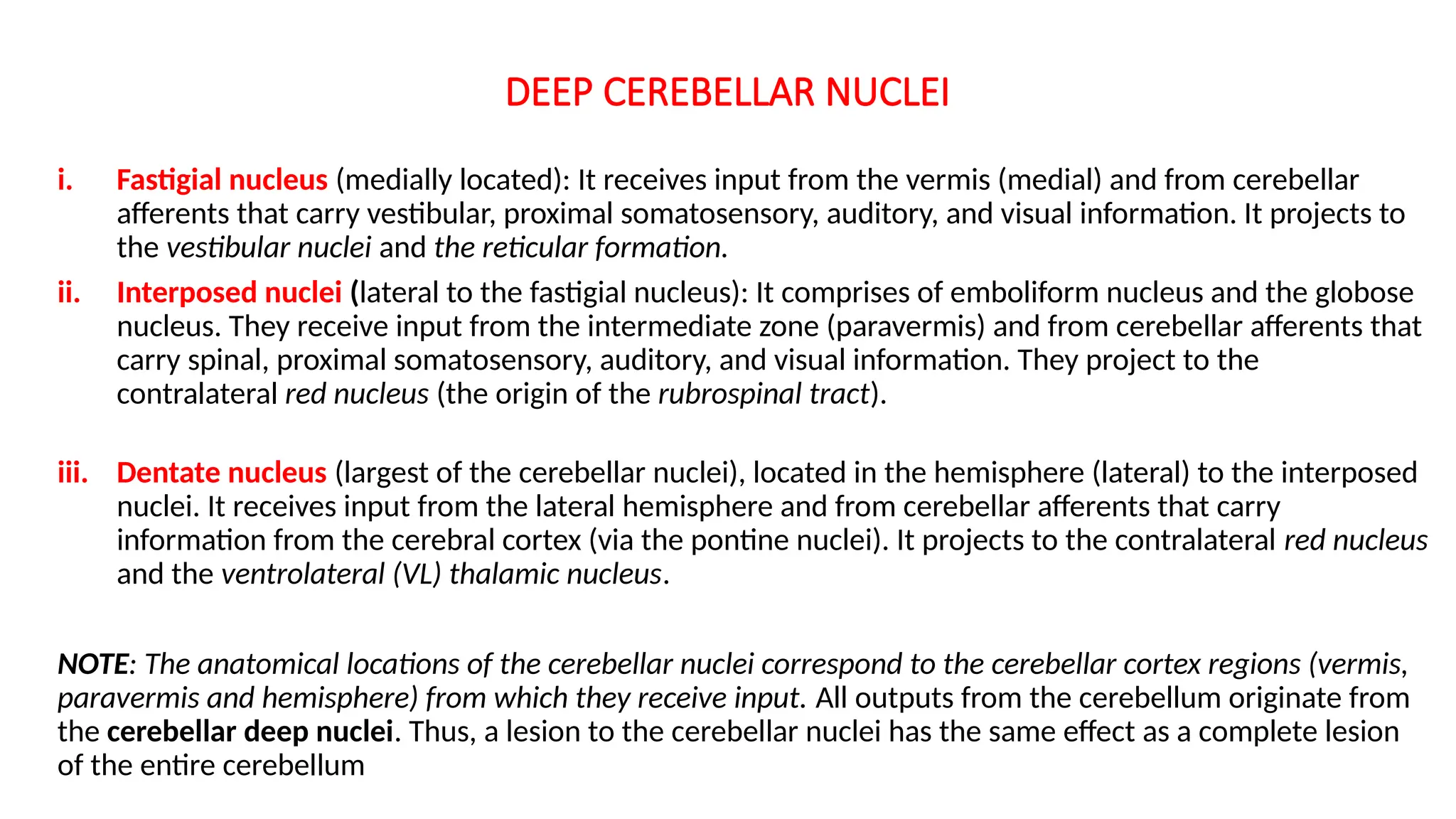
DEEP CEREBELLAR NUCLEI
i.Fastigial nucleus (medially located): It receives input from the vermis (medial) and from cerebellar
afferents that carry vestibular, proximal somatosensory, auditory, and visual information. It projects to
the vestibular nuclei and the reticular formation.
ii. Interposed nuclei (lateral to the fastigial nucleus): It comprises of emboliform nucleus and the globose
nucleus. They receive input from the intermediate zone (paravermis) and from cerebellar afferents that
carry spinal, proximal somatosensory, auditory, and visual information. They project to the
contralateral red nucleus (the origin of the rubrospinal tract).
iii. Dentate nucleus (largest of the cerebellar nuclei), located in the hemisphere (lateral) to the interposed
nuclei. It receives input from the lateral hemisphere and from cerebellar afferents that carry
information from the cerebral cortex (via the pontine nuclei). It projects to the contralateral red nucleus
and the ventrolateral (VL) thalamic nucleus.
NOTE: The anatomical locations of the cerebellar nuclei correspond to the cerebellar cortex regions (vermis,
paravermis and hemisphere) from which they receive input. All outputs from the cerebellum originate from
the cerebellar deep nuclei. Thus, a lesion to the cerebellar nuclei has the same effect as a complete lesion
of the entire cerebellum
15.
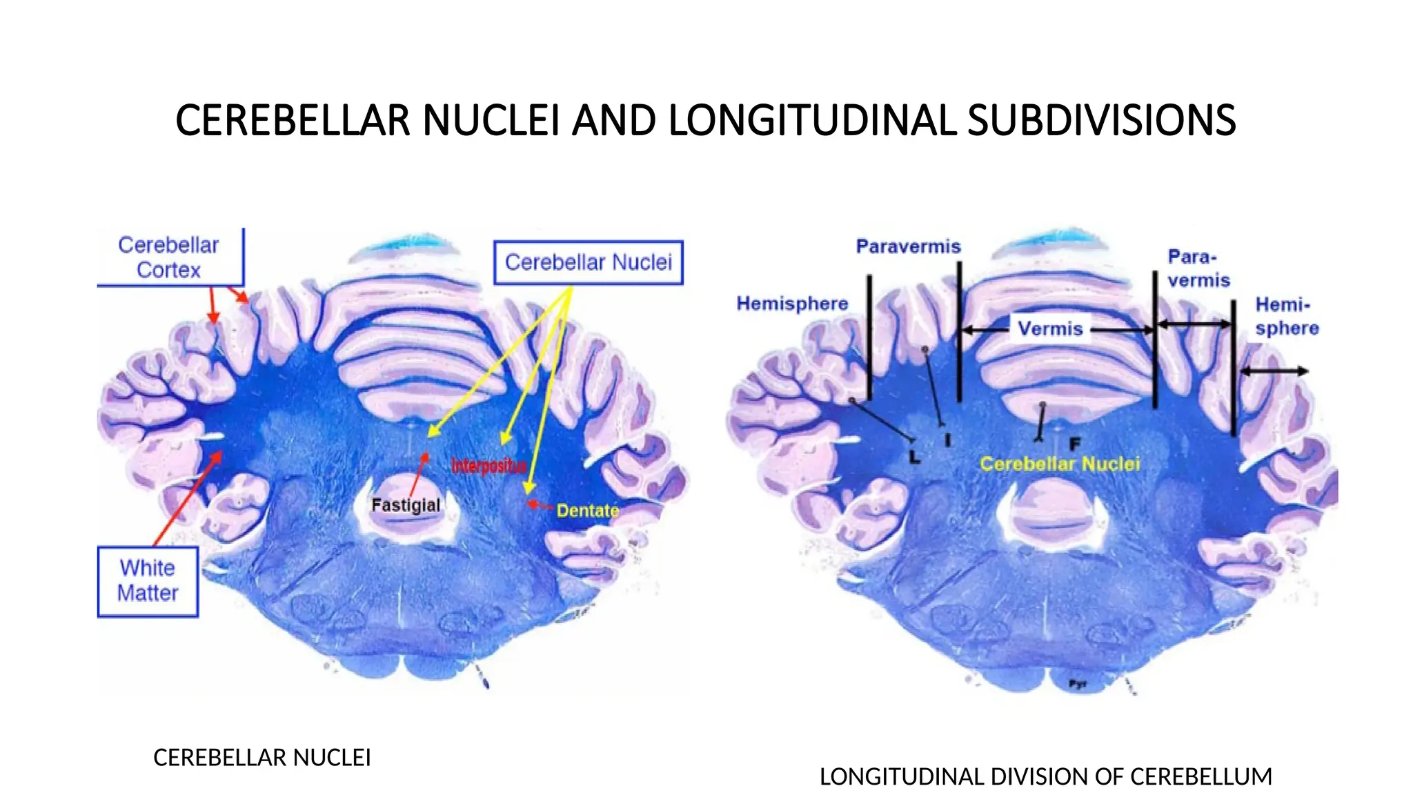
CEREBELLAR NUCLEI ANDLONGITUDINAL SUBDIVISIONS
CEREBELLAR NUCLEI
LONGITUDINAL DIVISION OF CEREBELLUM
16.
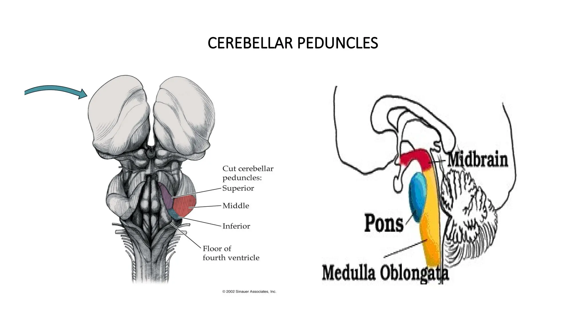
CEREBELLAR PEDUNCLES
Three fiberbundles carry the input and output of the cerebellum:
1. Inferior/Caudal cerebellar peduncle (A.K.A. Restiform body): It lies inferiorly
and connects the cerebellum with the medulla oblongata. Primarily contains
afferent fibers from the medulla, as well as efferent fibres to the vestibular
nuclei.
2. Middle cerebellar peduncle (A.K.A. Brachium pontis): Connects cerebellum with
the pons. Primarily contains afferents from the pontine nuclei.
3. Superior/Rostral cerebellar peduncle (A.K.A. Brachium conjunctivum): It lies
superiorly and connects cerebellum with the midbrain. Primarily contains
efferent fibers from the cerebellar nuclei, as well as some afferents from the
spinocerebellar tract.
NOTE: The damage to the cerebellum results in deficits to the ipsilateral side of the
body.
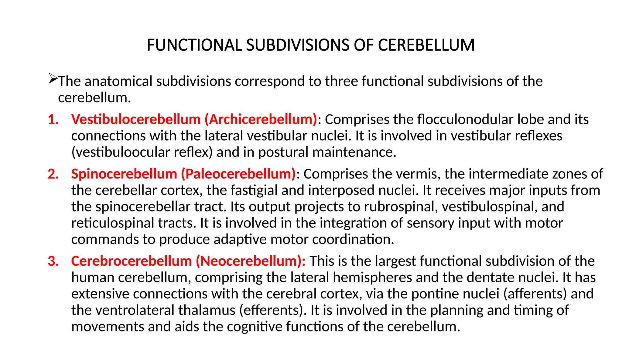
FUNCTIONAL SUBDIVISIONS OFCEREBELLUM
The anatomical subdivisions correspond to three functional subdivisions of the
cerebellum.
1. Vestibulocerebellum (Archicerebellum): Comprises the flocculonodular lobe and its
connections with the lateral vestibular nuclei. It is involved in vestibular reflexes
(vestibuloocular reflex) and in postural maintenance.
2. Spinocerebellum (Paleocerebellum): Comprises the vermis, the intermediate zones of
the cerebellar cortex, the fastigial and interposed nuclei. It receives major inputs from
the spinocerebellar tract. Its output projects to rubrospinal, vestibulospinal, and
reticulospinal tracts. It is involved in the integration of sensory input with motor
commands to produce adaptive motor coordination.
3. Cerebrocerebellum (Neocerebellum): This is the largest functional subdivision of the
human cerebellum, comprising the lateral hemispheres and the dentate nuclei. It has
extensive connections with the cerebral cortex, via the pontine nuclei (afferents) and
the ventrolateral thalamus (efferents). It is involved in the planning and timing of
movements and aids the cognitive functions of the cerebellum.
19.
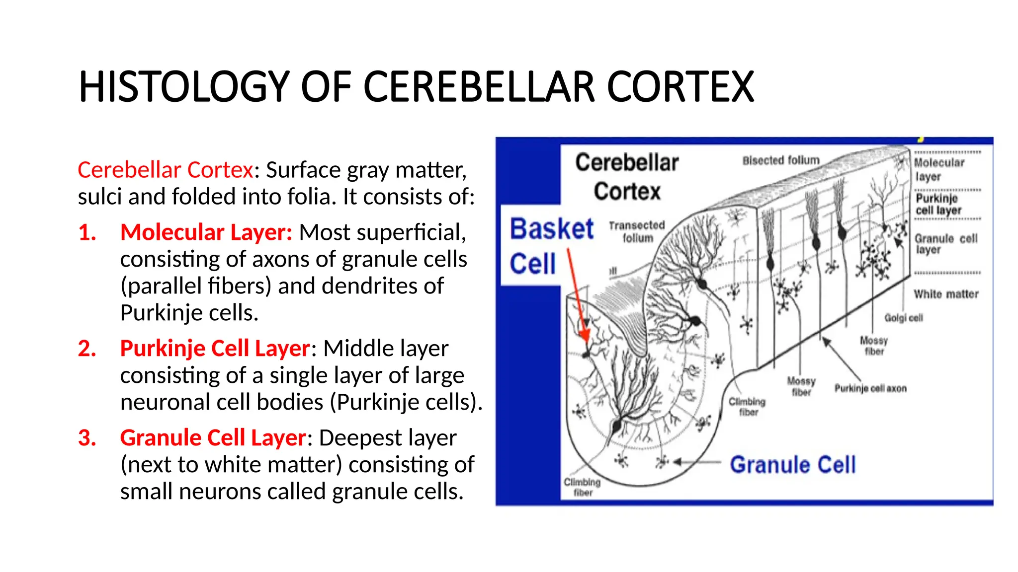
HISTOLOGY OF CEREBELLARCORTEX
Cerebellar Cortex: Surface gray matter,
sulci and folded into folia. It consists of:
1. Molecular Layer: Most superficial,
consisting of axons of granule cells
(parallel fibers) and dendrites of
Purkinje cells.
2. Purkinje Cell Layer: Middle layer
consisting of a single layer of large
neuronal cell bodies (Purkinje cells).
3. Granule Cell Layer: Deepest layer
(next to white matter) consisting of
small neurons called granule cells.
20.
CELL TYPES OFCEREBELLAR CORTEX
1. Granule cells: They are very small, densely packed intrinsic cells of cerebellar cortex that account
for the huge majority of neurons in the cerebellum (more than half of the neurons in the entire
brain). These cells receive input from mossy fibers and project to the Purkinje cells via parallel
fibres, use glutamate as an excitatory transmitter. It also excites Golgi, Basket and Stellate cells.
2. Purkinje cells: They are cells whose apical dendrites form a large fan of finely branched processes
and are the only output neuron from the cortex. They utilize GABA to inhibit neurons in deep
cerebellar nuclei.
3. Golgi cell
4. Stellate cell
5. Basket cell
The Golgi cell, stellate and the basket cell are mainly inhibitory interneuron. They utilize GABA
(Gamma Amino Butyric Acid) to inhibit Purkinje cells
NOTE: All these cells play a role in Neural sharpening (Control feed back mechanism within the
cerebellum).
21.
AFFERENT FIBRES INCEREBELLUM
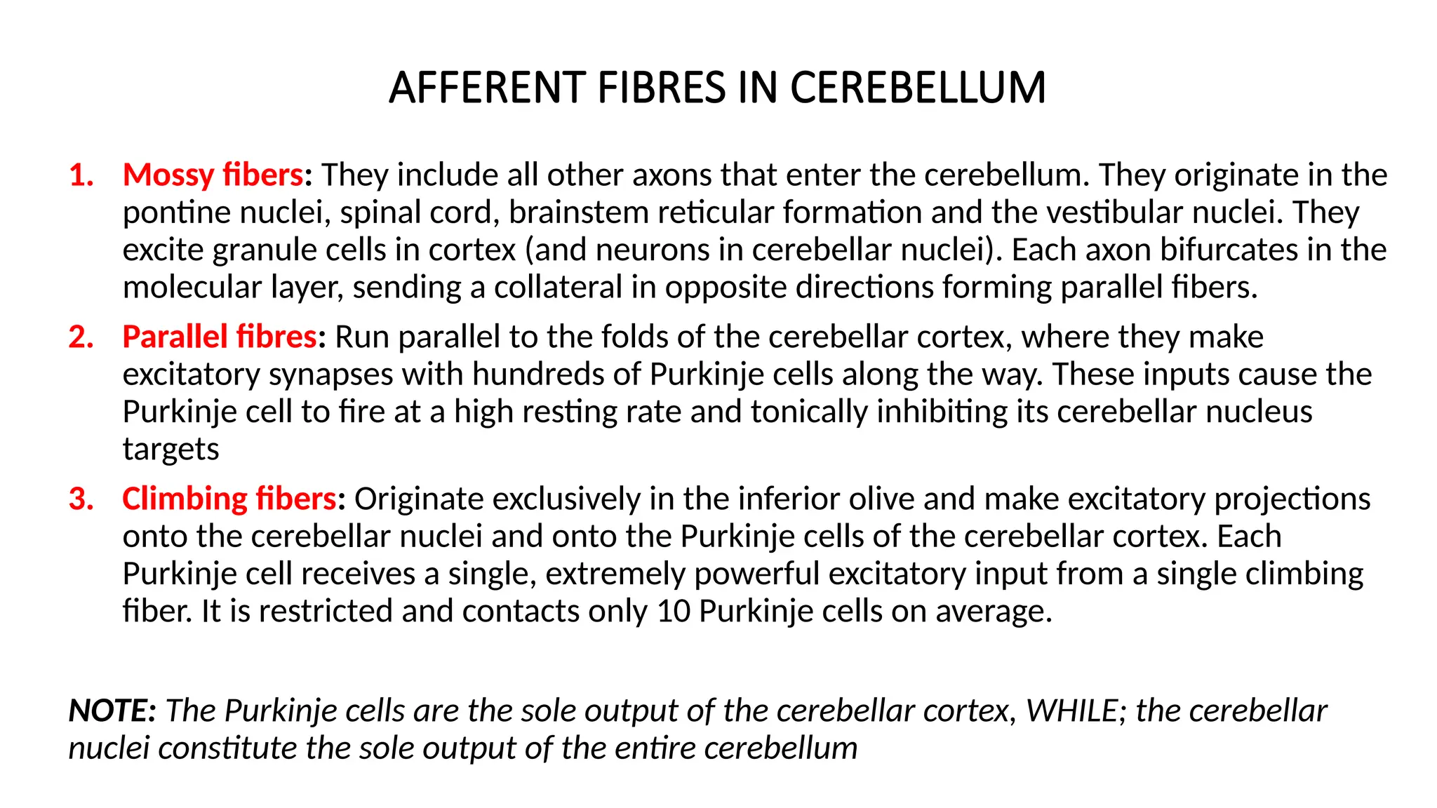
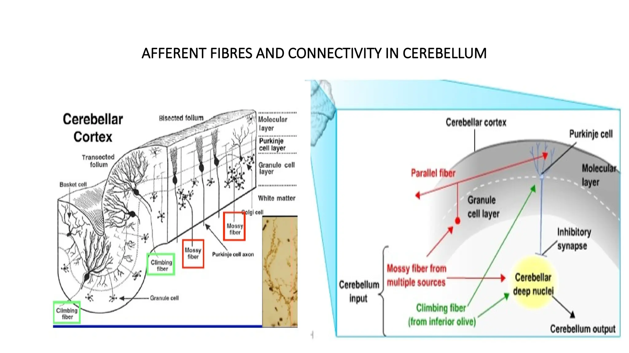
1. Mossy fibers: They include all other axons that enter the cerebellum. They originate in the
pontine nuclei, spinal cord, brainstem reticular formation and the vestibular nuclei. They
excite granule cells in cortex (and neurons in cerebellar nuclei). Each axon bifurcates in the
molecular layer, sending a collateral in opposite directions forming parallel fibers.
2. Parallel fibres: Run parallel to the folds of the cerebellar cortex, where they make
excitatory synapses with hundreds of Purkinje cells along the way. These inputs cause the
Purkinje cell to fire at a high resting rate and tonically inhibiting its cerebellar nucleus
targets
3. Climbing fibers: Originate exclusively in the inferior olive and make excitatory projections
onto the cerebellar nuclei and onto the Purkinje cells of the cerebellar cortex. Each
Purkinje cell receives a single, extremely powerful excitatory input from a single climbing
fiber. It is restricted and contacts only 10 Purkinje cells on average.
NOTE: The Purkinje cells are the sole output of the cerebellar cortex, WHILE; the cerebellar
nuclei constitute the sole output of the entire cerebellum
SYNAPTIC GLOMERULUS INGRANULAR LAYER
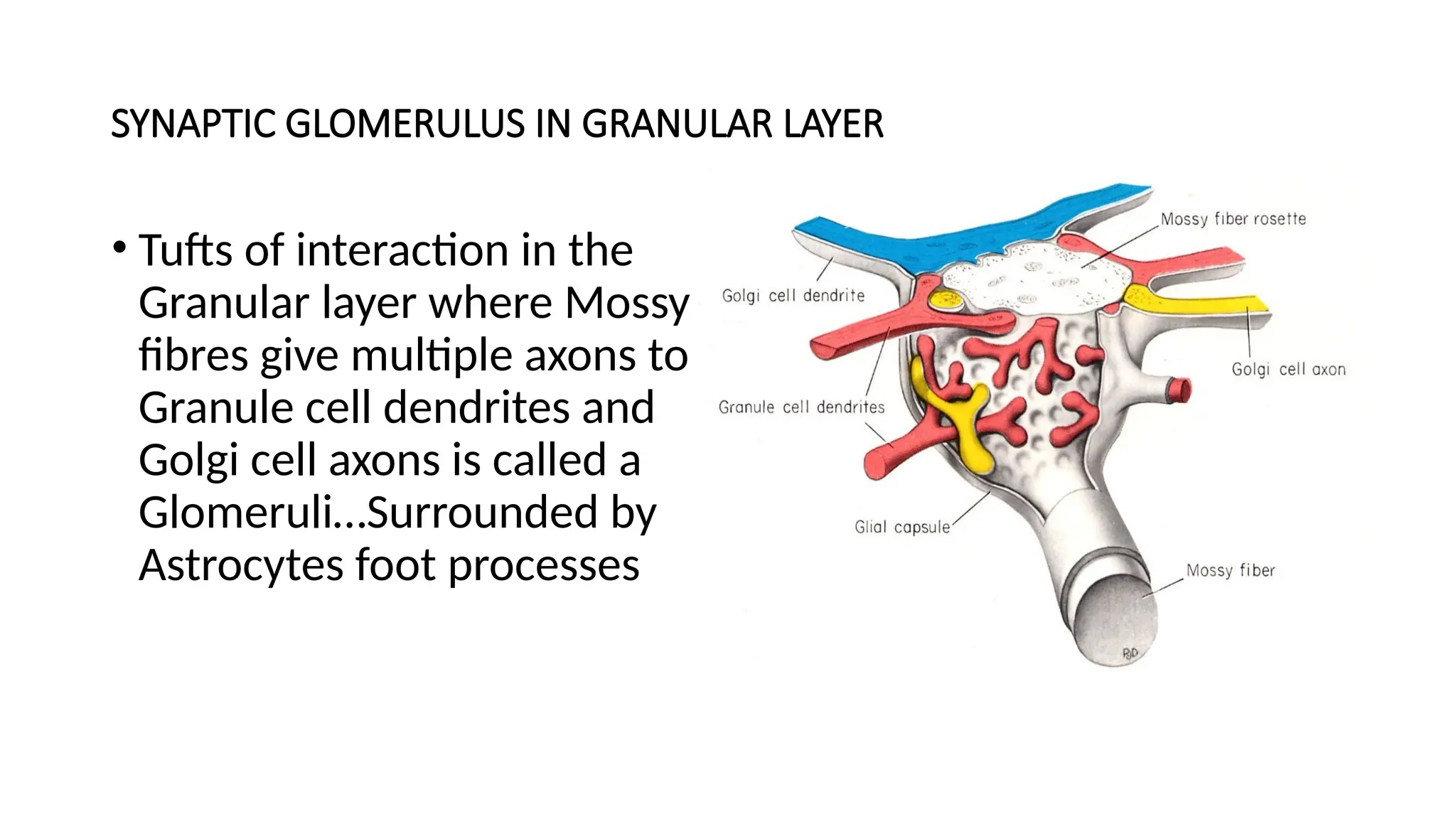
• Tufts of interaction in the
Granular layer where Mossy
fibres give multiple axons to
Granule cell dendrites and
Golgi cell axons is called a
Glomeruli…Surrounded by
Astrocytes foot processes
24.
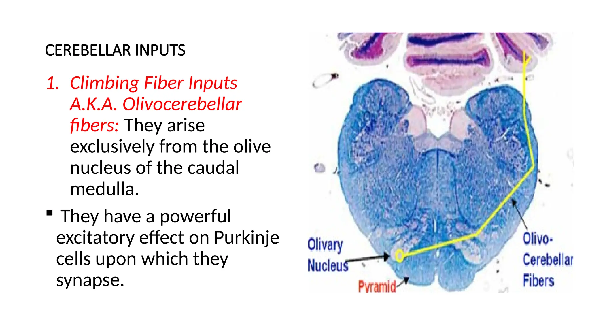
CEREBELLAR INPUTS
1. ClimbingFiber Inputs
A.K.A. Olivocerebellar
fibers: They arise
exclusively from the olive
nucleus of the caudal
medulla.
They have a powerful
excitatory effect on Purkinje
cells upon which they
synapse.
25.
CEREBELLAR INPUTS
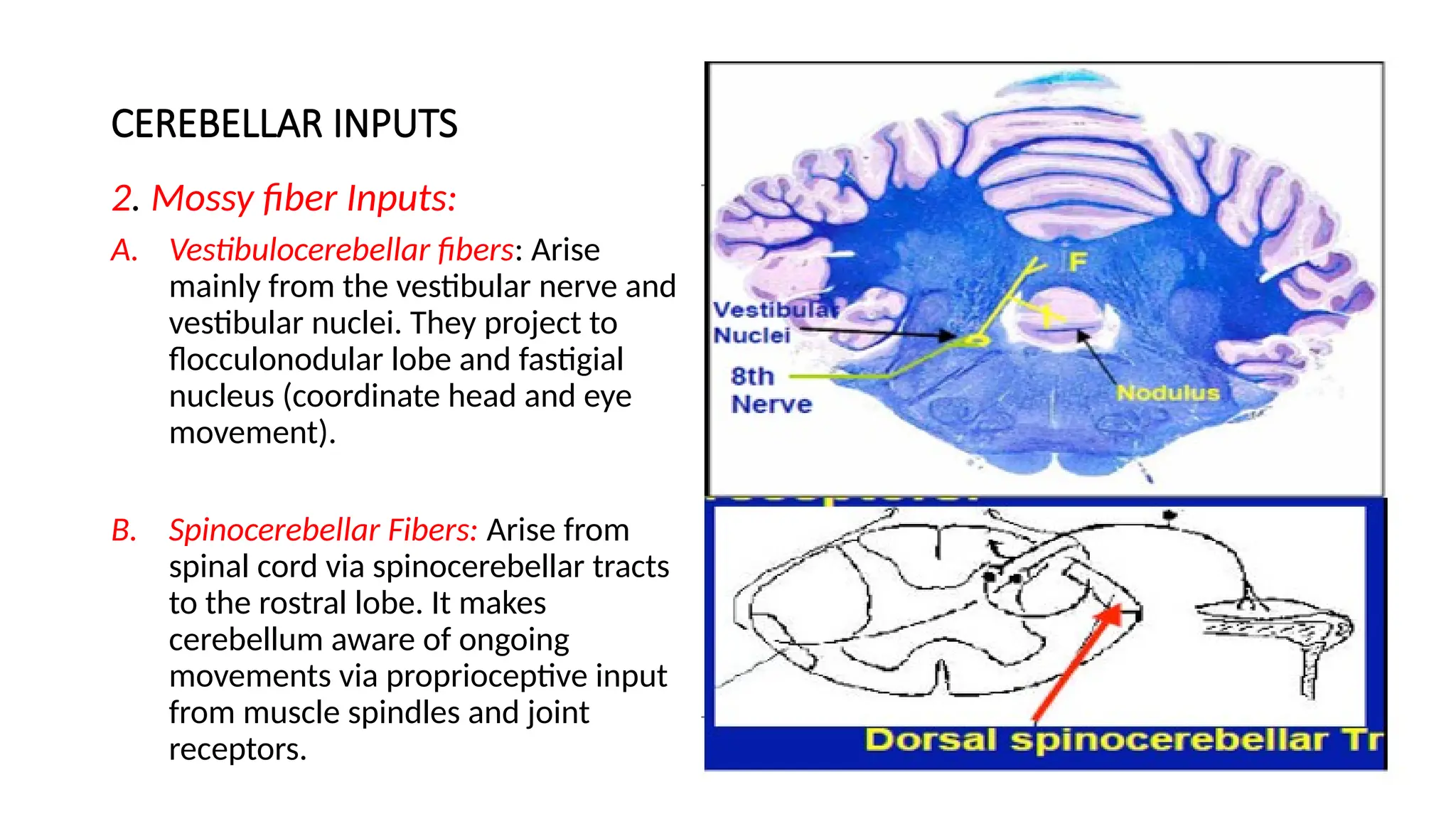
2. Mossyfiber Inputs:
A. Vestibulocerebellar fibers: Arise
mainly from the vestibular nerve and
vestibular nuclei. They project to
flocculonodular lobe and fastigial
nucleus (coordinate head and eye
movement).
B. Spinocerebellar Fibers: Arise from
spinal cord via spinocerebellar tracts
to the rostral lobe. It makes
cerebellum aware of ongoing
movements via proprioceptive input
from muscle spindles and joint
receptors.
26.
CEREBELLAR INPUTS
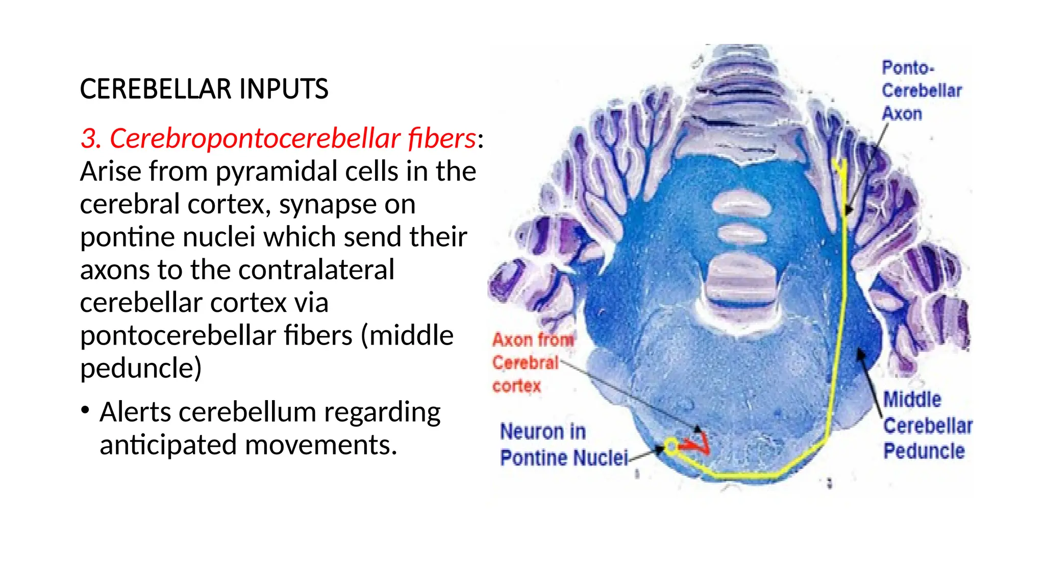
3. Cerebropontocerebellarfibers:
Arise from pyramidal cells in the
cerebral cortex, synapse on
pontine nuclei which send their
axons to the contralateral
cerebellar cortex via
pontocerebellar fibers (middle
peduncle)
• Alerts cerebellum regarding
anticipated movements.
27.
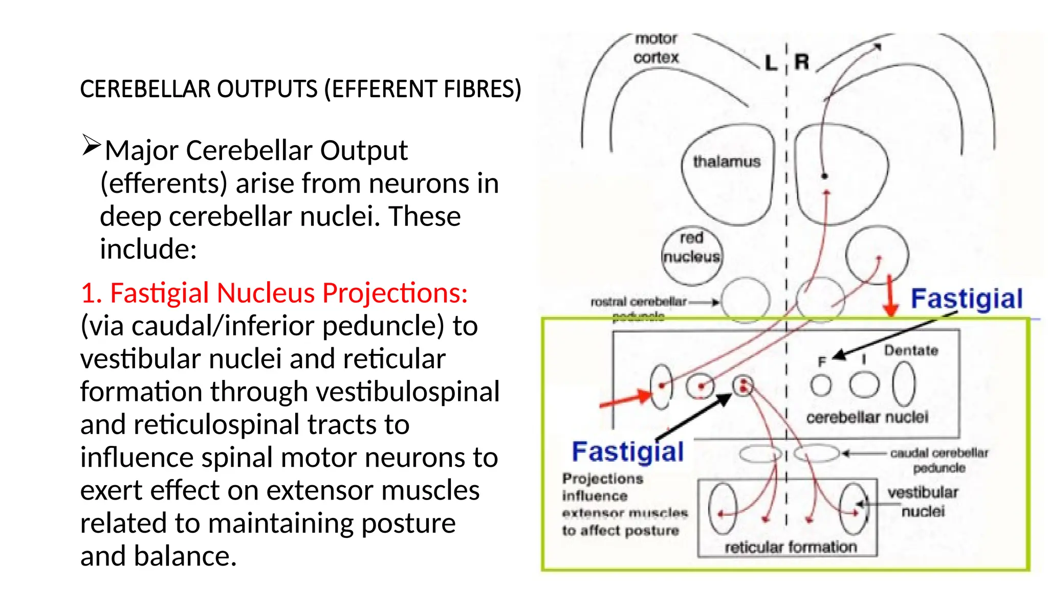
CEREBELLAR OUTPUTS (EFFERENTFIBRES)
Major Cerebellar Output
(efferents) arise from neurons in
deep cerebellar nuclei. These
include:
1. Fastigial Nucleus Projections:
(via caudal/inferior peduncle) to
vestibular nuclei and reticular
formation through vestibulospinal
and reticulospinal tracts to
influence spinal motor neurons to
exert effect on extensor muscles
related to maintaining posture
and balance.
28.
CEREBELLAR OUTPUTS (EFFERENTFIBRES)
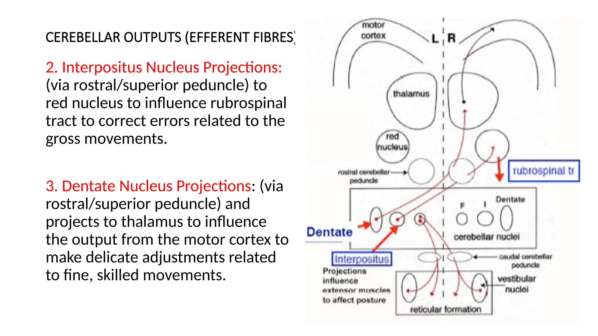
2. Interpositus Nucleus Projections:
(via rostral/superior peduncle) to
red nucleus to influence rubrospinal
tract to correct errors related to the
gross movements.
3. Dentate Nucleus Projections: (via
rostral/superior peduncle) and
projects to thalamus to influence
the output from the motor cortex to
make delicate adjustments related
to fine, skilled movements.
29.
BLOOD SUPPLY TOTHE CEREBELLUM
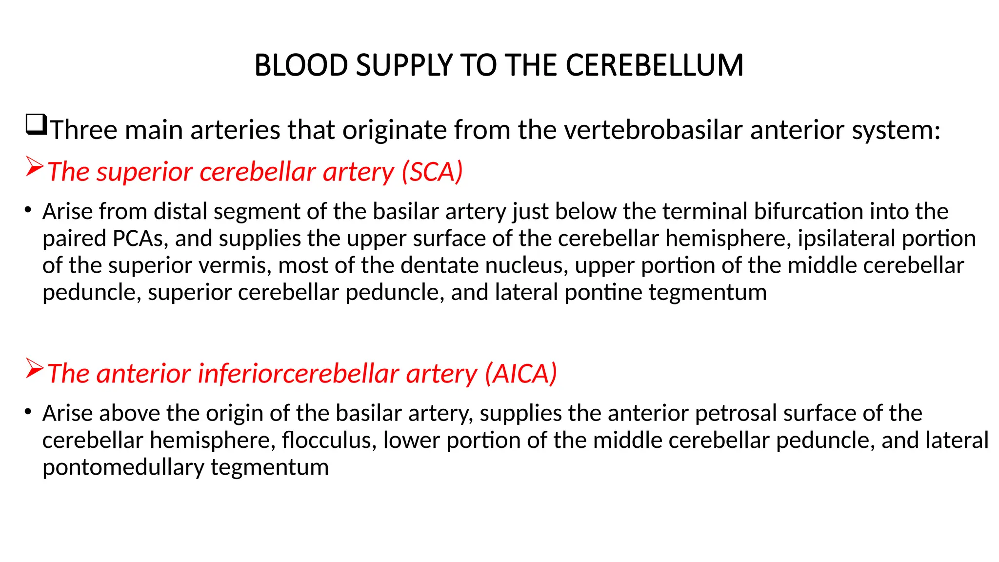
Three main arteries that originate from the vertebrobasilar anterior system:
The superior cerebellar artery (SCA)
• Arise from distal segment of the basilar artery just below the terminal bifurcation into the
paired PCAs, and supplies the upper surface of the cerebellar hemisphere, ipsilateral portion
of the superior vermis, most of the dentate nucleus, upper portion of the middle cerebellar
peduncle, superior cerebellar peduncle, and lateral pontine tegmentum
The anterior inferiorcerebellar artery (AICA)
• Arise above the origin of the basilar artery, supplies the anterior petrosal surface of the
cerebellar hemisphere, flocculus, lower portion of the middle cerebellar peduncle, and lateral
pontomedullary tegmentum
30.
BLOOD SUPPLY TOTHE CEREBELLUM
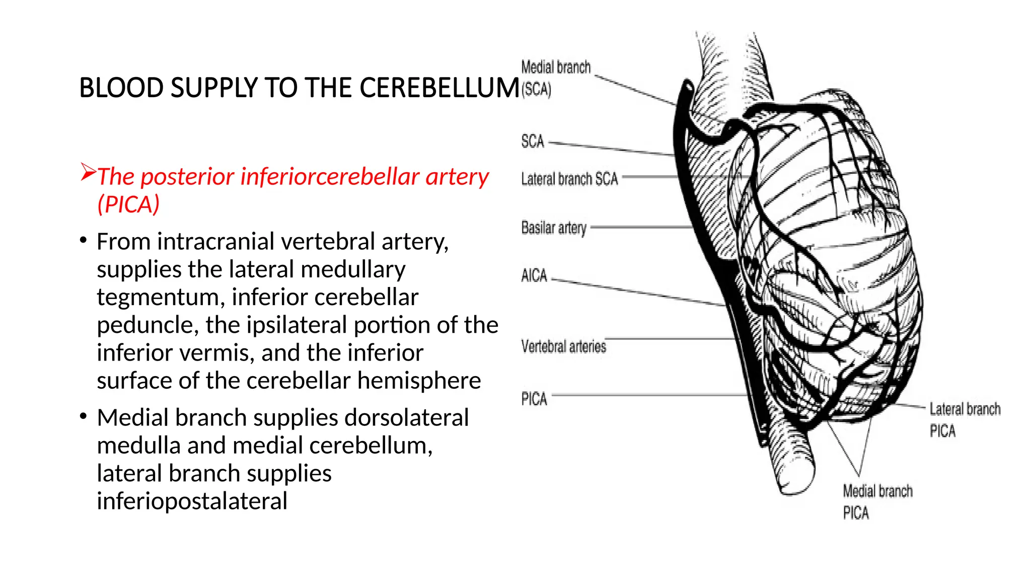
The posterior inferiorcerebellar artery
(PICA)
• From intracranial vertebral artery,
supplies the lateral medullary
tegmentum, inferior cerebellar
peduncle, the ipsilateral portion of the
inferior vermis, and the inferior
surface of the cerebellar hemisphere
• Medial branch supplies dorsolateral
medulla and medial cerebellum,
lateral branch supplies
inferiopostalateral
31.
DYSFUNCTION OF CEREBELLUM
1.Ataxia: A disturbance that alters the direction and extent of voluntary movements;
abnormal gait and uncoordinated movements
2. Dysmetria: Altered range of motion (misjudge distance)
3. Intention Tremor: Oscillating motion, especially of head, during movement
4. Deficits in motor learning in the execution of accurate, coordinated movements: One
prominent experimental model is the vestibuloocular reflex (VOR): This reflex
allows us to maintain gaze on an object when the head is rotated. Vestibular signals
detect the head movement, and send signals through the cerebellum to the eye
muscles to precisely counter the head rotation and maintain a stable center of gaze,
but this is lost in cerebellar damage.
5. Vestibular signs: nystagmus, head tilt
6. Dysdiadochokinesia: Patients have difficulty performing rapidly alternating
movements, such as hitting a surface rapidly and repeatedly with the palm and back
of the hand.
32.
CAUSES OF DISORDERSOF CEREBELLUM
1. Tumors (i.e., cerebellar cystic meningioma)
2. Viral Infections (encephalitis)
3. Heavy metal poisoning
4. Genetic Disorders: cerebellar degeneration