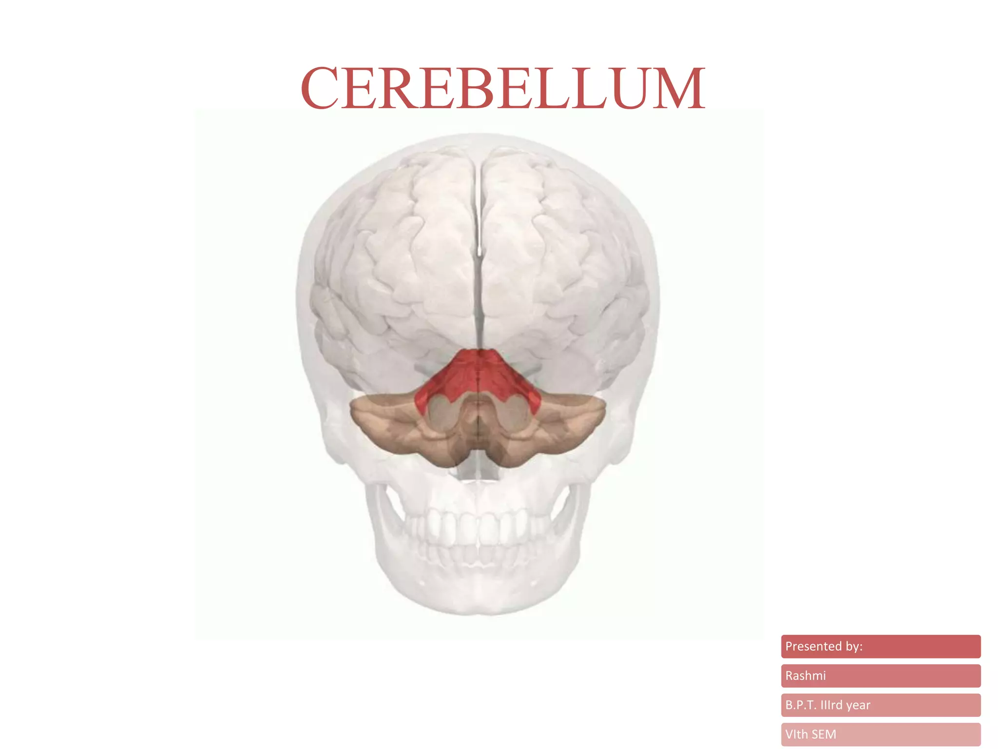
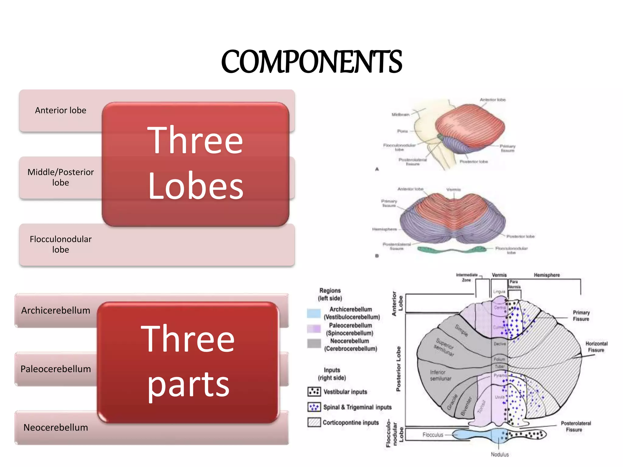
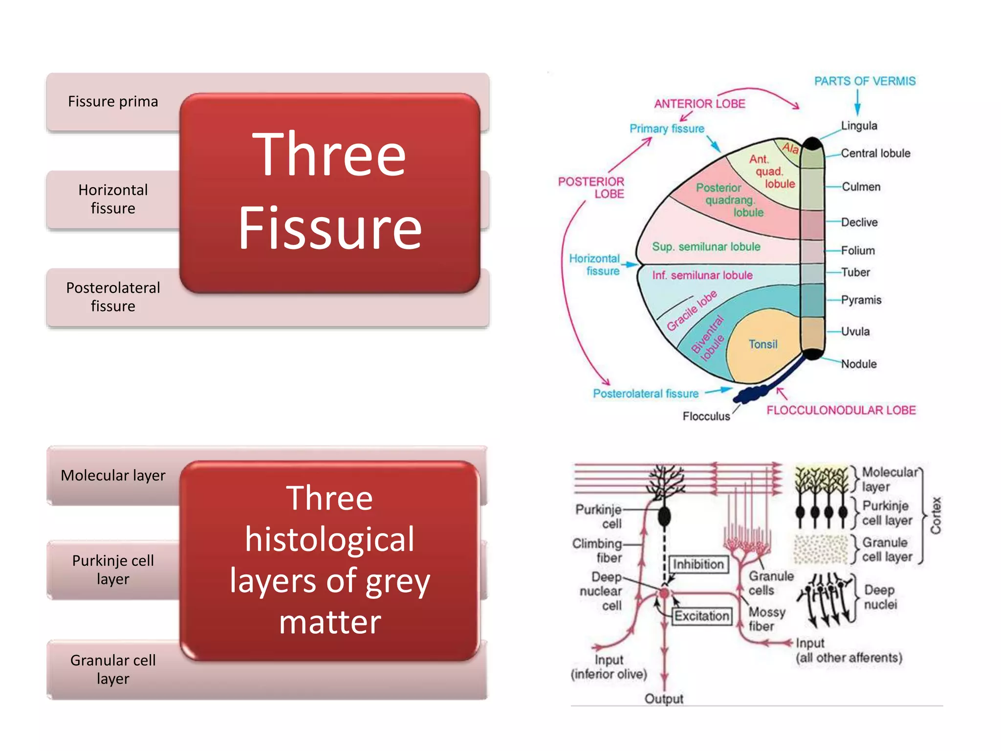
CEREBELLUM
Presented by:
Rashmi
B.P.T. IIIrd year
VIth SEM
CONTENT
o Introduction
o Location
o Components
o Connections
o Blood Supply
o Function
o Clinical Inplementation
INTRODUCTION
• Small brain
• Largest part of the hindbrain
• Two cerebellar hemisphere
joined by a narrow vermis
• Arbor vitae (vital tree of life)
• Ipsilateral body control
LOCATION
• Posterior cranial fossa
• Behind pons and medulla
COMPONENTS
Flocculonodular
lobe
Middle/Posterior
lobe
Anterior lobe
Three
Lobes
Neocerebellum
Paleocerebellum
Archicerebellum
Three
parts
Posterolateral
fissure
Horizontal
fissure
Fissure prima
Three
Fissure
Granular cell
layer
Purkinje cell
layer
Molecular layer
Three
histological
layers of grey
matter
CONNECTIONS
THREE
PEDUNCLES
Superior cerebellar
peduncle to
midbrain (efferent
fibres)
Middle cerebellar
peduncle to pons
(afferent fibres) .
Inferior cerebellar
peduncle to medulla
oblangata (afferent
fibres)
THREE
DEEP NUCLEI
Nucleus dentate with
neocerbellum.
Nucleus emboliformis
and nucleus globose
with paleocerebellum
Nucleus fastigii with
flocculonodular lobe
BLOOD SUPPLY
Superior
cerebellar artery
Anterior inferior
cerebellar artery
Posterior inferior
cerebellar artery
FUNCTION
THREE FUNCTION
body posture and equilibrium by flocculonodular lobe
Control of axial and limb muscles and postural reflexes by anterior lobe.
Smooth, accurate and balanced movements by middle lobe
THREE FUNCTIONAL ZONES
Vermal zone for trunk and girdle movement.
Intermediate zone for hands/feet.
lateral zone for planning and programming muscular activities
CLINICAL IMPLEMENTATION
• Each cerebellar hemishpere is connected by nervous pathways principally
with the same side of the body; it means lesions in one cerebellar
hemishpere gives rise to sign and symptoms that are limited to the same
side of the body.
• Hypotonia
• Dysdiadochokinesia
• Ataxia
• Nystagmus
• Disturbance of Reflexes – Pendullar
Knee Jerk
• Dysarthria
• Vermis Syndrome
• Cerebellar Hemisphere Syndrome
Cerebellum

Cerebellum