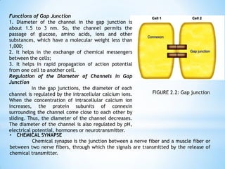
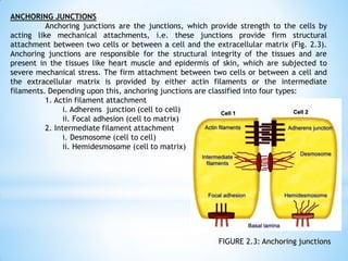
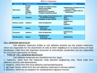
Chapter 2
Cell Junctions, Transport Membrane, and Homeostasis
* Section 1:
General Physiology
Prepared by: Doughlas C. Adona, MAHSE
Cell Junctions
DEFINITION AND CLASSIFICATION
Cell junction is the connection between the neighbouring cells or the contact
between the cell and extracellular matrix. It is also called membrane junction.
Cell junctions are classified into three types:
1. Occluding junctions
2. Communicating junctions
3. Anchoring junctions.
OCCLUDING JUNCTIONS
Cell junctions which prevent intercellular exchange of substances are called
occluding junctions, i.e. these junctions prevent the movement of ions and molecules from
one cell to another cell. Tight junctions belong to this category.
• TIGHT JUNCTION
Tight junction is the intercellular occluding junction that prevents the passage of
large molecules. It is also called zonula occludens. It is the region where the cell
membranes of the adjacent cells fuse together firmly. This type of junction is present in
the apical margins of epithelial and endothelial cells in intestinal mucosa, wall of renal
tubule, capillary wall and choroid plexus.
Structure of Tight Junction
Tight junction is made up of a ridge which has two halves. One half of the ridge
is from one cell and another half is from the other cell. Both halves of the ridge fuse with
each other very tightly and occupy the space between the two cells (Fig. 2.1). Each half of
the ridge consists of tight junction strands.
FIGURE 2.1: Tight junction
Proteins of tight junction
Proteins involved in the formation of
tight junctions are classified into two types:
1. Tight junction membrane proteins or integral
membrane proteins, such as occludin, claudin and
junctional adhesion molecules (JAMs)
2. Scaffold (framework or platform) proteins or
peripheral membrane proteins or cytoplasmic
plaque
proteins such as cingulin, symplekin and ZO1, 2,
3. Tight junction membrane protein molecules are
anchored in the strands of the ridge and attach
with their counterparts of neighboring cell, so that
both the cells are held together. The scaffold
(platform) proteins are attached with the tight
junction membrane proteins and strengthen the
anchoring in the ridges.
Functions of Tight Junction
1. Strength and stability: The tight junction holds the neighbouring cells of the tissues
firmly and thus
provides strength and stability to the tissues.
2. Selective permeability (gate function): The tight junction forms a selective barrier for
small molecules and a total barrier for large molecules.
3. Fencing function: Tight junction prevents the lateral movement of proteins (integral
membrane proteins) and lipids in cell membrane and thus acts as a fence.
4. Maintenance of cell polarity: Fencing function of the tight junction maintains the cell
polarity by keeping the proteins in the apical region of the cell membrane.
5. Blood-brain barrier: Tight junction in the brain capillaries forms the blood-brain barrier,
which prevents the entrance of many substances from capillary blood into brain tissues.
Only lipid-soluble substances like drugs and steroid hormones can pass through the blood-
brain barrier.
COMMUNICATING JUNCTIONS
Cell junctions which permit the intercellular exchange of substances are called
communicating junctions, i.e. these junctions permit the movement of ions and molecules
from one cell to another cell. Gap junction and chemical synapse are the communicating
junctions.
• GAP JUNCTION
Gap junction is the intercellular junction that allows passage of ions and smaller
molecules between the cells. It is also called nexus. It is present in heart, basal part of
epithelial cells of intestinal mucosa, etc.
Structure of Gap Junction
Membranes of the two adjacent cells lie very close to each other and the
intercellular space is reduced from the usual size of 2.5 to 3 nm. Cytoplasm of the two cells
is connected by the channels formed by the membranes of both cells. So, the molecules
move from one cell to another cell directly through these channels, without having contact
with extracellular fluid (ECF).
Each channel consists of two halves. Each half belongs to one of the two adjacent
cells. Each half of the channel is surrounded by 6 subunits of proteins which are called
connexins or connexons (Fig. 2.2).
FIGURE 2.2: Gap junction
Functions of Gap Junction
1. Diameter of the channel in the gap junction is
about 1.5 to 3 nm. So, the channel permits the
passage of glucose, amino acids, ions and other
substances, which have a molecular weight less than
1,000;
2. It helps in the exchange of chemical messengers
between the cells;
3. It helps in rapid propagation of action potential
from one cell to another cell.
Regulation of the Diameter of Channels in Gap
Junction
In the gap junctions, the diameter of each
channel is regulated by the intracellular calcium ions.
When the concentration of intracellular calcium ion
increases, the protein subunits of connexin
surrounding the channel come close to each other by
sliding. Thus, the diameter of the channel decreases.
The diameter of the channel is also regulated by pH,
electrical potential, hormones or neurotransmitter.
• CHEMICAL SYNAPSE
Chemical synapse is the junction between a nerve fiber and a muscle fiber or
between two nerve fibers, through which the signals are transmitted by the release of
chemical transmitter.
ANCHORING JUNCTIONS
Anchoring junctions are the junctions, which provide strength to the cells by
acting like mechanical attachments, i.e. these junctions provide firm structural
attachment between two cells or between a cell and the extracellular matrix (Fig. 2.3).
Anchoring junctions are responsible for the structural integrity of the tissues and are
present in the tissues like heart muscle and epidermis of skin, which are subjected to
severe mechanical stress. The firm attachment between two cells or between a cell and
the extracellular matrix is provided by either actin filaments or the intermediate
filaments. Depending upon this, anchoring junctions are classified into four types:
1. Actin filament attachment
i. Adherens junction (cell to cell)
ii. Focal adhesion (cell to matrix)
2. Intermediate filament attachment
i. Desmosome (cell to cell)
ii. Hemidesmosome (cell to matrix)
FIGURE 2.3: Anchoring junctions
• ADHERENS JUNCTION
Adherens junction is the cell to cell junction, which connects the actin filaments of one
cell to those of another cell. In some places like epithelial linings, this junction forms a
continuous adhesion (zonula adherens) just below the tight junctions. In adherens junction, the
membranes of the adjacent cells are held together by some transmembrane proteins called
cadherins. Adherens junction provides strong mechanical attachments of the adjacent cells.
Adherens junction is present in the intercalated disks between the branches of cardiac muscles.
During the contractions and relaxation of heart, the cardiac muscle fibers are held together
tightly by means of this junction. The adherens junction present in epidermis helps the skin to
withstand the mechanical stress.
• FOCAL ADHESION
Focal adhesion is the cell to matrix junctions, which connects the actin filaments of
the cell to the extracellular matrix. In epithelia of various organs, this junction connects the cells
with their basal lamina. The transmembrane proteins, which hold the cell membrane and the
matrix are called integrins.
• DESMOSOME
Desmosome is a cell to cell junction, where the intermediate filaments connect two
adjacent cells. Desmosome is also called macula adherens. The membranes of two adjacent cells,
which oppose each other, are thickened and become spot-like patches. Intermediate filaments are
attached with the thickened patches. Some of these filaments are parallel to the membrane and
others are arranged in radiating fashion. Desmosomes function like tight junctions. The
transmembrane proteins involved in desmosome are mainly cadherins.
• HEMIDESMOSOME
Hemidesmosome is a cell to matrix junction, which connects the intermediate
filaments of the cell to the extracellular matrix. This type of cell junction is like half desmosome
and the thickening of membrane of only one cell occurs. So, this is known as hemidesmosome or
half desmosome. Mostly, the hemidesmosome connects the cells with their basal lamina. The
proteins involved in this are integrins (Table 2.1).
CELL ADHESION MOLECULES
Cell adhesion molecules (CAMs) or cell adhesion proteins are the protein molecules,
which are responsible for the attachment of cells to their neighbours or to basal lamina (or basal
membrane). CAMs form the important structures of intercellular connections and are responsible
for structural organization of tissues.
• TYPES OF CELL ADHESION MOLECULES
Cell adhesion molecules are classified into four types:
1. Cadherins, which form the molecular limbs between neighboring cells. These CAMs form
adherens junction and desomosome;
2. Integrins, which form the focal adhesion and hemidesmosome;
3. IgG super family, which form the cell adhesion molecules in nervous system;
4. Selectins, which act as receptors for carbohydrates (ligand or mucin).
Transport through Cell Membrane
INTRODUCTION
All the cells in the body must be supplied with essential substances like nutrients,
water, electrolytes, etc. Cells also must get rid of many unwanted substances like waste
materials, carbon dioxide, etc. The cells achieve these by means of transport mechanisms across
the cell membrane. Structure of the cell membrane is well suited for the transport of substances
in and out of the cell. Lipids and proteins of cell membrane play an important role in the
transport of various substances between extracellular fluid (ECF) and intracellular fluid (ICF).
BASIC MECHANISM OF TRANSPORT
Two types of basic mechanisms are involved in the transport of substances across the cell
membrane:
1. Passive transport mechanism
2. Active transport mechanism
PASSIVE TRANSPORT
Passive transport is the transport of substances along the concentration gradient or
electrical gradient or both (electrochemical gradient). It is also known as diffusion or downhill
movement. It does not need energy. Passive transport is like swimming in the direction of water
flow in a river. Here, the substances move from region of higher concentration to the region of
lower concentration. Diffusion is of two types, namely simple diffusion and facilitated diffusion.
Simple diffusion of substances occurs either through lipid layer or protein layer of the cell
membrane. Facilitated diffusion occurs with the help of the carrier proteins of the cell
membrane. Thus, the diffusion can be discussed under three headings:
1. Simple diffusion through lipid layer
2. Simple diffusion through protein layer
3. Facilitated or carrier-mediated diffusion
• SIMPLE DIFFUSION THROUGH LIPID LAYER
Lipid layer of the cell membrane is permeable only to lipid-soluble substances like
oxygen, carbon dioxide and alcohol. The diffusion through the lipid layer is directly proportional to
the solubility of the substances in lipids (Fig. 3.1A).
• SIMPLE DIFFUSION THROUGH PROTEIN LAYER
Protein layer of the cell membrane is permeable to water-soluble substances. Mainly,
electrolytes diffuse through the protein layer.
FIGURE 3.1: Hypothetical diagram of simple diffusion through the cell membrane.
A. Diffusion through lipid layer; B. Diffusion through ungated channel; C. Diffusion
through gated channel.
Protein Channels or Ion Channels
Throughout the central lipid layer of the cell membrane, there are some pores. Integral
protein molecules of protein layer invaginate into these pores from either surface of the cell
membrane. Thus, the pores present in the central lipid layer are entirely lined up by the integral
protein molecules. These pores are the hypothetical pores and form the channels for the diffusion
of water, electrolytes and other substances, which cannot pass through the lipid layer. As the
channels are lined by protein molecules, these are called protein channels for water-soluble
substances.
Types of Protein Channels or Ion Channels
Characteristic feature of the protein channels is the selective permeability. That is, each
channel can permit only one type of ion to pass through it. Accordingly, the channels are named
after the ions which diffuse through these channels such as sodium channels, potassium channels,
etc.
Regulation of the Channels
Some of the protein channels are continuously opened and most of the channels are
always closed. Continuously opened channels are called ungated channels (Fig. 3.1B). Closed
channels are called gated channels. These channels are opened only when required (Fig. 3.1C).
Gated Channels
Gated channels are divided into three categories:
i. Voltage-gated channels
Voltage-gated channels are the channels which open whenever there is a change in the
electrical potential. For example, in the neuromuscular junction, when action potential reaches
axon terminal, the calcium channels are opened and calcium ions diffuse into the interior of the
axon terminal from ECF.
ii. Ligand-gated channels
Ligand-gated channels are the type of channels which open in the presence of some
hormonal substances. The hormonal substances are called ligands and the channels are called
ligand-gated channels. During the transmission of impulse through the neuromuscular junction,
acetylcholine is released from the vesicles.
iii. Mechanically gated channels
Mechanically gated channels are the channels which are opened by some
mechanical factors. Examples are, channels present in the pressure receptors (Pacinian
corpuscles) and the receptor cells (hair cells) of organ of Corti and vestibular apparatus.
When a Pacinian corpuscle is subjected to pressure, it is compressed resulting in
deformation of its core fiber. This deformation causes opening of sodium channel and
development of receptor potential.
FACILITATED OR CARRIER MEDIATED DIFFUSION
Facilitated or carrier-mediated diffusion is the type of diffusion by which the
water-soluble substances having larger molecules are transported through the cell
membrane with the help of a carrier protein. By this process, the substances are transported
across the cell membrane faster than the transport by simple diffusion. Glucose and amino
acids are transported by facilitated diffusion. Glucose or amino acid molecules cannot
diffuse through the channels because the diameter of these molecules is larger than the
diameter of the channels. Molecule of these substances binds with carrier protein. Now,
some conformational change occurs in the carrier protein. Due to this change, the molecule
reaches the other side of the cell membrane (Fig. 3.2).
FACTORS AFFECTING RATE OF DIFFUSION
Rate of diffusion of substances through the cell membrane is affected by the
following factors:
1. Permeability of the Cell Membrane
Rate of diffusion is directly proportional to the permeability of cell membrane.
Since the cell membrane is selectively permeable, only limited number of substances can
diffuse through the membrane.
2. Temperature
Rate of diffusion is directly proportional
to the body temperature. Increase in temperature
increases the rate of diffusion. This is because of
the thermal motion of molecules during increased
temperature.
3. Concentration Gradient or Electrical
Gradient of the Substance across the Cell
Membrane
Rate of diffusion is directly proportional
to the concentration gradient or electrical
gradient of the diffusing substances across the
cell membrane. However, facilitated diffusion has
some limitation beyond certain level of
concentration gradient.
4. Solubility of the Substance
Diffusion rate is directly proportional to
the solubility of substances, particularly the lipid-
soluble substances. Since oxygen is highly soluble
in lipids, it diffuses very rapidly through the lipid
layer.
5. Thickness of the Cell Membrane
Rate of diffusion is inversely
proportional to the thickness of the cell
membrane. If the cell membrane is thick,
diffusion of the substances is very slow.
FIGURE 3.2: Hypothetical diagram of
facilitated diffusion from higher
concentration (ECF) to lower concentration
(ICF). Stage 1. Glucose binds with carrier
protein. Stage 2. Conformational change
occurs in the carrier protein and glucose is
released into ICF.
6. Size of the Molecules
Rate of diffusion is inversely proportional to the size of the molecules. Thus, the
substances with smaller molecules diffuse rapidly than the substances with larger molecules.
7. Size of the Ions
Generally, rate of diffusion is inversely proportional to the size of the ions. Smaller
ions can pass through the membrane more easily than larger ions with the same charge.
However, it is not applicable always. For instance, sodium ions are smaller in size than
potassium ions. Still, sodium ions cannot pass through the membrane as easily as potassium
ions because sodium ions have got the tendency to gather water molecules around them. This
makes it difficult for sodium ions to diffuse through the membrane.
8. Charge of the Ions
Rate of diffusion is inversely proportional to the charge of the ions. Greater the
charge of the ions, lesser is the rate of diffusion. For example, diffusion of calcium (Ca++)
ions is slower than the sodium (Na+) ions.
SPECIAL TYPES OF PASSIVE TRANSPORT
In addition to diffusion, there are some special types of passive transport, viz.
1. Bulk flow
2. Filtration
3. Osmosis
• BULK FLOW
Bulk flow is the diffusion of large quantity of substances from a region of high
pressure to the region of low pressure. It is due to the pressure gradient of the substance
across the cell membrane.
Best example for bulk flow is the exchange of gases across the respiratory
membrane in lungs. Partial pressure of oxygen is greater in the alveolar air than in the
alveolar capillary blood. So, oxygen moves from alveolar air into the blood through the
respiratory membrane. Partial pressure of carbon dioxide is more in blood than in the
alveoli. So, it moves from the blood into the alveoli through the respiratory membrane.
• FILTRATION
Movement of water and solutes from an area of high hydrostatic pressure to an
area of low hydrostatic pressure is called filtration. Hydrostatic pressure is developed by the
weight of the fluid. Filtration process is seen at arterial end of the capillaries, where
movement of fluid occurs along with dissolved substances from blood into the interstitial
fluid. It also occurs in glomeruli of kidneys.
• OSMOSIS
Osmosis is the special type of
diffusion. It is defined as the movement of
water or any other solvent from an area of
lower concentration to an area of higher
concentration of a solute, through a
semipermeable membrane (Fig. 3.3). The
semipermeable membrane permits the
passage of only water or other solvents but
not the solutes.
FIGURE 3.3: Osmosis. Red objects = solute, Yellow shade = water, Green dotted line = semipermeable membrane. In (I),
concentration of solute is high in compartment B and low in compartment A. So, water moves from A to B through
semipermeable membrane. In (II), entrance of water into B exerts osmotic pressure.
• Osmotic Pressure
Osmotic pressure is the pressure created by the solutes in a fluid. During osmosis,
when water or any other solvent moves from the area of lower concentration to the area of
higher concentration, the solutes in the area of higher concentration get dissolved in the
solvent. This creates a pressure which is known as osmotic pressure. Normally, the osmotic
pressure prevents further movement of water or other solvent during osmosis.
• Reverse Osmotic Pressure
Reverse osmosis is a process in which water or other solvent flows in reverse
direction (from the area of higher concentration to the area of lower concentration of the
solute), if an external pressure is applied on the area of higher concentration.
• Colloidal Osmotic Pressure and Oncotic Pressure
The osmotic pressure exerted by the colloidal substances in the body is called the
colloidal osmotic pressure. And, the osmotic pressure exerted by the colloidal substances
(proteins) of the plasma is known as oncotic pressure
and it is about 25 mm Hg.
Types of Osmosis
Osmosis across the cell membrane is of two types:
1. Endosmosis: Movement of water into the cell
2. Exosmosis: Movement of water out of the cell
ACTIVE TRANSPORT
Active transport is the movement of substances against the chemical or electrical
or electrochemical gradient. It is like swimming against the water tide in a river. It is also
called uphill transport. Active transport requires energy, which is obtained mainly by
breakdown of high energy compounds like adenosine triphosphate (ATP).
TYPES OF ACTIVE TRANSPORT
Active transport is of two types:
1. Primary active transport
2. Secondary active transport
• Primary Active Transport
Primary active transport is the type of transport mechanism in which the energy is
liberated directly from the breakdown of ATP. By this method, the substances like sodium,
potassium, calcium, hydrogen and chloride are transported across the cell membrane.
Primary Active Transport of Sodium and Potassium: Sodium-Potassium Pump
Sodium and potassium ions are transported across the cell membrane by means of
a common carrier protein called sodium-potassium (Na+-K+) pump. It is also called Na+-K+
ATPase pump or Na+-K+ ATPase. This pump transports sodium from inside to outside the cell
and potassium from outside to inside the cell. This pump is present in all the cells of the
body. Na+-K+ pump is responsible for the distribution of sodium and potassium ions across
the cell membrane and the development of resting membrane potential.
Mechanism of action of Na+-K+ pump
Three sodium ions from the cell get attached to the receptor sites of sodium ions
on the inner surface of the carrier protein. Two potassium ions outside the cell bind to the
receptor sites of potassium ions located on the outer surface of the carrier protein (Fig. 3.4,
Stage 1).
Binding of sodium and potassium ions to carrier protein activates the enzyme
ATPase. ATPase causes breakdown of ATP into adenosine diphosphate (ADP) with the release
of one high energy phosphate. Now, the energy liberated causes some sort of conformational
change in the molecule of the carrier protein. Because of this, the outer surface of the
molecule (with potassium ions) now faces the inner side of the cell. And, the inner surface
of the protein molecule (with sodium ions)
faces the outer side of the cell (Fig. 3.4, Stage
2). Now, dissociation and release of the ions
take place so that the sodium ions are released
outside the cell (ECF) and the potassium ions
are released inside the cell (ICF). Exact
mechanisms involved in the dissociation and
release of ions are not yet known.
FIGURE 3.4: Hypothetical diagram of sodium-
potassium pump. C = carrier protein. Stage 1: Three
Na+ from ICF and two K+ from ECF bind with ‘C’. Stage
2: Conformational change occurs in ‘C’ followed by
release of Na+ into ECF and K+ into ICF.
• Secondary Active Transport
Secondary active transport is the
transport of a substance with sodium ion, by
means of a common carrier protein. When
sodium is transported by a carrier protein,
another substance is also transported by the
same protein simultaneously, either in the same
direction (of sodium movement) or in the
opposite direction. Thus, the transport of sodium
is coupled with transport of another substance.
Secondary active transport is of two types:
1. Cotransport
2. Counter transport
Sodium Cotransport
Sodium cotransport is the process in which, along with sodium, another substance
is transported by a carrier protein called symport. Energy for movement of sodium is
obtained by breakdown of ATP
. And the energy released by the movement of sodium is
utilized for movement of another substance. Substances carried by sodium cotransport are
glucose, amino acids, chloride, iodine, iron and urate.
Sodium cotransport of glucose
One sodium ion and one glucose molecule from the ECF bind with the respective
receptor sites of carrier protein of the cell membrane. Now, the carrier protein is activated.
It causes conformational changes in the carrier protein, so that sodium and glucose are
released into the cell (Fig. 3.5). Sodium cotransport of glucose occurs during absorption of
glucose from the intestine and reabsorption of glucose from the renal tubule.
Sodium cotransport of amino acids
Carrier proteins for the
transport of amino acids are different
from the carrier proteins for the
transport of glucose. For the transport of
amino acids, there are five sets of carrier
proteins in the cell membrane. Each one
carries different amino acids depending
upon the molecular weight of the amino
acids. Sodium cotransport of amino acids
also occurs during the absorption of
amino acids from the intestine and
reabsorption from renal tubule.
FIGURE 3.5: Sodium cotransport. A. Na+ and glucose from ECF
bind with carrier protein; B. Conformational change occurs in
the carrier protein; C. Na+ and glucose are released into ICF.
SPECIAL TYPES OF ACTIVE TRANSPORT
In addition to primary and secondary active transport systems, there are some
special categories of active transport which are generally called the vesicular transport.
Special categories of active transport:
1. Endocytosis
2. Exocytosis
3. Transcytosis
• ENDOCYTOSIS
Endocytosis is defined as a transport mechanism by which the macromolecules
enter the cell. Macromolecules (substances with larger molecules) cannot pass through the
cell membrane either by active or by passive transport mechanism. Such substances are
transported into the cell by endocytosis.
Endocytosis is of three types:
1. Pinocytosis
2. Phagocytosis
3. Receptor-mediated endocytosis.
1. Pinocytosis
Pinocytosis is a process by which macromolecules like bacteria and antigens are
taken into the cells. It is otherwise called the cell drinking.
Mechanism of pinocytosis
Pinocytosis involves following events:
i. Macromolecules (in the form of droplets of fluid) bind to the outer surface of the cell
membrane
ii. Now, the cell membrane evaginates around the droplets
iii. Droplets are engulfed by the membrane
iv. Engulfed droplets are converted into vesicles and vacuoles, which are called endosomes
(Fig. 3.8).
v. Endosome travels into the interior of the cell
vi. Primary lysosome in the cytoplasm fuses with endosome and forms secondary lysosome
vii. Now, hydrolytic enzymes present in the secondary lysosome are activated resulting in
digestion and degradation of the endosomal contents.
FIGURE 3.8: Process of pinocytosis
2. Phagocytosis
Phagocytosis is the process by which particles larger than the macromolecules are
engulfed into the cells. It is also called cell eating. Larger bacteria, larger antigens and
other larger foreign bodies are taken inside the cell by means of phagocytosis. Only few
cells in the body like neutrophils, monocytes and the tissue macrophages show phagocytosis.
Among these cells, the macrophages are the largest phagocytic cells.
Mechanism of phagocytosis
i. When bacteria or foreign body enters the body, first the phagocytic cell sends cytoplasmic
extension (pseudopodium) around bacteria or foreign body
ii. Then, these particles are engulfed and are converted into endosome like vacuole. Vacuole
is very large and it is usually called the phagosome
iii. Phagosome travels into the interior of cell
iv. Primary lysosome fuses with this phagosome and forms secondary lysosome
v. Hydrolytic enzymes present in the secondary lysosome are activated resulting in digestion
and degradation of the phagosomal contents (Fig. 3.9).
FIGURE 3.9: Process of phagocytosis
3. Receptor-mediated Endocytosis
Receptor-mediated endocytosis is the transport of macromolecules with the help of
a receptor protein. Surface of cell membrane has some pits which contain a receptor protein
called clathrin. Together with a receptor protein (clathrin), each pit is called receptor-
coated pit. These receptor-coated pits are involved in the receptor-mediated endocytosis
(Fig. 3.10).
Mechanism of receptor-mediated endocytosis
i. Receptor-mediated endocytosis is induced by substances like ligands (Fig. 3.10-i)
ii. Ligand molecules approach the cell and bind to receptors in the coated pits and form
ligand receptor complex (Fig. 3.10-ii)
iii. Ligand-receptor complex gets aggregated in the coated pits. Then, the pit is detached
from cell membrane and becomes the coated vesicle. This coated vesicle forms the
endosome (Fig. 3.10-iii)
iv. Endosome travels into the interior of the cell. Primary lysosome in the cytoplasm fuses
with endosome and forms secondary lysosome (Fig. 3.10-iv)
v. Now, the hydrolytic enzymes present in secondary lysosome are activated resulting in
release of ligands into the cytoplasm (Fig. 3.10-v)
vi. Receptor may move to a new pit of the cell membrane (Fig. 3.10-vi).
FIGURE 3.10: Mechanism of receptor-mediated endocytosis.
The numbering of each figure corresponds with the numbers
used in the text.
Receptor-mediated endocytosis play an important role in the transport of several
types of macromolecules into the cells, viz.
i. Hormones: Growth hormone, thyroid stimulating hormone, luteinizing hormone, prolactin,
insulin, glucagon, calcitonin and catecholamines
ii. Lipids: Cholesterol and low-density lipoproteins (LDL)
iii. Growth factors (GF): Nerve GF, epidermal GF, platelet-derived GF, interferon
iv. Toxins and bacteria: Cholera toxin, diphtheria toxin, pseudomonas toxin
v. Viruses: Rous sarcoma virus, forest virus, vesicular stomatitis virus and adenovirus
vi. Transport proteins: Transferrin and transcobalamine
vii. Antibodies: IgE, polymeric IgG and maternal IgG
• EXOCYTOSIS
Exocytosis is the process by which
the substances are expelled from the cell. In
this process, the substances are extruded from
cell without passing through the cell
membrane. This is the reverse of endocytosis.
Mechanism of Exocytosis
Exocytosis is involved in the release
of secretory substances from cells. Secretory
substances of the cell are stored in the form of
secretory vesicles in the cytoplasm. When
required, the vesicles approach the cell
membrane and get fused with the cell
membrane. Later, the contents of the vesicles
are released out of the cell (Fig. 3.11).
FIGURE 3.11: Process of exocytosis
• TRANSCYTOSIS
Transcytosis is a transport mechanism in which an extracellular macromolecule
enters through one side of a cell, migrates across cytoplasm of the cell and exits through the
other side.
Mechanism of Transcytosis
Cell encloses the extracellular substance by invagination of the cell membrane to
form a vesicle. Vesicle then moves across the cell and thrown out through opposite cell
membrane by means of exocytosis. Transcytosis involves the receptor-coated pits as in
receptor-mediated endocytosis. Receptor protein coating the pits in this process is caveolin
and not clathrin. Transcytosis is also called, vesicle trafficking or cytopempsis.
Homeostasis
INTRODUCTION
‘Homeostasis’ refers to the maintenance of constant internal environment of the
body (homeo = same; stasis = standing). Importance of internal environment was notified by
the great biologist of 19th century Claude Bernard. He enlightened the fact that
multicellular organisms including man live in a perfectly organized and controlled internal
environment, which he called ‘milieu interieur’. The word ‘homeostasis’ was introduced by
Harvard Professor, Walter B Cannon in 1930. Internal environment in the body is the
extracellular fluid (ECF) in which the cells live. It is the fluid outside the cell and it
constantly moves throughout the body. It includes blood, which circulates in the vascular
system and fluid present in between the cells called interstitial fluid. ECF contains
nutrients, ions and all other substances necessary for the survival of the cells.
Normal healthy living of large organisms including human beings depends upon
the constant maintenance of internal environment within the physiological limits. If the
internal environment deviates beyond the set limits, body suffers from malfunction or
dysfunction. Therefore, the ultimate goal of an organism is to have a normal healthy living,
which is achieved by the maintenance of internal environment within set limits.
For the functioning of homeostatic mechanism, the body must recognize the
deviation of any physiological activity from the normal limits. Fortunately, body is provided
with appropriate detectors or sensors, which recognize the deviation. These detectors
sense the deviation and alert the integrating center. The integrating center immediately
sends information to the concerned effectors to either accelerate or inhibit the activity so
that the normalcy is restored.
COMPONENTS OF HOMEOSTATIC SYSTEM
Homeostatic system in the body acts
through self-regulating devices, which operate
in a cyclic manner (Fig. 4.1). This cycle
includes four components:
1. Sensors or detectors, which recognize the
deviation;
2. Transmission of this message to a control
center;
3. Transmission of information from the control
center to the effectors for correcting the
deviation. Transmission of the message or
information may be an electrical process in the
form of impulses through nerves or a chemical
process mainly in the form of hormones
through blood and body fluids;
4. Effectors, which correct the deviation.
FIGURE 4.1: Components of homeostatic system
• MECHANISM OF ACTION OF HOMEOSTATIC SYSTEM
Homeostatic mechanism in the body is responsible for maintaining the normalcy of
various body systems. Whenever there is any change in behavioral pattern of any system, the
effectors bring back the normalcy either by inhibiting and reversing the change or by
supporting and accelerating the change depending upon requirement of the situation. This is
achieved by means of feedback signals.
Feedback is a process in which some proportion of the output signal of a system is
fed (passed) back to the input. This is done more often intentionally in order to control the
behavior pattern of the system. Whenever any change occurs, system receives and reacts to
two types of feedback:
1. Negative feedback
2. Positive feedback
NEGATIVE FEEDBACK
Negative feedback is the one to which the system reacts in such a way as to arrest
the change or reverse the direction of change. After receiving a message, effectors send
negative feedback signals back to the system. Now, the system stabilizes its own function
and makes an attempt to maintain homeostasis.
Many homeostatic mechanisms in the body function through negative feedback. For
example, thyroid-stimulating hormone (TSH) released from pituitary gland stimulates thyroid
gland to secrete thyroxine. When thyroxine level increases in blood, it inhibits the secretion
of TSH from pituitary so that, the secretion of thyroxin from thyroid gland decreases (Fig.
4.2). On the other hand, if thyroxine secretion is less, its low blood level induces pituitary
gland to release TSH. Now, TSH stimulates thyroid gland to secrete thyroxine. Another
example for negative feedback mechanism is maintenance of water balance in the body (Fig.
4.3).
FIGURE 4.2:
Negative feedback
mechanism – secretion of
thyroxine. TSH = Thyroid-
stimulating hormone.
FIGURE 4.3:
Negative feedback
mechanism –maintenance of
water balance. ADH =
Antidiuretic hormone.
POSITIVE FEEDBACK
Positive feedback is the one to which the system reacts in such a way as to
increase the intensity of the change in the same direction. Positive feedback is less common
than the negative feedback. However, it has its own significance particularly during
emergency conditions. One of the positive feedbacks occurs during the blood clotting. Blood
clotting is necessary to arrest bleeding during injury and it occurs in three stages.
The three stages are:
i. Formation of prothrombin activator
ii. Conversion of prothrombin into thrombin
iii. Conversion of fibrinogen into fibrin.
Thrombin formed in the second stage stimulates the formation of more
prothrombin activator in addition to converting fibrinogen into fibrin (Fig. 4.4). It causes
formation of more and more amount of prothrombin activator so that the blood clotting
process is accelerated and blood loss is prevented quickly.
FIGURE 4.4:
Positive feedback mechanism
– coagulation of blood. Once formed,
thrombin induces the formation of more
prothrombin activator.
Other processes where positive
feedback occurs are milk ejection reflex and
parturition (Fig. 4.5) and both the processes
involve oxytocin secretion.
FIGURE 4.5:
Positive feedback mechanism – parturition
• ROLE OF VARIOUS SYSTEMS OF THE BODY IN HOMEOSTASIS
One or more systems are involved in homeostatic mechanism of each function.
Some of the functions in which the homeostatic mechanism is well established are given
below:
1. The pH of the ECF has to be maintained at the critical value of 7.4. The tissues cannot
survive if it is altered. Thus, the decrease in pH (acidosis) or increase in pH (alkalosis)
affects the tissues markedly. The respiratory system, blood and kidney help in the
regulation of pH.
2. Body temperature must be maintained at 37.5°C. Increase or decrease in temperature
alters the metabolic activities of the cells. The skin, respiratory system, digestive system,
excretory system, skeletal muscles and nervous system are involve in maintaining the
temperature within normal limits.
3. Adequate amount of nutrients must be supplied to the cells. Nutrients are essential for
various activities of the cell and growth of the tissues. These substances also form the
source of energy required for various activities of the cells. Nutrients must be digested,
absorbed into the blood and supplied to the cells. Digestive system and circulatory system
play major roles in the supply of nutrients.
4. Adequate amount of oxygen should be made available to the cells for the metabolism of
the nutrients. Simultaneously, the carbon dioxide and other metabolic end products must
be removed. Respiratory system is concerned with the supply of oxygen and removal of
carbon dioxide. Kidneys and other excretory organs are involved in the excretion of waste
products.
5. Many hormones are essential for the metabolism of nutrients and other substances
necessary for the cells. Hormones are to be synthesized and released from the endocrine
glands in appropriate quantities and these hormones must act on the body cells
appropriately. Otherwise, it leads to abnormal signs and symptoms.
6. Water and electrolyte balance should be maintained optimally. Otherwise it leads to
dehydration or water toxicity and alteration in the osmolality of the body fluids. Kidneys,
skin, salivary glands and gastrointestinal tract take care of this.
7. For all these functions, the blood, which forms the major part of internal environment,
must be normal. It should contain required number of normal red blood cells and adequate
amount of plasma with normal composition. Only then, it can transport the nutritive
substances, respiratory gases, metabolic and other waste products.
8. Skeletal muscles are also involved in homeostasis. This system helps the organism to move
around in search of food. It also helps to protect the organism from adverse surroundings,
thus preventing damage or destruction.
9. Central nervous system, which includes brain and spinal cord also, plays an important role
in homeostasis. Sensory system detects the state of the body or surroundings. Brain
integrates and interprets the pros and cons of these information and commands the body to
act accordingly through motor system so that, the body can avoid the damage.
10. Autonomic nervous system regulates all the vegetative functions of the body essential
for homeostasis.
EVALUATION

Section 1 Chapter 2 - Cell Junction, Membrane Transport, Homeostasis.pdf

  • 1.
    Chapter 2 Cell Junctions,Transport Membrane, and Homeostasis * Section 1: General Physiology Prepared by: Doughlas C. Adona, MAHSE
  • 2.
    Cell Junctions DEFINITION ANDCLASSIFICATION Cell junction is the connection between the neighbouring cells or the contact between the cell and extracellular matrix. It is also called membrane junction. Cell junctions are classified into three types: 1. Occluding junctions 2. Communicating junctions 3. Anchoring junctions. OCCLUDING JUNCTIONS Cell junctions which prevent intercellular exchange of substances are called occluding junctions, i.e. these junctions prevent the movement of ions and molecules from one cell to another cell. Tight junctions belong to this category. • TIGHT JUNCTION Tight junction is the intercellular occluding junction that prevents the passage of large molecules. It is also called zonula occludens. It is the region where the cell membranes of the adjacent cells fuse together firmly. This type of junction is present in the apical margins of epithelial and endothelial cells in intestinal mucosa, wall of renal tubule, capillary wall and choroid plexus. Structure of Tight Junction Tight junction is made up of a ridge which has two halves. One half of the ridge is from one cell and another half is from the other cell. Both halves of the ridge fuse with each other very tightly and occupy the space between the two cells (Fig. 2.1). Each half of the ridge consists of tight junction strands.
  • 3.
    FIGURE 2.1: Tightjunction Proteins of tight junction Proteins involved in the formation of tight junctions are classified into two types: 1. Tight junction membrane proteins or integral membrane proteins, such as occludin, claudin and junctional adhesion molecules (JAMs) 2. Scaffold (framework or platform) proteins or peripheral membrane proteins or cytoplasmic plaque proteins such as cingulin, symplekin and ZO1, 2, 3. Tight junction membrane protein molecules are anchored in the strands of the ridge and attach with their counterparts of neighboring cell, so that both the cells are held together. The scaffold (platform) proteins are attached with the tight junction membrane proteins and strengthen the anchoring in the ridges. Functions of Tight Junction 1. Strength and stability: The tight junction holds the neighbouring cells of the tissues firmly and thus provides strength and stability to the tissues. 2. Selective permeability (gate function): The tight junction forms a selective barrier for small molecules and a total barrier for large molecules. 3. Fencing function: Tight junction prevents the lateral movement of proteins (integral membrane proteins) and lipids in cell membrane and thus acts as a fence.
  • 4.
    4. Maintenance ofcell polarity: Fencing function of the tight junction maintains the cell polarity by keeping the proteins in the apical region of the cell membrane. 5. Blood-brain barrier: Tight junction in the brain capillaries forms the blood-brain barrier, which prevents the entrance of many substances from capillary blood into brain tissues. Only lipid-soluble substances like drugs and steroid hormones can pass through the blood- brain barrier. COMMUNICATING JUNCTIONS Cell junctions which permit the intercellular exchange of substances are called communicating junctions, i.e. these junctions permit the movement of ions and molecules from one cell to another cell. Gap junction and chemical synapse are the communicating junctions. • GAP JUNCTION Gap junction is the intercellular junction that allows passage of ions and smaller molecules between the cells. It is also called nexus. It is present in heart, basal part of epithelial cells of intestinal mucosa, etc. Structure of Gap Junction Membranes of the two adjacent cells lie very close to each other and the intercellular space is reduced from the usual size of 2.5 to 3 nm. Cytoplasm of the two cells is connected by the channels formed by the membranes of both cells. So, the molecules move from one cell to another cell directly through these channels, without having contact with extracellular fluid (ECF). Each channel consists of two halves. Each half belongs to one of the two adjacent cells. Each half of the channel is surrounded by 6 subunits of proteins which are called connexins or connexons (Fig. 2.2).
  • 5.
    FIGURE 2.2: Gapjunction Functions of Gap Junction 1. Diameter of the channel in the gap junction is about 1.5 to 3 nm. So, the channel permits the passage of glucose, amino acids, ions and other substances, which have a molecular weight less than 1,000; 2. It helps in the exchange of chemical messengers between the cells; 3. It helps in rapid propagation of action potential from one cell to another cell. Regulation of the Diameter of Channels in Gap Junction In the gap junctions, the diameter of each channel is regulated by the intracellular calcium ions. When the concentration of intracellular calcium ion increases, the protein subunits of connexin surrounding the channel come close to each other by sliding. Thus, the diameter of the channel decreases. The diameter of the channel is also regulated by pH, electrical potential, hormones or neurotransmitter. • CHEMICAL SYNAPSE Chemical synapse is the junction between a nerve fiber and a muscle fiber or between two nerve fibers, through which the signals are transmitted by the release of chemical transmitter.
  • 6.
    ANCHORING JUNCTIONS Anchoring junctionsare the junctions, which provide strength to the cells by acting like mechanical attachments, i.e. these junctions provide firm structural attachment between two cells or between a cell and the extracellular matrix (Fig. 2.3). Anchoring junctions are responsible for the structural integrity of the tissues and are present in the tissues like heart muscle and epidermis of skin, which are subjected to severe mechanical stress. The firm attachment between two cells or between a cell and the extracellular matrix is provided by either actin filaments or the intermediate filaments. Depending upon this, anchoring junctions are classified into four types: 1. Actin filament attachment i. Adherens junction (cell to cell) ii. Focal adhesion (cell to matrix) 2. Intermediate filament attachment i. Desmosome (cell to cell) ii. Hemidesmosome (cell to matrix) FIGURE 2.3: Anchoring junctions
  • 7.
    • ADHERENS JUNCTION Adherensjunction is the cell to cell junction, which connects the actin filaments of one cell to those of another cell. In some places like epithelial linings, this junction forms a continuous adhesion (zonula adherens) just below the tight junctions. In adherens junction, the membranes of the adjacent cells are held together by some transmembrane proteins called cadherins. Adherens junction provides strong mechanical attachments of the adjacent cells. Adherens junction is present in the intercalated disks between the branches of cardiac muscles. During the contractions and relaxation of heart, the cardiac muscle fibers are held together tightly by means of this junction. The adherens junction present in epidermis helps the skin to withstand the mechanical stress. • FOCAL ADHESION Focal adhesion is the cell to matrix junctions, which connects the actin filaments of the cell to the extracellular matrix. In epithelia of various organs, this junction connects the cells with their basal lamina. The transmembrane proteins, which hold the cell membrane and the matrix are called integrins. • DESMOSOME Desmosome is a cell to cell junction, where the intermediate filaments connect two adjacent cells. Desmosome is also called macula adherens. The membranes of two adjacent cells, which oppose each other, are thickened and become spot-like patches. Intermediate filaments are attached with the thickened patches. Some of these filaments are parallel to the membrane and others are arranged in radiating fashion. Desmosomes function like tight junctions. The transmembrane proteins involved in desmosome are mainly cadherins. • HEMIDESMOSOME Hemidesmosome is a cell to matrix junction, which connects the intermediate filaments of the cell to the extracellular matrix. This type of cell junction is like half desmosome and the thickening of membrane of only one cell occurs. So, this is known as hemidesmosome or half desmosome. Mostly, the hemidesmosome connects the cells with their basal lamina. The proteins involved in this are integrins (Table 2.1).
  • 8.
    CELL ADHESION MOLECULES Celladhesion molecules (CAMs) or cell adhesion proteins are the protein molecules, which are responsible for the attachment of cells to their neighbours or to basal lamina (or basal membrane). CAMs form the important structures of intercellular connections and are responsible for structural organization of tissues. • TYPES OF CELL ADHESION MOLECULES Cell adhesion molecules are classified into four types: 1. Cadherins, which form the molecular limbs between neighboring cells. These CAMs form adherens junction and desomosome; 2. Integrins, which form the focal adhesion and hemidesmosome; 3. IgG super family, which form the cell adhesion molecules in nervous system; 4. Selectins, which act as receptors for carbohydrates (ligand or mucin).
  • 9.
    Transport through CellMembrane INTRODUCTION All the cells in the body must be supplied with essential substances like nutrients, water, electrolytes, etc. Cells also must get rid of many unwanted substances like waste materials, carbon dioxide, etc. The cells achieve these by means of transport mechanisms across the cell membrane. Structure of the cell membrane is well suited for the transport of substances in and out of the cell. Lipids and proteins of cell membrane play an important role in the transport of various substances between extracellular fluid (ECF) and intracellular fluid (ICF). BASIC MECHANISM OF TRANSPORT Two types of basic mechanisms are involved in the transport of substances across the cell membrane: 1. Passive transport mechanism 2. Active transport mechanism PASSIVE TRANSPORT Passive transport is the transport of substances along the concentration gradient or electrical gradient or both (electrochemical gradient). It is also known as diffusion or downhill movement. It does not need energy. Passive transport is like swimming in the direction of water flow in a river. Here, the substances move from region of higher concentration to the region of lower concentration. Diffusion is of two types, namely simple diffusion and facilitated diffusion. Simple diffusion of substances occurs either through lipid layer or protein layer of the cell membrane. Facilitated diffusion occurs with the help of the carrier proteins of the cell membrane. Thus, the diffusion can be discussed under three headings: 1. Simple diffusion through lipid layer 2. Simple diffusion through protein layer 3. Facilitated or carrier-mediated diffusion
  • 10.
    • SIMPLE DIFFUSIONTHROUGH LIPID LAYER Lipid layer of the cell membrane is permeable only to lipid-soluble substances like oxygen, carbon dioxide and alcohol. The diffusion through the lipid layer is directly proportional to the solubility of the substances in lipids (Fig. 3.1A). • SIMPLE DIFFUSION THROUGH PROTEIN LAYER Protein layer of the cell membrane is permeable to water-soluble substances. Mainly, electrolytes diffuse through the protein layer. FIGURE 3.1: Hypothetical diagram of simple diffusion through the cell membrane. A. Diffusion through lipid layer; B. Diffusion through ungated channel; C. Diffusion through gated channel.
  • 11.
    Protein Channels orIon Channels Throughout the central lipid layer of the cell membrane, there are some pores. Integral protein molecules of protein layer invaginate into these pores from either surface of the cell membrane. Thus, the pores present in the central lipid layer are entirely lined up by the integral protein molecules. These pores are the hypothetical pores and form the channels for the diffusion of water, electrolytes and other substances, which cannot pass through the lipid layer. As the channels are lined by protein molecules, these are called protein channels for water-soluble substances. Types of Protein Channels or Ion Channels Characteristic feature of the protein channels is the selective permeability. That is, each channel can permit only one type of ion to pass through it. Accordingly, the channels are named after the ions which diffuse through these channels such as sodium channels, potassium channels, etc. Regulation of the Channels Some of the protein channels are continuously opened and most of the channels are always closed. Continuously opened channels are called ungated channels (Fig. 3.1B). Closed channels are called gated channels. These channels are opened only when required (Fig. 3.1C). Gated Channels Gated channels are divided into three categories: i. Voltage-gated channels Voltage-gated channels are the channels which open whenever there is a change in the electrical potential. For example, in the neuromuscular junction, when action potential reaches axon terminal, the calcium channels are opened and calcium ions diffuse into the interior of the axon terminal from ECF. ii. Ligand-gated channels Ligand-gated channels are the type of channels which open in the presence of some hormonal substances. The hormonal substances are called ligands and the channels are called ligand-gated channels. During the transmission of impulse through the neuromuscular junction, acetylcholine is released from the vesicles.
  • 12.
    iii. Mechanically gatedchannels Mechanically gated channels are the channels which are opened by some mechanical factors. Examples are, channels present in the pressure receptors (Pacinian corpuscles) and the receptor cells (hair cells) of organ of Corti and vestibular apparatus. When a Pacinian corpuscle is subjected to pressure, it is compressed resulting in deformation of its core fiber. This deformation causes opening of sodium channel and development of receptor potential. FACILITATED OR CARRIER MEDIATED DIFFUSION Facilitated or carrier-mediated diffusion is the type of diffusion by which the water-soluble substances having larger molecules are transported through the cell membrane with the help of a carrier protein. By this process, the substances are transported across the cell membrane faster than the transport by simple diffusion. Glucose and amino acids are transported by facilitated diffusion. Glucose or amino acid molecules cannot diffuse through the channels because the diameter of these molecules is larger than the diameter of the channels. Molecule of these substances binds with carrier protein. Now, some conformational change occurs in the carrier protein. Due to this change, the molecule reaches the other side of the cell membrane (Fig. 3.2). FACTORS AFFECTING RATE OF DIFFUSION Rate of diffusion of substances through the cell membrane is affected by the following factors: 1. Permeability of the Cell Membrane Rate of diffusion is directly proportional to the permeability of cell membrane. Since the cell membrane is selectively permeable, only limited number of substances can diffuse through the membrane.
  • 13.
    2. Temperature Rate ofdiffusion is directly proportional to the body temperature. Increase in temperature increases the rate of diffusion. This is because of the thermal motion of molecules during increased temperature. 3. Concentration Gradient or Electrical Gradient of the Substance across the Cell Membrane Rate of diffusion is directly proportional to the concentration gradient or electrical gradient of the diffusing substances across the cell membrane. However, facilitated diffusion has some limitation beyond certain level of concentration gradient. 4. Solubility of the Substance Diffusion rate is directly proportional to the solubility of substances, particularly the lipid- soluble substances. Since oxygen is highly soluble in lipids, it diffuses very rapidly through the lipid layer. 5. Thickness of the Cell Membrane Rate of diffusion is inversely proportional to the thickness of the cell membrane. If the cell membrane is thick, diffusion of the substances is very slow. FIGURE 3.2: Hypothetical diagram of facilitated diffusion from higher concentration (ECF) to lower concentration (ICF). Stage 1. Glucose binds with carrier protein. Stage 2. Conformational change occurs in the carrier protein and glucose is released into ICF.
  • 14.
    6. Size ofthe Molecules Rate of diffusion is inversely proportional to the size of the molecules. Thus, the substances with smaller molecules diffuse rapidly than the substances with larger molecules. 7. Size of the Ions Generally, rate of diffusion is inversely proportional to the size of the ions. Smaller ions can pass through the membrane more easily than larger ions with the same charge. However, it is not applicable always. For instance, sodium ions are smaller in size than potassium ions. Still, sodium ions cannot pass through the membrane as easily as potassium ions because sodium ions have got the tendency to gather water molecules around them. This makes it difficult for sodium ions to diffuse through the membrane. 8. Charge of the Ions Rate of diffusion is inversely proportional to the charge of the ions. Greater the charge of the ions, lesser is the rate of diffusion. For example, diffusion of calcium (Ca++) ions is slower than the sodium (Na+) ions. SPECIAL TYPES OF PASSIVE TRANSPORT In addition to diffusion, there are some special types of passive transport, viz. 1. Bulk flow 2. Filtration 3. Osmosis • BULK FLOW Bulk flow is the diffusion of large quantity of substances from a region of high pressure to the region of low pressure. It is due to the pressure gradient of the substance across the cell membrane.
  • 15.
    Best example forbulk flow is the exchange of gases across the respiratory membrane in lungs. Partial pressure of oxygen is greater in the alveolar air than in the alveolar capillary blood. So, oxygen moves from alveolar air into the blood through the respiratory membrane. Partial pressure of carbon dioxide is more in blood than in the alveoli. So, it moves from the blood into the alveoli through the respiratory membrane. • FILTRATION Movement of water and solutes from an area of high hydrostatic pressure to an area of low hydrostatic pressure is called filtration. Hydrostatic pressure is developed by the weight of the fluid. Filtration process is seen at arterial end of the capillaries, where movement of fluid occurs along with dissolved substances from blood into the interstitial fluid. It also occurs in glomeruli of kidneys. • OSMOSIS Osmosis is the special type of diffusion. It is defined as the movement of water or any other solvent from an area of lower concentration to an area of higher concentration of a solute, through a semipermeable membrane (Fig. 3.3). The semipermeable membrane permits the passage of only water or other solvents but not the solutes. FIGURE 3.3: Osmosis. Red objects = solute, Yellow shade = water, Green dotted line = semipermeable membrane. In (I), concentration of solute is high in compartment B and low in compartment A. So, water moves from A to B through semipermeable membrane. In (II), entrance of water into B exerts osmotic pressure.
  • 16.
    • Osmotic Pressure Osmoticpressure is the pressure created by the solutes in a fluid. During osmosis, when water or any other solvent moves from the area of lower concentration to the area of higher concentration, the solutes in the area of higher concentration get dissolved in the solvent. This creates a pressure which is known as osmotic pressure. Normally, the osmotic pressure prevents further movement of water or other solvent during osmosis. • Reverse Osmotic Pressure Reverse osmosis is a process in which water or other solvent flows in reverse direction (from the area of higher concentration to the area of lower concentration of the solute), if an external pressure is applied on the area of higher concentration. • Colloidal Osmotic Pressure and Oncotic Pressure The osmotic pressure exerted by the colloidal substances in the body is called the colloidal osmotic pressure. And, the osmotic pressure exerted by the colloidal substances (proteins) of the plasma is known as oncotic pressure and it is about 25 mm Hg. Types of Osmosis Osmosis across the cell membrane is of two types: 1. Endosmosis: Movement of water into the cell 2. Exosmosis: Movement of water out of the cell ACTIVE TRANSPORT Active transport is the movement of substances against the chemical or electrical or electrochemical gradient. It is like swimming against the water tide in a river. It is also called uphill transport. Active transport requires energy, which is obtained mainly by breakdown of high energy compounds like adenosine triphosphate (ATP).
  • 17.
    TYPES OF ACTIVETRANSPORT Active transport is of two types: 1. Primary active transport 2. Secondary active transport • Primary Active Transport Primary active transport is the type of transport mechanism in which the energy is liberated directly from the breakdown of ATP. By this method, the substances like sodium, potassium, calcium, hydrogen and chloride are transported across the cell membrane. Primary Active Transport of Sodium and Potassium: Sodium-Potassium Pump Sodium and potassium ions are transported across the cell membrane by means of a common carrier protein called sodium-potassium (Na+-K+) pump. It is also called Na+-K+ ATPase pump or Na+-K+ ATPase. This pump transports sodium from inside to outside the cell and potassium from outside to inside the cell. This pump is present in all the cells of the body. Na+-K+ pump is responsible for the distribution of sodium and potassium ions across the cell membrane and the development of resting membrane potential. Mechanism of action of Na+-K+ pump Three sodium ions from the cell get attached to the receptor sites of sodium ions on the inner surface of the carrier protein. Two potassium ions outside the cell bind to the receptor sites of potassium ions located on the outer surface of the carrier protein (Fig. 3.4, Stage 1). Binding of sodium and potassium ions to carrier protein activates the enzyme ATPase. ATPase causes breakdown of ATP into adenosine diphosphate (ADP) with the release of one high energy phosphate. Now, the energy liberated causes some sort of conformational change in the molecule of the carrier protein. Because of this, the outer surface of the molecule (with potassium ions) now faces the inner side of the cell. And, the inner surface
  • 18.
    of the proteinmolecule (with sodium ions) faces the outer side of the cell (Fig. 3.4, Stage 2). Now, dissociation and release of the ions take place so that the sodium ions are released outside the cell (ECF) and the potassium ions are released inside the cell (ICF). Exact mechanisms involved in the dissociation and release of ions are not yet known. FIGURE 3.4: Hypothetical diagram of sodium- potassium pump. C = carrier protein. Stage 1: Three Na+ from ICF and two K+ from ECF bind with ‘C’. Stage 2: Conformational change occurs in ‘C’ followed by release of Na+ into ECF and K+ into ICF. • Secondary Active Transport Secondary active transport is the transport of a substance with sodium ion, by means of a common carrier protein. When sodium is transported by a carrier protein, another substance is also transported by the same protein simultaneously, either in the same direction (of sodium movement) or in the opposite direction. Thus, the transport of sodium is coupled with transport of another substance. Secondary active transport is of two types: 1. Cotransport 2. Counter transport
  • 19.
    Sodium Cotransport Sodium cotransportis the process in which, along with sodium, another substance is transported by a carrier protein called symport. Energy for movement of sodium is obtained by breakdown of ATP . And the energy released by the movement of sodium is utilized for movement of another substance. Substances carried by sodium cotransport are glucose, amino acids, chloride, iodine, iron and urate. Sodium cotransport of glucose One sodium ion and one glucose molecule from the ECF bind with the respective receptor sites of carrier protein of the cell membrane. Now, the carrier protein is activated. It causes conformational changes in the carrier protein, so that sodium and glucose are released into the cell (Fig. 3.5). Sodium cotransport of glucose occurs during absorption of glucose from the intestine and reabsorption of glucose from the renal tubule. Sodium cotransport of amino acids Carrier proteins for the transport of amino acids are different from the carrier proteins for the transport of glucose. For the transport of amino acids, there are five sets of carrier proteins in the cell membrane. Each one carries different amino acids depending upon the molecular weight of the amino acids. Sodium cotransport of amino acids also occurs during the absorption of amino acids from the intestine and reabsorption from renal tubule. FIGURE 3.5: Sodium cotransport. A. Na+ and glucose from ECF bind with carrier protein; B. Conformational change occurs in the carrier protein; C. Na+ and glucose are released into ICF.
  • 20.
    SPECIAL TYPES OFACTIVE TRANSPORT In addition to primary and secondary active transport systems, there are some special categories of active transport which are generally called the vesicular transport. Special categories of active transport: 1. Endocytosis 2. Exocytosis 3. Transcytosis • ENDOCYTOSIS Endocytosis is defined as a transport mechanism by which the macromolecules enter the cell. Macromolecules (substances with larger molecules) cannot pass through the cell membrane either by active or by passive transport mechanism. Such substances are transported into the cell by endocytosis. Endocytosis is of three types: 1. Pinocytosis 2. Phagocytosis 3. Receptor-mediated endocytosis. 1. Pinocytosis Pinocytosis is a process by which macromolecules like bacteria and antigens are taken into the cells. It is otherwise called the cell drinking. Mechanism of pinocytosis Pinocytosis involves following events: i. Macromolecules (in the form of droplets of fluid) bind to the outer surface of the cell membrane ii. Now, the cell membrane evaginates around the droplets iii. Droplets are engulfed by the membrane
  • 21.
    iv. Engulfed dropletsare converted into vesicles and vacuoles, which are called endosomes (Fig. 3.8). v. Endosome travels into the interior of the cell vi. Primary lysosome in the cytoplasm fuses with endosome and forms secondary lysosome vii. Now, hydrolytic enzymes present in the secondary lysosome are activated resulting in digestion and degradation of the endosomal contents. FIGURE 3.8: Process of pinocytosis 2. Phagocytosis Phagocytosis is the process by which particles larger than the macromolecules are engulfed into the cells. It is also called cell eating. Larger bacteria, larger antigens and other larger foreign bodies are taken inside the cell by means of phagocytosis. Only few cells in the body like neutrophils, monocytes and the tissue macrophages show phagocytosis. Among these cells, the macrophages are the largest phagocytic cells.
  • 22.
    Mechanism of phagocytosis i.When bacteria or foreign body enters the body, first the phagocytic cell sends cytoplasmic extension (pseudopodium) around bacteria or foreign body ii. Then, these particles are engulfed and are converted into endosome like vacuole. Vacuole is very large and it is usually called the phagosome iii. Phagosome travels into the interior of cell iv. Primary lysosome fuses with this phagosome and forms secondary lysosome v. Hydrolytic enzymes present in the secondary lysosome are activated resulting in digestion and degradation of the phagosomal contents (Fig. 3.9). FIGURE 3.9: Process of phagocytosis 3. Receptor-mediated Endocytosis Receptor-mediated endocytosis is the transport of macromolecules with the help of a receptor protein. Surface of cell membrane has some pits which contain a receptor protein called clathrin. Together with a receptor protein (clathrin), each pit is called receptor- coated pit. These receptor-coated pits are involved in the receptor-mediated endocytosis (Fig. 3.10).
  • 23.
    Mechanism of receptor-mediatedendocytosis i. Receptor-mediated endocytosis is induced by substances like ligands (Fig. 3.10-i) ii. Ligand molecules approach the cell and bind to receptors in the coated pits and form ligand receptor complex (Fig. 3.10-ii) iii. Ligand-receptor complex gets aggregated in the coated pits. Then, the pit is detached from cell membrane and becomes the coated vesicle. This coated vesicle forms the endosome (Fig. 3.10-iii) iv. Endosome travels into the interior of the cell. Primary lysosome in the cytoplasm fuses with endosome and forms secondary lysosome (Fig. 3.10-iv) v. Now, the hydrolytic enzymes present in secondary lysosome are activated resulting in release of ligands into the cytoplasm (Fig. 3.10-v) vi. Receptor may move to a new pit of the cell membrane (Fig. 3.10-vi). FIGURE 3.10: Mechanism of receptor-mediated endocytosis. The numbering of each figure corresponds with the numbers used in the text.
  • 24.
    Receptor-mediated endocytosis playan important role in the transport of several types of macromolecules into the cells, viz. i. Hormones: Growth hormone, thyroid stimulating hormone, luteinizing hormone, prolactin, insulin, glucagon, calcitonin and catecholamines ii. Lipids: Cholesterol and low-density lipoproteins (LDL) iii. Growth factors (GF): Nerve GF, epidermal GF, platelet-derived GF, interferon iv. Toxins and bacteria: Cholera toxin, diphtheria toxin, pseudomonas toxin v. Viruses: Rous sarcoma virus, forest virus, vesicular stomatitis virus and adenovirus vi. Transport proteins: Transferrin and transcobalamine vii. Antibodies: IgE, polymeric IgG and maternal IgG • EXOCYTOSIS Exocytosis is the process by which the substances are expelled from the cell. In this process, the substances are extruded from cell without passing through the cell membrane. This is the reverse of endocytosis. Mechanism of Exocytosis Exocytosis is involved in the release of secretory substances from cells. Secretory substances of the cell are stored in the form of secretory vesicles in the cytoplasm. When required, the vesicles approach the cell membrane and get fused with the cell membrane. Later, the contents of the vesicles are released out of the cell (Fig. 3.11). FIGURE 3.11: Process of exocytosis
  • 25.
    • TRANSCYTOSIS Transcytosis isa transport mechanism in which an extracellular macromolecule enters through one side of a cell, migrates across cytoplasm of the cell and exits through the other side. Mechanism of Transcytosis Cell encloses the extracellular substance by invagination of the cell membrane to form a vesicle. Vesicle then moves across the cell and thrown out through opposite cell membrane by means of exocytosis. Transcytosis involves the receptor-coated pits as in receptor-mediated endocytosis. Receptor protein coating the pits in this process is caveolin and not clathrin. Transcytosis is also called, vesicle trafficking or cytopempsis.
  • 26.
    Homeostasis INTRODUCTION ‘Homeostasis’ refers tothe maintenance of constant internal environment of the body (homeo = same; stasis = standing). Importance of internal environment was notified by the great biologist of 19th century Claude Bernard. He enlightened the fact that multicellular organisms including man live in a perfectly organized and controlled internal environment, which he called ‘milieu interieur’. The word ‘homeostasis’ was introduced by Harvard Professor, Walter B Cannon in 1930. Internal environment in the body is the extracellular fluid (ECF) in which the cells live. It is the fluid outside the cell and it constantly moves throughout the body. It includes blood, which circulates in the vascular system and fluid present in between the cells called interstitial fluid. ECF contains nutrients, ions and all other substances necessary for the survival of the cells. Normal healthy living of large organisms including human beings depends upon the constant maintenance of internal environment within the physiological limits. If the internal environment deviates beyond the set limits, body suffers from malfunction or dysfunction. Therefore, the ultimate goal of an organism is to have a normal healthy living, which is achieved by the maintenance of internal environment within set limits. For the functioning of homeostatic mechanism, the body must recognize the deviation of any physiological activity from the normal limits. Fortunately, body is provided with appropriate detectors or sensors, which recognize the deviation. These detectors sense the deviation and alert the integrating center. The integrating center immediately sends information to the concerned effectors to either accelerate or inhibit the activity so that the normalcy is restored.
  • 27.
    COMPONENTS OF HOMEOSTATICSYSTEM Homeostatic system in the body acts through self-regulating devices, which operate in a cyclic manner (Fig. 4.1). This cycle includes four components: 1. Sensors or detectors, which recognize the deviation; 2. Transmission of this message to a control center; 3. Transmission of information from the control center to the effectors for correcting the deviation. Transmission of the message or information may be an electrical process in the form of impulses through nerves or a chemical process mainly in the form of hormones through blood and body fluids; 4. Effectors, which correct the deviation. FIGURE 4.1: Components of homeostatic system
  • 28.
    • MECHANISM OFACTION OF HOMEOSTATIC SYSTEM Homeostatic mechanism in the body is responsible for maintaining the normalcy of various body systems. Whenever there is any change in behavioral pattern of any system, the effectors bring back the normalcy either by inhibiting and reversing the change or by supporting and accelerating the change depending upon requirement of the situation. This is achieved by means of feedback signals. Feedback is a process in which some proportion of the output signal of a system is fed (passed) back to the input. This is done more often intentionally in order to control the behavior pattern of the system. Whenever any change occurs, system receives and reacts to two types of feedback: 1. Negative feedback 2. Positive feedback NEGATIVE FEEDBACK Negative feedback is the one to which the system reacts in such a way as to arrest the change or reverse the direction of change. After receiving a message, effectors send negative feedback signals back to the system. Now, the system stabilizes its own function and makes an attempt to maintain homeostasis. Many homeostatic mechanisms in the body function through negative feedback. For example, thyroid-stimulating hormone (TSH) released from pituitary gland stimulates thyroid gland to secrete thyroxine. When thyroxine level increases in blood, it inhibits the secretion of TSH from pituitary so that, the secretion of thyroxin from thyroid gland decreases (Fig. 4.2). On the other hand, if thyroxine secretion is less, its low blood level induces pituitary gland to release TSH. Now, TSH stimulates thyroid gland to secrete thyroxine. Another example for negative feedback mechanism is maintenance of water balance in the body (Fig. 4.3).
  • 29.
    FIGURE 4.2: Negative feedback mechanism– secretion of thyroxine. TSH = Thyroid- stimulating hormone. FIGURE 4.3: Negative feedback mechanism –maintenance of water balance. ADH = Antidiuretic hormone.
  • 30.
    POSITIVE FEEDBACK Positive feedbackis the one to which the system reacts in such a way as to increase the intensity of the change in the same direction. Positive feedback is less common than the negative feedback. However, it has its own significance particularly during emergency conditions. One of the positive feedbacks occurs during the blood clotting. Blood clotting is necessary to arrest bleeding during injury and it occurs in three stages. The three stages are: i. Formation of prothrombin activator ii. Conversion of prothrombin into thrombin iii. Conversion of fibrinogen into fibrin. Thrombin formed in the second stage stimulates the formation of more prothrombin activator in addition to converting fibrinogen into fibrin (Fig. 4.4). It causes formation of more and more amount of prothrombin activator so that the blood clotting process is accelerated and blood loss is prevented quickly. FIGURE 4.4: Positive feedback mechanism – coagulation of blood. Once formed, thrombin induces the formation of more prothrombin activator.
  • 31.
    Other processes wherepositive feedback occurs are milk ejection reflex and parturition (Fig. 4.5) and both the processes involve oxytocin secretion. FIGURE 4.5: Positive feedback mechanism – parturition
  • 32.
    • ROLE OFVARIOUS SYSTEMS OF THE BODY IN HOMEOSTASIS One or more systems are involved in homeostatic mechanism of each function. Some of the functions in which the homeostatic mechanism is well established are given below: 1. The pH of the ECF has to be maintained at the critical value of 7.4. The tissues cannot survive if it is altered. Thus, the decrease in pH (acidosis) or increase in pH (alkalosis) affects the tissues markedly. The respiratory system, blood and kidney help in the regulation of pH. 2. Body temperature must be maintained at 37.5°C. Increase or decrease in temperature alters the metabolic activities of the cells. The skin, respiratory system, digestive system, excretory system, skeletal muscles and nervous system are involve in maintaining the temperature within normal limits. 3. Adequate amount of nutrients must be supplied to the cells. Nutrients are essential for various activities of the cell and growth of the tissues. These substances also form the source of energy required for various activities of the cells. Nutrients must be digested, absorbed into the blood and supplied to the cells. Digestive system and circulatory system play major roles in the supply of nutrients. 4. Adequate amount of oxygen should be made available to the cells for the metabolism of the nutrients. Simultaneously, the carbon dioxide and other metabolic end products must be removed. Respiratory system is concerned with the supply of oxygen and removal of carbon dioxide. Kidneys and other excretory organs are involved in the excretion of waste products. 5. Many hormones are essential for the metabolism of nutrients and other substances necessary for the cells. Hormones are to be synthesized and released from the endocrine glands in appropriate quantities and these hormones must act on the body cells appropriately. Otherwise, it leads to abnormal signs and symptoms.
  • 33.
    6. Water andelectrolyte balance should be maintained optimally. Otherwise it leads to dehydration or water toxicity and alteration in the osmolality of the body fluids. Kidneys, skin, salivary glands and gastrointestinal tract take care of this. 7. For all these functions, the blood, which forms the major part of internal environment, must be normal. It should contain required number of normal red blood cells and adequate amount of plasma with normal composition. Only then, it can transport the nutritive substances, respiratory gases, metabolic and other waste products. 8. Skeletal muscles are also involved in homeostasis. This system helps the organism to move around in search of food. It also helps to protect the organism from adverse surroundings, thus preventing damage or destruction. 9. Central nervous system, which includes brain and spinal cord also, plays an important role in homeostasis. Sensory system detects the state of the body or surroundings. Brain integrates and interprets the pros and cons of these information and commands the body to act accordingly through motor system so that, the body can avoid the damage. 10. Autonomic nervous system regulates all the vegetative functions of the body essential for homeostasis.
  • 34.