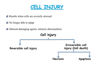
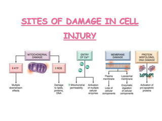
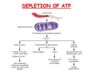
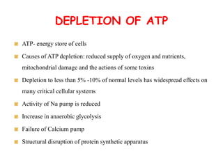
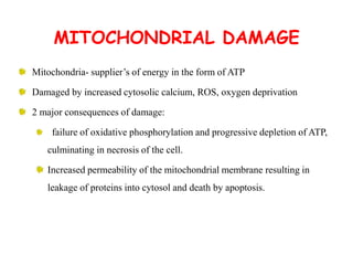
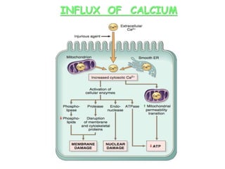
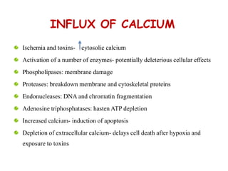
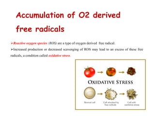
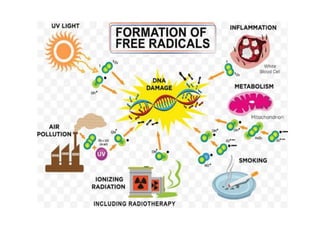
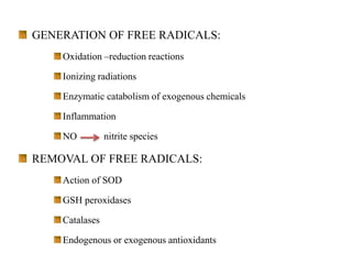
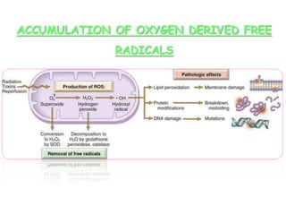
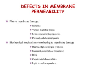
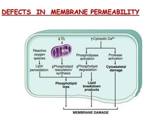
Prepared by
Swaliha C K
Assistant Professor
Senghundhar College of Pharmacy
CELL INJURY
Results when cells are severely stressed
No longer able to adapt
Inherent damaging agents, intrinsic abnormalities
Cell injury
Reversible cell injury
Irreversible cell
injury (Cell death)
Necrosis Apoptosis
REVERSIBLE CELL INJURY:
Removal of damaging stimulus_ reversible functional and
morphologic changes.
NECROSIS
Irreversible cell injury characterised by loss of membrane
integrity, enzymatic digestion of cells, leakage of cellular contents and
frequently a host reaction. Always pathologic.
APOPTOSIS
Programmed cell death or regulated mechanism of cell death
that serves to eliminate unwanted and irreparably damaged cells, with the
least possible host reaction. It may be pathologic or physiologic
SITES OF DAMAGE IN CELL
INJURY
MECHANISMS OF CELL INJURY
Depletion of ATP
Mitochondrial damage
Influx of calcium
Accumulation of O2 derived free radicals
Defects in membrane permeability
DEPLETION OF ATP
DEPLETION OF ATP
ATP- energy store of cells
Causes of ATP depletion: reduced supply of oxygen and nutrients,
mitochondrial damage and the actions of some toxins
Depletion to less than 5% -10% of normal levels has widespread effects on
many critical cellular systems
Activity of Na pump is reduced
Increase in anaerobic glycolysis
Failure of Calcium pump
Structural disruption of protein synthetic apparatus
MITOCHONDRIAL DAMAGE
Mitochondria- supplier’s of energy in the form of ATP
Damaged by increased cytosolic calcium, ROS, oxygen deprivation
2 major consequences of damage:
failure of oxidative phosphorylation and progressive depletion of ATP,
culminating in necrosis of the cell.
Increased permeability of the mitochondrial membrane resulting in
leakage of proteins into cytosol and death by apoptosis.
INFLUX OF CALCIUM
INFLUX OF CALCIUM
Ischemia and toxins- cytosolic calcium
Activation of a number of enzymes- potentially deleterious cellular effects
Phospholipases: membrane damage
Proteases: breakdown membrane and cytoskeletal proteins
Endonucleases: DNA and chromatin fragmentation
Adenosine triphosphatases: hasten ATP depletion
Increased calcium- induction of apoptosis
Depletion of extracellular calcium- delays cell death after hypoxia and
exposure to toxins
Reactive oxygen species (ROS) are a type of oxygen derived free radical.
Increased production or decreased scavenging of ROS may lead to an excess of these free
radicals, a condition called oxidative stress.
Accumulation of O2 derived
free radicals
OXIDATIVE STRESS
ROS- produced in cells during ETC, degraded and removed by cellular defense
systems
Increased ROS production- excess of free radicals- oxidative stress
Cell injury involves damage by free radicals in situations like:
Ischemia reperfusion
Chemical and radiation injury
Toxicity from oxygen and other gases
Cellular aging
Microbial killing by phagocytic cells
Tissue injury caused by inflammatory cells
GENERATION OF FREE RADICALS:
Oxidation –reduction reactions
Ionizing radiations
Enzymatic catabolism of exogenous chemicals
Inflammation
NO nitrite species
REMOVAL OF FREE RADICALS:
Action of SOD
GSH peroxidases
Catalases
Endogenous or exogenous antioxidants
ACCUMULATION OF OXYGEN DERIVED FREE
RADICALS
DEFECTS IN MEMBRANE
PERMEABILITY
Plasma membrane damage:
Ischemia
Various microbial toxins
Lytic complement components
Physical and chemical agents
Biochemical mechanisms contributing to membrane damage
Decreased phospholipid synthesis
Increased phospholipid breakdown
ROS
Cytoskeletal abnormalities
Lipid breakdown products
DEFECTS IN MEMBRANE PERMEABILITY
Cell injury -  mechanism

Cell injury - mechanism

  • 1.
    Prepared by Swaliha CK Assistant Professor Senghundhar College of Pharmacy
  • 2.
    CELL INJURY Results whencells are severely stressed No longer able to adapt Inherent damaging agents, intrinsic abnormalities Cell injury Reversible cell injury Irreversible cell injury (Cell death) Necrosis Apoptosis
  • 3.
    REVERSIBLE CELL INJURY: Removalof damaging stimulus_ reversible functional and morphologic changes. NECROSIS Irreversible cell injury characterised by loss of membrane integrity, enzymatic digestion of cells, leakage of cellular contents and frequently a host reaction. Always pathologic. APOPTOSIS Programmed cell death or regulated mechanism of cell death that serves to eliminate unwanted and irreparably damaged cells, with the least possible host reaction. It may be pathologic or physiologic
  • 4.
    SITES OF DAMAGEIN CELL INJURY
  • 5.
    MECHANISMS OF CELLINJURY Depletion of ATP Mitochondrial damage Influx of calcium Accumulation of O2 derived free radicals Defects in membrane permeability
  • 6.
  • 7.
    DEPLETION OF ATP ATP-energy store of cells Causes of ATP depletion: reduced supply of oxygen and nutrients, mitochondrial damage and the actions of some toxins Depletion to less than 5% -10% of normal levels has widespread effects on many critical cellular systems Activity of Na pump is reduced Increase in anaerobic glycolysis Failure of Calcium pump Structural disruption of protein synthetic apparatus
  • 8.
  • 9.
    MITOCHONDRIAL DAMAGE Mitochondria- supplier’sof energy in the form of ATP Damaged by increased cytosolic calcium, ROS, oxygen deprivation 2 major consequences of damage: failure of oxidative phosphorylation and progressive depletion of ATP, culminating in necrosis of the cell. Increased permeability of the mitochondrial membrane resulting in leakage of proteins into cytosol and death by apoptosis.
  • 10.
  • 11.
    INFLUX OF CALCIUM Ischemiaand toxins- cytosolic calcium Activation of a number of enzymes- potentially deleterious cellular effects Phospholipases: membrane damage Proteases: breakdown membrane and cytoskeletal proteins Endonucleases: DNA and chromatin fragmentation Adenosine triphosphatases: hasten ATP depletion Increased calcium- induction of apoptosis Depletion of extracellular calcium- delays cell death after hypoxia and exposure to toxins
  • 12.
    Reactive oxygen species(ROS) are a type of oxygen derived free radical. Increased production or decreased scavenging of ROS may lead to an excess of these free radicals, a condition called oxidative stress. Accumulation of O2 derived free radicals
  • 13.
    OXIDATIVE STRESS ROS- producedin cells during ETC, degraded and removed by cellular defense systems Increased ROS production- excess of free radicals- oxidative stress Cell injury involves damage by free radicals in situations like: Ischemia reperfusion Chemical and radiation injury Toxicity from oxygen and other gases Cellular aging Microbial killing by phagocytic cells Tissue injury caused by inflammatory cells
  • 15.
    GENERATION OF FREERADICALS: Oxidation –reduction reactions Ionizing radiations Enzymatic catabolism of exogenous chemicals Inflammation NO nitrite species REMOVAL OF FREE RADICALS: Action of SOD GSH peroxidases Catalases Endogenous or exogenous antioxidants
  • 16.
    ACCUMULATION OF OXYGENDERIVED FREE RADICALS
  • 17.
    DEFECTS IN MEMBRANE PERMEABILITY Plasmamembrane damage: Ischemia Various microbial toxins Lytic complement components Physical and chemical agents Biochemical mechanisms contributing to membrane damage Decreased phospholipid synthesis Increased phospholipid breakdown ROS Cytoskeletal abnormalities Lipid breakdown products
  • 18.
    DEFECTS IN MEMBRANEPERMEABILITY