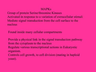
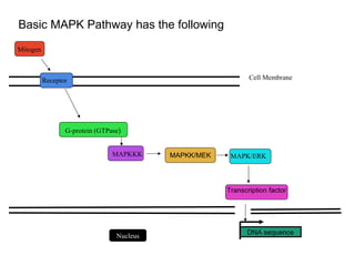
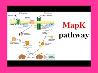
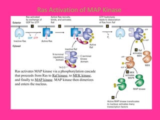
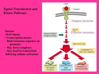
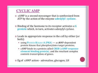
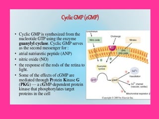
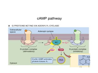
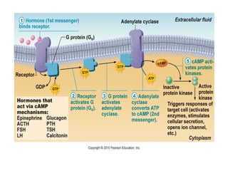
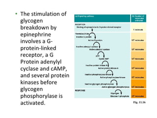
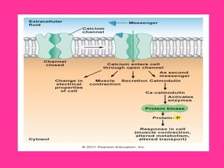
Mitogen-activated (stimulates mitosis)
Protein Kinase (MAPK) Pathway
Mohammed Kaleem Khan (Ph.D. Candidate)
ID:1802147
Department of Biochemistry
Faculty of Life Science
King Abdulaziz University,
KSA
Second messengers
•cAMP second messenger
• Phosphatidylinositol cycle
•Sphingolipid
•Eicosanoids
•Calcium as a second messenger
•Others
MAP kinase pathway
Activity of MAP kinase
Role in signaling
Cross functions
MAPKs
Group of protein Serine/threonine Kinases
Activated in response to a variation of extracellular stimuli
Mediate signal transduction from the cell surface to the
nucleus
Found inside many cellular compartments
Provide a physical link in the signal transduction pathway
from the cytoplasm to the nucleus
Regulate various transcriptional actions in Eukaryotic
organism.
Controls cell growth, to cell division (mating in haploid
yeast).
Basic MAPK Pathway has the following
Mitogen
Receptor
MAPKKK MAPKK/MEK MAPK/ERK
Transcription factor
DNA sequence
Cell Membrane
G-protein (GTPase)
Nucleus
Ras Activation of MAP Kinase
Ras activates MAP kinase via a phosphorylation cascade
that proceeds from Ras to Raf kinase, to MEK kinase,
and finally to MAP kinase. MAP kinase then dimerizes
and enters the nucleus.
Regulation of gene expression through MAPK signaling
pathways
Phosphorylation of transcription factors, thereby enhancing their activity:
Gene Expression
Metabolism
Cell Division
Cell Morphology
Cell Survival
Regulate Cancer, Inflammation and Cardiovascular disease.
Negatively regulated transcription factors by promoting their retention in the
cytoplasm upon phosphorylation active dephosphorylation of these factors is
needed for their migration to the nucleus
Translational regulation
Regulation of protein degradation
phosphorylation of transcription factors impedes their ubiquitination and thus their
degradation by the Proteasome
Recognition of a reaction partner by a kinase or
phosphatase
Signal Transduction and
Kinase Pathways
Nucleus
MAP kinase,
• Transcription factors
– Bind consensus sequence on
promoter
– May form complexes
– May itself be transcribed
following cellular activation
Adaptor proteins
Effector enzymes
Second Messenger
An extracellular signaling molecule is termed “first messenger”
First messenger → production of small and transient signaling
molecule
on inside of the cell → referred to as second messenger →
intracellular response
The second messenger is used to define “signalling molecules
which are found to be produced by cells in response to the
perception of the first message” (extracellular ligand).
Feature of Intracellular Messenger
need amplification
For example, one molecule bind to the exterior surface of the
cells →activation of hundreds or thousands of enzyme molecules
inside the cell→ catalyze many rounds of reaction → produced
amplification of signal
Mechanism of Action of Hydrophilic Messengers
Hydrophilic messengers cannot cross the cell membrane.
This restriction forces them into a 'classical' method of
messenger action, namely the production of intracellular second
messengers which are responsible for the effects of the various
messengers.
serve to amplify a signal
Molecules whose presence is a signal
Synthesized or released from storage
Act as intracellular ligands
This second messenger then is responsible for
the displays of all of the effects of the primary
messenger inside the cell.
Cascades have certain advantages to
include signal divergence, convergence
and amplification.
The resultant ligand receptor complex is
coupled to an enzyme found on the internal
surface of the cell membrane and stimulates the
conversion of some metabolite into a second
messenger.
Mostly, the second messenger acts by
stimulating or inhibiting protein
phosphorylation/dephosphorylation cascades.
Ligand binds to receptors located on the
external surface of the plasma membrane.
Primary amplifier enzymes:
adenylyl cyclase → cyclic AMP from ATP
guanylyl cyclase → cyclic GMP from GTP
phospholipase C (PLC)
cleaves phosphatidylinositol bisphosphate (PIP2) →
diacylglycerol (DAG) — diffuses in membrane
inositol trisphosphate (IP3) — released into cytosol
Secondary amplifier enzymes:
protein kinase A (PKA)
protein kinase G (PKG)
protein kinase C (PKC)
calcium-calmodulin kinase (Ca-CM-K)
Phosphatidylinositol Cycle
The PI-cycle is a series of enzyme-catalyzed biochemical
reactions that form a cyclical process, which the
intermediates pathway of second messenger.
Found in the plasma membrane.
It is an vital constituent of a signalling pathway in the cell
nucleus
Produces inositol triphosphate that is a vital ligand in opening
calcium channels Functions
Growth, cell survival, proliferation, cytoskeletal
rearrangement, intracellular vesicle trafficking, and cell
metabolism
Regulates intracellular calcium levels
EicosanoidsEicosanoids
((Prostaglandins,, Thromboxanes,, Leukotrienes))
Anti inflammatory Drugs inhibit Eicosanoid SynthesisAnti inflammatory Drugs inhibit Eicosanoid Synthesis
LeukotrienesProstaglandins,
thromboxanes
NSAIDs
Cyclo-
oxygenase
Lipoxygenase
Membrane lipids
Arachidonic Acid
Steroids
Phospholipase A2
Properties
Induction of inflammation
Mediation of pain signals
Induction of fever
Smooth muscle contraction (including uterus)
Smooth muscle relaxation
Protection of stomach lining
Simulation of platelet aggregation
Inhibition of platelet aggregation
Sodium and water retention
Phosphatidyl choline
Arachidonic acid
Phospholipase A2
Ca++
Phosphatidylinositol bisphosphate
Phospholipase C
1,2 Diacylglycerol
Arachidonic acid Monoacylglycerol
DAG
lipase
Arachidonic acid
MAG lipase
Arachidonic acid release from membrane lipidsArachidonic acid release from membrane lipids
Stimulus
Pathways for Arachidonic Acid MetabolismPathways for Arachidonic Acid Metabolism
Arachidonic acid
Cyclo-oxygenase
Pathway
PGG2
Prostaglandins
Thromboxanes
lipoxygenase
Pathway
HPETE
Leukotrienes
HETE
Lipoxins
Function of PGI2, PGE2, PGD2
↑ Vasodilation, cAMP
↓ Platelet and leukocyte aggregation, IL1 and IL2, T-cell
proliferation, lymphocyte migration
PGF2a
↑ Vasoconstriction, Bronchoconstriction, smooth muscle
contraction
TXA2
↑ Vasoconstriction, Platelet aggregation, lymphocyte
proliferation, bronchoconstriction
Calcium Systems
Regulator of cell function.
Involved in the control of motile/fusion process and can act as a
second messenger (IP3/DAG system).
A different cellular proteins act to control intracellular Ca+2
levels which comprise:
A) gated Ca+2
channels
B) Ca+2
Pumps
Voltage-gated Ca+2
channel (VGCC):
VGCC in the synaptic bulb, which allows for Ca+2
entry which in
turn leads to vesicle fusion and neurotransmitter release into the
synaptic cleft
Ligand-gated Ca+2
channels (LGCC):
LGCC of the SER and mitochondria which recognize IP3
Furthermore, there are a diversity of 'external' ligand-gated
channels which respond to neurotransmitters, and a special
channel, the ryanodine channel, which is activated by Ca+2
.
The significance of the ryanodine channel is that it allows small
changes in Ca+2
concentration to be magnified by release on
'large' intracellular stores.
Two types of Ca+2
pumps:
Utilize a Na+
gradient to move Ca+2
Use ATP directly
Calmodulin
The intracellular effects of Ca+2
are
exhibit through a different Ca+2
binding proteins: Calmodulin.
This small protein binds four Ca+2
ions in a cooperative manner and
then interacts with a different
proteins to modify their activity.
Mapk modified
Mapk modified
Mapk modified
Mapk modified
Mapk modified
Mapk modified

Mapk modified

  • 1.
    Mitogen-activated (stimulates mitosis) ProteinKinase (MAPK) Pathway Mohammed Kaleem Khan (Ph.D. Candidate) ID:1802147 Department of Biochemistry Faculty of Life Science King Abdulaziz University, KSA
  • 2.
    Second messengers •cAMP secondmessenger • Phosphatidylinositol cycle •Sphingolipid •Eicosanoids •Calcium as a second messenger •Others MAP kinase pathway Activity of MAP kinase Role in signaling Cross functions
  • 3.
    MAPKs Group of proteinSerine/threonine Kinases Activated in response to a variation of extracellular stimuli Mediate signal transduction from the cell surface to the nucleus Found inside many cellular compartments Provide a physical link in the signal transduction pathway from the cytoplasm to the nucleus Regulate various transcriptional actions in Eukaryotic organism. Controls cell growth, to cell division (mating in haploid yeast).
  • 4.
    Basic MAPK Pathwayhas the following Mitogen Receptor MAPKKK MAPKK/MEK MAPK/ERK Transcription factor DNA sequence Cell Membrane G-protein (GTPase) Nucleus
  • 6.
    Ras Activation ofMAP Kinase Ras activates MAP kinase via a phosphorylation cascade that proceeds from Ras to Raf kinase, to MEK kinase, and finally to MAP kinase. MAP kinase then dimerizes and enters the nucleus.
  • 7.
    Regulation of geneexpression through MAPK signaling pathways Phosphorylation of transcription factors, thereby enhancing their activity: Gene Expression Metabolism Cell Division Cell Morphology Cell Survival Regulate Cancer, Inflammation and Cardiovascular disease. Negatively regulated transcription factors by promoting their retention in the cytoplasm upon phosphorylation active dephosphorylation of these factors is needed for their migration to the nucleus Translational regulation Regulation of protein degradation phosphorylation of transcription factors impedes their ubiquitination and thus their degradation by the Proteasome
  • 8.
    Recognition of areaction partner by a kinase or phosphatase
  • 9.
    Signal Transduction and KinasePathways Nucleus MAP kinase, • Transcription factors – Bind consensus sequence on promoter – May form complexes – May itself be transcribed following cellular activation Adaptor proteins Effector enzymes
  • 10.
    Second Messenger An extracellularsignaling molecule is termed “first messenger” First messenger → production of small and transient signaling molecule on inside of the cell → referred to as second messenger → intracellular response The second messenger is used to define “signalling molecules which are found to be produced by cells in response to the perception of the first message” (extracellular ligand).
  • 11.
    Feature of IntracellularMessenger need amplification For example, one molecule bind to the exterior surface of the cells →activation of hundreds or thousands of enzyme molecules inside the cell→ catalyze many rounds of reaction → produced amplification of signal
  • 12.
    Mechanism of Actionof Hydrophilic Messengers Hydrophilic messengers cannot cross the cell membrane. This restriction forces them into a 'classical' method of messenger action, namely the production of intracellular second messengers which are responsible for the effects of the various messengers. serve to amplify a signal Molecules whose presence is a signal Synthesized or released from storage Act as intracellular ligands
  • 14.
    This second messengerthen is responsible for the displays of all of the effects of the primary messenger inside the cell. Cascades have certain advantages to include signal divergence, convergence and amplification. The resultant ligand receptor complex is coupled to an enzyme found on the internal surface of the cell membrane and stimulates the conversion of some metabolite into a second messenger. Mostly, the second messenger acts by stimulating or inhibiting protein phosphorylation/dephosphorylation cascades. Ligand binds to receptors located on the external surface of the plasma membrane.
  • 15.
    Primary amplifier enzymes: adenylylcyclase → cyclic AMP from ATP guanylyl cyclase → cyclic GMP from GTP phospholipase C (PLC) cleaves phosphatidylinositol bisphosphate (PIP2) → diacylglycerol (DAG) — diffuses in membrane inositol trisphosphate (IP3) — released into cytosol Secondary amplifier enzymes: protein kinase A (PKA) protein kinase G (PKG) protein kinase C (PKC) calcium-calmodulin kinase (Ca-CM-K)
  • 23.
    Phosphatidylinositol Cycle The PI-cycleis a series of enzyme-catalyzed biochemical reactions that form a cyclical process, which the intermediates pathway of second messenger. Found in the plasma membrane. It is an vital constituent of a signalling pathway in the cell nucleus Produces inositol triphosphate that is a vital ligand in opening calcium channels Functions Growth, cell survival, proliferation, cytoskeletal rearrangement, intracellular vesicle trafficking, and cell metabolism Regulates intracellular calcium levels
  • 27.
  • 28.
    Anti inflammatory Drugsinhibit Eicosanoid SynthesisAnti inflammatory Drugs inhibit Eicosanoid Synthesis LeukotrienesProstaglandins, thromboxanes NSAIDs Cyclo- oxygenase Lipoxygenase Membrane lipids Arachidonic Acid Steroids Phospholipase A2
  • 29.
    Properties Induction of inflammation Mediationof pain signals Induction of fever Smooth muscle contraction (including uterus) Smooth muscle relaxation Protection of stomach lining Simulation of platelet aggregation Inhibition of platelet aggregation Sodium and water retention
  • 30.
    Phosphatidyl choline Arachidonic acid PhospholipaseA2 Ca++ Phosphatidylinositol bisphosphate Phospholipase C 1,2 Diacylglycerol Arachidonic acid Monoacylglycerol DAG lipase Arachidonic acid MAG lipase Arachidonic acid release from membrane lipidsArachidonic acid release from membrane lipids Stimulus
  • 31.
    Pathways for ArachidonicAcid MetabolismPathways for Arachidonic Acid Metabolism Arachidonic acid Cyclo-oxygenase Pathway PGG2 Prostaglandins Thromboxanes lipoxygenase Pathway HPETE Leukotrienes HETE Lipoxins
  • 32.
    Function of PGI2,PGE2, PGD2 ↑ Vasodilation, cAMP ↓ Platelet and leukocyte aggregation, IL1 and IL2, T-cell proliferation, lymphocyte migration PGF2a ↑ Vasoconstriction, Bronchoconstriction, smooth muscle contraction TXA2 ↑ Vasoconstriction, Platelet aggregation, lymphocyte proliferation, bronchoconstriction
  • 33.
    Calcium Systems Regulator ofcell function. Involved in the control of motile/fusion process and can act as a second messenger (IP3/DAG system). A different cellular proteins act to control intracellular Ca+2 levels which comprise: A) gated Ca+2 channels B) Ca+2 Pumps Voltage-gated Ca+2 channel (VGCC): VGCC in the synaptic bulb, which allows for Ca+2 entry which in turn leads to vesicle fusion and neurotransmitter release into the synaptic cleft
  • 34.
    Ligand-gated Ca+2 channels (LGCC): LGCCof the SER and mitochondria which recognize IP3 Furthermore, there are a diversity of 'external' ligand-gated channels which respond to neurotransmitters, and a special channel, the ryanodine channel, which is activated by Ca+2 . The significance of the ryanodine channel is that it allows small changes in Ca+2 concentration to be magnified by release on 'large' intracellular stores. Two types of Ca+2 pumps: Utilize a Na+ gradient to move Ca+2 Use ATP directly
  • 35.
    Calmodulin The intracellular effectsof Ca+2 are exhibit through a different Ca+2 binding proteins: Calmodulin. This small protein binds four Ca+2 ions in a cooperative manner and then interacts with a different proteins to modify their activity.