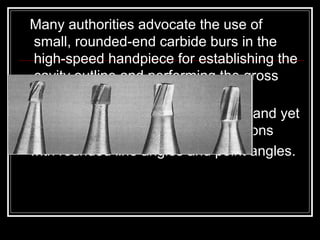
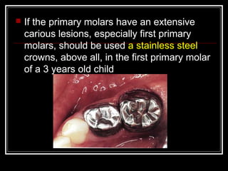
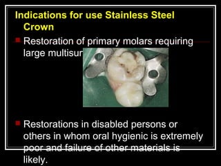
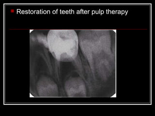
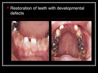
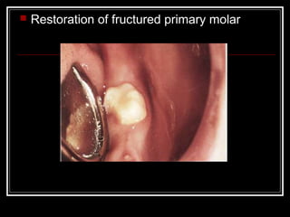
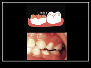
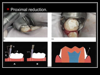
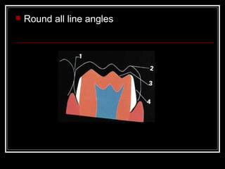
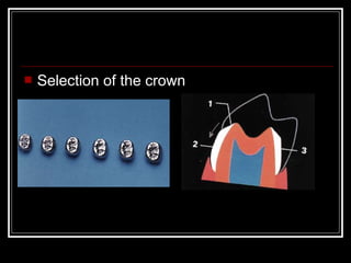
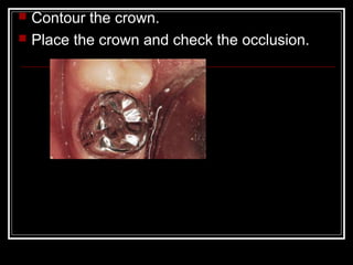
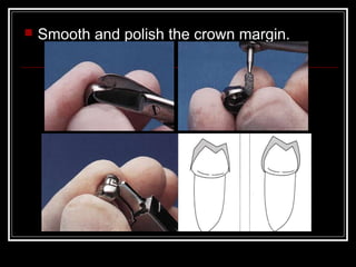
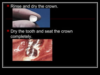
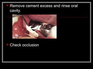
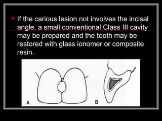
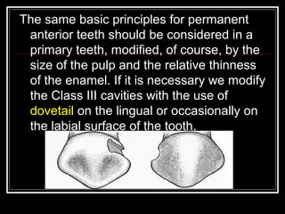
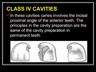
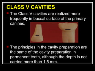
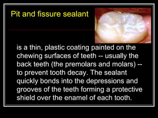
CAVITY 
PREPARATION IN 
PRIMARY TEETH 
Dr. Masar Mohammed
 INTRODUCTION 
 BASIC PRINCIPLES IN THE 
PREPARATION OF CAVITIES IN 
PRIMARY TEETH 
 CLASS I CAVITIES 
 CLASS II CAVITIES 
 CLASS III CAVITIES 
 CLASS IV CAVITIES 
 CLASS V CAVITIES 
 RECENT CONCEPTS IN RESTORATIVE 
DENTISTRY
Operative dentistry: 
Is the art and science of the diagnosis, 
treatment and prognosis of defects of teeth 
that do not require full coverage restorations 
for correction. 
The aim of pediatric operative dentistry is to 
maintain the tooth in the dental arch in a 
healthy state, so as to prevent its loss and 
the subsequent problems that will result.
Main reasons to control caries in primary 
Dentition 
 Prevent pain and discomfort. 
 Prevent local infections. 
 Prevent general infections 
 Prevent negative attitudes and promote 
keeping good oral health 
 Maintenance good mastication, aesthetic 
and overall well-being 
 Prevent caries in permanent teeth 
 Prevent malocclusion.
ANATOMIC CONSIDERATIONS OF 
PRIMARY TEETH 
1. Shorter crown. 
2. Thinner enamel and dentin. 
3. Larger pulp and higher 
pulp horns. 
4. Enamel rods in cervical 
area directed occlusally. 
5. Greater cervical constriction. 
6. Broad, flat proximal contacts. 
7. Narrow occlusal table. 
8. Lighter in color.
 Prevention is the cornerstone of good 
management of dental caries in children 
 History taking is fundamental to the 
execution of restorative care in the primary 
and mixed dentition. 
 Communication skills are essential in 
obtaining a child’s co-operation in 
completing treatment.
In the restoration of primary teeth, we 
should consider the following 
factors: 
 The child: age, physical condition, and 
cooperation among others. 
 Caries degree. 
 Degree of radicular reabsorption of the 
tooth. 
 Condition of the bone support. 
 Dental material
 BASIC PRINCIPLES IN THE 
PREPARATION OF CAVITIES IN 
PRIMARY TEETH. 
The steps in the preparation of a cavity in a 
primary tooth are not difficult but do 
require precise operator control
Many authorities advocate the use of 
small, rounded-end carbide burs in the 
high-speed handpiece for establishing the 
cavity outline and performing the gross 
preparation. 
they are designed to cut efficiently and yet 
allow conservative cavity preparations 
with rounded line angles and point angles.
 The Black’s principles with some 
modification are basic principles in 
the preparation of the cavities in the 
primary teeth. There are three 
operative steps with the use of the 
high-speed handpiece:
 Opening and conformation of the cavity 
with the use of the high-speed handpiece. 
 Eliminating the caries of the buccal, lingual, 
mesial and distal walls with the use of the 
high-speed handpiece. Eliminate the caries 
of the pulpal wall with the use of the lower-speed 
handpiece. 
 The third step will include dentine 
sterilization and the cement base.
CLASS I CAVITIES 
 Incipient carious lesion in child under 2 
years old should be eliminated. Small cavity 
preparation may be made with a No.329 or 
No. 330 pear-shaped bur. We should 
open the decayed area and extend the 
cavosurface margin only to the extent of the 
carious lesion. The preparation can be 
completed in a few seconds.
 The outline form should include all pits, 
fissures and grooves into which a sharp 
explorer can penetrate.
 The pulpal floor should be flat or slightly 
concave throughout to allow for greater 
depth of the filling material, for better 
distribution of stress in the restoration and 
to avoid endangering the high pulpal 
horns. 
 The depth of pulpal floor should be 
established just beneath the 
dentinoenamel junction (0.5 mm) to avoid 
pulp exposure.
 All the internal line angles should be 
rounded. 
 The side walls should slightly converge 
towards occlusal so that the preparation 
will follow the outer form of the crown. 
 Beside the regular class I cavity 
preparations done in primary molars, 
occlusal spot preparations have been 
recommended.
In such preparations only the carious pits or 
groove is prepared and the tooth is 
restored in the usual manner. These 
preparations are applicable in any of the 
primary molars with exception of the lower 
second primary molars in which extension 
for prevention including all deep pits and 
fissures is recommended above all, if the 
child has high caries index
 cavity should be covered with calcium 
hydroxide . A base of polycarboxlate, glass 
ionomer or rapid-setting zinc-oxide-eugenol 
cement may then be placed over the 
calcium hydroxide material to provide 
adequate thermal pulp protection.
 Do not cross the oblique ridge in the upper 
second primary or first permanent molars 
and the transverse ridge of the lower first 
primary molar unless they are undermined 
with caries. These heavy ridges add 
support to the tooth.
CLASS II CAVITIES. 
 These preparations include an occlusal, an 
isthmus and proximal portion. The outline 
form of the occlusal step 
should be dovetail-shaped 
including all carious pits, 
fissures, and developmental 
grooves.
 The side walls of the occlusal step should 
converge from the pulpal wall to the 
occlusal surface. 
 The pulpal floor should be established just 
beneath the dentinoenamel junction.
 Angles between the side walls and the 
pulpal floor should be gently rounded. 
 The width of the isthmus should be 
approximately one-third of the intercuspal 
dimension of the tooth.
 The axio-pulpal line angle should be 
beveled to reduce the concentration of 
stresses and provide grater bulk of material 
in the isthmus area, which is liable to 
fracture
 The greater constriction of primary teeth 
increases the danger of damaging the 
interproximal soft tissues during cavity 
preparation. 
 Extreme care must be taken when 
breaking through the marginal ridge to 
prevent damage to the adjacent proximal 
surface, especially when the bur is 
revolving at high speed.
 The proximal box line angles and walls 
should converge towards the occlusal. 
When viewed from the occlusal aspect 
the resulting axial wall should follow the 
outline of the original proximal surface.
 An axiobuccal and axiolingual retentive 
groove may be included in the preparation.
 The bur is used in a pendulum-swinging 
fashion to undermine the marginal ridge 
and at the same time to establish the 
gingival depth. 
 The gingival seat should be of sufficient 
depth to break contact with the adjacent 
tooth. 
 A liner or intermediate base should be 
placed before the insertion of the silver 
amalgam.
 The amalgam restoration in the Class II 
cavity needs the use of a matrix retainer. 
The matrix should be rigid enough to allow 
adequate packing pressure, ensuring a 
well-condensed restoration free from an 
excess of residual mercury.
 If the primary molars have an extensive 
carious lesions, especially first primary 
molars, should be used a stainless steel 
crowns, above all, in the first primary molar 
of a 3 years old child
Indications for use Stainless Steel 
Crown 
 Restoration of primary molars requiring 
large multisurface restoration. 
 Restorations in disabled persons or 
others in whom oral hygienic is extremely 
poor and failure of other materials is 
likely.
 Restorations of teeth in children with 
rampant caries.
 Restoration of teeth after pulp therapy
 Restoration of teeth with developmental 
defects
 Restoration of fructured primary molar
 As abutment for space maintainer
 In children with bruxism
 Restoration of hypoplastic young 
permanent molars
Steps of preparation and placement of 
Stainless Steel Crown. 
 Evaluate the preoperative occlusion. 
 Administer appropriate anesthesia. 
 Establish access. 
 Reduction of the occlusal surface.
 Proximal reduction.
 Round all line angles
 Selection of the crown
 Contour the crown. 
 Place the crown and check the occlusion.
 Smooth and polish the crown margin.
 Rinse and dry the crown. 
 Dry the tooth and seat the crown 
completely.
 Remove cement excess and rinse oral 
cavity. 
 Check occlusion
CLASS III CAVITIES 
 Carious lesions on the proximal surfaces of 
anterior primary teeth sometimes occur in 
children whose teeth are in contact and in 
those children who have evidence of arch 
inadequacy or crowding. 
 If caries is not extensive, disking by sand 
paper disc is performed to remove the 
decay, and then fluoride is applied topically
 If the carious lesion not involves the incisal 
angle, a small conventional Class III cavity 
may be prepared and the tooth may be 
restored with glass ionomer or composite 
resin.
The same basic principles for permanent 
anterior teeth should be considered in a 
primary teeth, modified, of course, by the 
size of the pulp and the relative thinness 
of the enamel. If it is necessary we modify 
the Class III cavities with the use of 
dovetail on the lingual or occasionally on 
the labial surface of the tooth.
Because of the narrow labiolingual width of 
the primary incisor teeth, the Class III 
preparation is very difficult to perform and 
often needs a labial or lingual dovetail to 
gain access and aid in retention of the 
restoration.
The distal surface of the primary canine 
is a frequent site of caries attack
CLASS IV CAVITIES 
 In these cavities caries involves the incisal 
proximal angle of the anterior teeth. The 
principles in the cavity preparation are the 
same of the cavity preparation in 
permanent teeth
 In regular class IV cavity preparations, 
composite resin material can be used for 
restoration.
CLASS V CAVITIES 
 The Class V cavities are realized more 
frequently in buccal surface of the primary 
canines. 
 The principles in the cavity preparation are 
the same of the cavity preparation in 
permanent teeth, although the depth is not 
carried more than 1.5 mm.
 Walls of preparation converge toward 
buccal surface of tooth for retention of 
restoration. 
 When a necessary, retentive groove can be 
placed along the gingivoaxial and 
occlusoaxial line angles. Use a No. 1/2 
round bur at slow speed. 
 Glass ionomer cement could be used 
effectively for restoring these cavities.
Pit and fissure sealant 
is a thin, plastic coating painted on the 
chewing surfaces of teeth -- usually the 
back teeth (the premolars and molars) -- 
to prevent tooth decay. The sealant 
quickly bonds into the depressions and 
grooves of the teeth forming a protective 
shield over the enamel of each tooth.
Indicaations of sealant placement: 
1. Deep retintive pits and fissures. 
2. Stained pits and fissures with minmum 
decalcification. 
3. No radiographic evidence of proximal 
caries. 
4. Factores associated with increased caries 
incidence.
5. Caries free. 
6. Possibility of adequate isolation. 
How Are Sealants Applied?
Cavitypreparation 130320103634-phpapp01

Cavitypreparation 130320103634-phpapp01

  • 1.
    CAVITY PREPARATION IN PRIMARY TEETH Dr. Masar Mohammed
  • 2.
     INTRODUCTION BASIC PRINCIPLES IN THE PREPARATION OF CAVITIES IN PRIMARY TEETH  CLASS I CAVITIES  CLASS II CAVITIES  CLASS III CAVITIES  CLASS IV CAVITIES  CLASS V CAVITIES  RECENT CONCEPTS IN RESTORATIVE DENTISTRY
  • 3.
    Operative dentistry: Isthe art and science of the diagnosis, treatment and prognosis of defects of teeth that do not require full coverage restorations for correction. The aim of pediatric operative dentistry is to maintain the tooth in the dental arch in a healthy state, so as to prevent its loss and the subsequent problems that will result.
  • 4.
    Main reasons tocontrol caries in primary Dentition  Prevent pain and discomfort.  Prevent local infections.  Prevent general infections  Prevent negative attitudes and promote keeping good oral health  Maintenance good mastication, aesthetic and overall well-being  Prevent caries in permanent teeth  Prevent malocclusion.
  • 5.
    ANATOMIC CONSIDERATIONS OF PRIMARY TEETH 1. Shorter crown. 2. Thinner enamel and dentin. 3. Larger pulp and higher pulp horns. 4. Enamel rods in cervical area directed occlusally. 5. Greater cervical constriction. 6. Broad, flat proximal contacts. 7. Narrow occlusal table. 8. Lighter in color.
  • 6.
     Prevention isthe cornerstone of good management of dental caries in children  History taking is fundamental to the execution of restorative care in the primary and mixed dentition.  Communication skills are essential in obtaining a child’s co-operation in completing treatment.
  • 7.
    In the restorationof primary teeth, we should consider the following factors:  The child: age, physical condition, and cooperation among others.  Caries degree.  Degree of radicular reabsorption of the tooth.  Condition of the bone support.  Dental material
  • 8.
     BASIC PRINCIPLESIN THE PREPARATION OF CAVITIES IN PRIMARY TEETH. The steps in the preparation of a cavity in a primary tooth are not difficult but do require precise operator control
  • 9.
    Many authorities advocatethe use of small, rounded-end carbide burs in the high-speed handpiece for establishing the cavity outline and performing the gross preparation. they are designed to cut efficiently and yet allow conservative cavity preparations with rounded line angles and point angles.
  • 10.
     The Black’sprinciples with some modification are basic principles in the preparation of the cavities in the primary teeth. There are three operative steps with the use of the high-speed handpiece:
  • 11.
     Opening andconformation of the cavity with the use of the high-speed handpiece.  Eliminating the caries of the buccal, lingual, mesial and distal walls with the use of the high-speed handpiece. Eliminate the caries of the pulpal wall with the use of the lower-speed handpiece.  The third step will include dentine sterilization and the cement base.
  • 12.
    CLASS I CAVITIES  Incipient carious lesion in child under 2 years old should be eliminated. Small cavity preparation may be made with a No.329 or No. 330 pear-shaped bur. We should open the decayed area and extend the cavosurface margin only to the extent of the carious lesion. The preparation can be completed in a few seconds.
  • 13.
     The outlineform should include all pits, fissures and grooves into which a sharp explorer can penetrate.
  • 14.
     The pulpalfloor should be flat or slightly concave throughout to allow for greater depth of the filling material, for better distribution of stress in the restoration and to avoid endangering the high pulpal horns.  The depth of pulpal floor should be established just beneath the dentinoenamel junction (0.5 mm) to avoid pulp exposure.
  • 15.
     All theinternal line angles should be rounded.  The side walls should slightly converge towards occlusal so that the preparation will follow the outer form of the crown.  Beside the regular class I cavity preparations done in primary molars, occlusal spot preparations have been recommended.
  • 16.
    In such preparationsonly the carious pits or groove is prepared and the tooth is restored in the usual manner. These preparations are applicable in any of the primary molars with exception of the lower second primary molars in which extension for prevention including all deep pits and fissures is recommended above all, if the child has high caries index
  • 17.
     cavity shouldbe covered with calcium hydroxide . A base of polycarboxlate, glass ionomer or rapid-setting zinc-oxide-eugenol cement may then be placed over the calcium hydroxide material to provide adequate thermal pulp protection.
  • 18.
     Do notcross the oblique ridge in the upper second primary or first permanent molars and the transverse ridge of the lower first primary molar unless they are undermined with caries. These heavy ridges add support to the tooth.
  • 19.
    CLASS II CAVITIES.  These preparations include an occlusal, an isthmus and proximal portion. The outline form of the occlusal step should be dovetail-shaped including all carious pits, fissures, and developmental grooves.
  • 20.
     The sidewalls of the occlusal step should converge from the pulpal wall to the occlusal surface.  The pulpal floor should be established just beneath the dentinoenamel junction.
  • 21.
     Angles betweenthe side walls and the pulpal floor should be gently rounded.  The width of the isthmus should be approximately one-third of the intercuspal dimension of the tooth.
  • 22.
     The axio-pulpalline angle should be beveled to reduce the concentration of stresses and provide grater bulk of material in the isthmus area, which is liable to fracture
  • 23.
     The greaterconstriction of primary teeth increases the danger of damaging the interproximal soft tissues during cavity preparation.  Extreme care must be taken when breaking through the marginal ridge to prevent damage to the adjacent proximal surface, especially when the bur is revolving at high speed.
  • 24.
     The proximalbox line angles and walls should converge towards the occlusal. When viewed from the occlusal aspect the resulting axial wall should follow the outline of the original proximal surface.
  • 25.
     An axiobuccaland axiolingual retentive groove may be included in the preparation.
  • 26.
     The buris used in a pendulum-swinging fashion to undermine the marginal ridge and at the same time to establish the gingival depth.  The gingival seat should be of sufficient depth to break contact with the adjacent tooth.  A liner or intermediate base should be placed before the insertion of the silver amalgam.
  • 27.
     The amalgamrestoration in the Class II cavity needs the use of a matrix retainer. The matrix should be rigid enough to allow adequate packing pressure, ensuring a well-condensed restoration free from an excess of residual mercury.
  • 28.
     If theprimary molars have an extensive carious lesions, especially first primary molars, should be used a stainless steel crowns, above all, in the first primary molar of a 3 years old child
  • 29.
    Indications for useStainless Steel Crown  Restoration of primary molars requiring large multisurface restoration.  Restorations in disabled persons or others in whom oral hygienic is extremely poor and failure of other materials is likely.
  • 30.
     Restorations ofteeth in children with rampant caries.
  • 31.
     Restoration ofteeth after pulp therapy
  • 32.
     Restoration ofteeth with developmental defects
  • 33.
     Restoration offructured primary molar
  • 34.
     As abutmentfor space maintainer
  • 35.
     In childrenwith bruxism
  • 36.
     Restoration ofhypoplastic young permanent molars
  • 37.
    Steps of preparationand placement of Stainless Steel Crown.  Evaluate the preoperative occlusion.  Administer appropriate anesthesia.  Establish access.  Reduction of the occlusal surface.
  • 39.
  • 40.
     Round allline angles
  • 41.
  • 42.
     Contour thecrown.  Place the crown and check the occlusion.
  • 43.
     Smooth andpolish the crown margin.
  • 44.
     Rinse anddry the crown.  Dry the tooth and seat the crown completely.
  • 45.
     Remove cementexcess and rinse oral cavity.  Check occlusion
  • 46.
    CLASS III CAVITIES  Carious lesions on the proximal surfaces of anterior primary teeth sometimes occur in children whose teeth are in contact and in those children who have evidence of arch inadequacy or crowding.  If caries is not extensive, disking by sand paper disc is performed to remove the decay, and then fluoride is applied topically
  • 47.
     If thecarious lesion not involves the incisal angle, a small conventional Class III cavity may be prepared and the tooth may be restored with glass ionomer or composite resin.
  • 48.
    The same basicprinciples for permanent anterior teeth should be considered in a primary teeth, modified, of course, by the size of the pulp and the relative thinness of the enamel. If it is necessary we modify the Class III cavities with the use of dovetail on the lingual or occasionally on the labial surface of the tooth.
  • 49.
    Because of thenarrow labiolingual width of the primary incisor teeth, the Class III preparation is very difficult to perform and often needs a labial or lingual dovetail to gain access and aid in retention of the restoration.
  • 50.
    The distal surfaceof the primary canine is a frequent site of caries attack
  • 51.
    CLASS IV CAVITIES  In these cavities caries involves the incisal proximal angle of the anterior teeth. The principles in the cavity preparation are the same of the cavity preparation in permanent teeth
  • 52.
     In regularclass IV cavity preparations, composite resin material can be used for restoration.
  • 53.
    CLASS V CAVITIES  The Class V cavities are realized more frequently in buccal surface of the primary canines.  The principles in the cavity preparation are the same of the cavity preparation in permanent teeth, although the depth is not carried more than 1.5 mm.
  • 54.
     Walls ofpreparation converge toward buccal surface of tooth for retention of restoration.  When a necessary, retentive groove can be placed along the gingivoaxial and occlusoaxial line angles. Use a No. 1/2 round bur at slow speed.  Glass ionomer cement could be used effectively for restoring these cavities.
  • 55.
    Pit and fissuresealant is a thin, plastic coating painted on the chewing surfaces of teeth -- usually the back teeth (the premolars and molars) -- to prevent tooth decay. The sealant quickly bonds into the depressions and grooves of the teeth forming a protective shield over the enamel of each tooth.
  • 56.
    Indicaations of sealantplacement: 1. Deep retintive pits and fissures. 2. Stained pits and fissures with minmum decalcification. 3. No radiographic evidence of proximal caries. 4. Factores associated with increased caries incidence.
  • 57.
    5. Caries free. 6. Possibility of adequate isolation. How Are Sealants Applied?