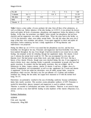
Maggie Kilbride
1
Maggie Kilbride
3/28/16
Assessment:
History of present illness:
This patient is a 56-year-old female. She presented to the Roudebush VA on 1/11/16 due to her
nasojejunal tube (NJ) falling out, as well as worsening lower extremity swelling accompanied by
redness and blisters. She had been recently diagnosed with anastomotic gastric ulcer with
secondary partial gastric outlet obstruction and malnutrition which resulted in the placement of a
NJ tube. This malnutrition is likely secondary to gastric bypass and gastric outlet obstruction.
Gastric outlet obstruction is known as a blockage between the stomach and small intestine, and
can be accompanied by abdominal pain, early satiation, or bloating. It is important to note that
this patient has a history of a Roux-en-Y Gastric Bypass (RYGB) in 2004. It is known that long-
term complications involving micronutrient deficiencies do occur after a RYGB. The
gastrojejunostomy is also more susceptible to anastomotic ulcers. These ulcers following a
Roux-en-Y Gastric Bypass surgery occur in around 16% of patients (Steinemann et al., 2013).
The patient’s initial visit had been from 12/29/15-1/2/16 for abdominal pain, nausea, vomiting
and significant weight loss in the past year. During that admission an EGD revealed a non-
bleeding ulcer just beyond the gastrojejunal anastomosis with angulation of the jejunal limb. A
nasojejunal tube was then placed via endoscopy past this anastomosis. Up until 4 days prior to
1/11/16 when her NJ tube fell out, she had been receiving tube feeds as well as a full liquid diet.
After complications replacing the NJ tube, it was decided she would no longer use a tube feeding
and will only eat a clear liquid diet. The patient admitted for a third time on 1/19/16 due to chest
pain. Up to this final visit, she had been following a clear liquid diet at home.
Current complaints:
On the patient’s initial admit on 12/29/15 she had come in due to ongoing symptoms including
weight loss totaling 50 pounds as well as PO intolerance over the past few days. Complaints of
PO intolerance included abdominal pain, nausea, and early satiety. The patient then readmitted to
the hospital on 1/11/16 for bilateral lower extremity swelling accompanied by redness and
blisters. She reported recent blistering and weeping. Due to a fall, her NJ tube had fallen out. She
also had complaints of abdominal pain and nausea. Her last admission on 1/19/16 was from
complaints of chest pain.
Past Medical History:
This patient’s past medical history consists of a gastric ulcer, partial gastric outlet obstruction,
Roux-en-Y Gastric Bypass in 2004, cholecystectomy in 2001, malnutrition, hypertension,
hypothyroidism, depression, GERD, anemia and urinary incontinence, vitamin D deficiency.
Anthropometrics:
Maggie Kilbride
2
This patient’s height is 67in (170cm). Her weight on 12/29/15 was 105.5lb (48kg), giving her a
BMI of 15. Her ideal body weight is 135lb (61.3kg) and at this weight her percent ideal body
weight was 77%. At this BMI she was classified with severe malnutrition.
Her weight on her second admit (1/11) after the NJ tube was placed had gone up. The rise in this
weight was attributed to bilateral lower extremity edema and is not reflective of her dry weight.
This weight was 120lb (54.7). A BMI of 18.9 and 88% of ideal body weight. The weight taken
on a follow up (1/15) showed her closer to a dry weight at 110lb (50kg). With that body weight
her BMI was 17.3 and percent ideal body weight was 81%. At this BMI she was classified with
moderate malnutrition.
Her final admission weight on 1/19/16 was 115lb (52 kg). This gave her a BMI of 18 and
resulted in being 85% of her ideal body weight. With this BMI she was classified with mild
malnutrition.
Date lb.(kg) BMI
12/16/15 104 (47.5) 16
12/30/15 101 (46) 15
1/11/16 108.9 (49) 17
1/12/16 120.4 (54.6) 18
1/15/16 110 (50) 17
1/19/16 115 (52) 18
Estimated Energy Needs:
These needs were estimated using 30-35 calories per kilogram of actual body weight. Actual
body weight was used rather than ideal body weight since the patient was considered
underweight with mild-severe malnutrition based on her BMI. The equations used on “healthy”
individuals are generally within 10% of measured values. These equations are not applicable
when dealing with patients who are at extremes in their weight. Malnutrition feeding can
decrease resting energy expenditures by about15-20%. Estimated energy requirements for
hospitalized patients based on their BMI suggests a patient with a BMI ranging from 15-19
should need 30-35 calories per kilogram of their actual body weight (Klein & Jeejeebhoy, 2002,
p.256). Given this information, her estimated energy needs were 1430-1670 kcal/kg on her initial
visit (12/29/15). The protein needs suggested were 1.2-1.5 grams of protein per kilogram of
actual body weight. Her protein needs on this visit were 57-72 grams of protein.
Her second admission on 1/11/16 came to require 1635-1900 kcal per kg of current weight (30-
25 kcal/kg). This weight calculating her needs was not thought to be a dry weight. The protein
requirement was 83 g (1.5 gm/kg actual body weight). Her fluid needs were 1635 mL fluid (1
mL/kcal). A follow-up appointment on 1/15/16 estimated calorie needs at 1500-1750 kcal (30-35
kcal/kg) although weight was still not dry weight. Protein needs were 75 gm (1.5 gm/kg actual
body weight). Fluid needs were >1500-1750 mL (1 mL/kcal).
Maggie Kilbride
3
Her final admission on 1/19/16 estimated calorie needs at 1560-1834 kcal (30-35 kcal/kg actual
body weight). Protein needs at 78 gm (1.5 gm/kg). Fluid needs >1500 mL (1 mL/kcal).
Nutrition related physical findings:
On all three admits, the patient was noted to have significant physical signs of nutrient deficits.
Subcutaneous fat loss was observed on the orbital fat pad, triceps and chest. Bilateral muscle
depletion was found in the temples, clavicle, shoulders and interosseous muscle. During the first
admit on 12/29, her skin remained intact. However, on following admission her skin integrity
was noted with bilateral lower extremity edema, lesions on BLE and blisters. In a malnutrition
assessment, mild to moderate inflammation was found on all three admissions.
Food and nutrition related history:
During the first admission the patient had a nasojejunal tube placed. Her unintentional weight
loss had totaled 50 pounds in one year. It is believed this malnutrition was due to her history of a
gastric bypass as well as gastric outlet obstruction. The patient had poor food intake for a few
days prior to admission, as well as abdominal pain, nausea and early satiety. After obtaining her
food intake history it was found that her typical meals consisted of soup, toast, or Sprite. She did
not try to follow any special therapeutic diet at home. The patient was discharged with her tube
feeds and a clear liquid diet. In-between the initial visit and the second visit the patient had
tripped over her NJ tube causing it to fall out. The tube was replaced on 1/12/16. The
replacement tube was seen to have a leak at the head of the tube during flushing. The tube was
removed and the patient was sent home on 1/15/16 to resume a diet solely of oral nutrition
supplements. During the brief time at home before her third admit, she reported eating foods
such as applesauce, mashed potatoes, and a little meat. She had been attempting to drink 2-3 cans
of Boost Plus a day. It was advised the doctors advanced her diet from clear liquid when
appropriate.
According to the guidelines of The National Institute for Health and Clinical Excellence (NICE),
they recommend refeeding is started at no more than 50% of energy requirements. These
guidelines can be used in patients who have eaten little or nothing for more than 5 days. Once
they are cleared of risk for refeeding syndrome, the rate can be increased to meet the needs over
a four to seven span. Electrolyte and fluid imbalances should be corrected both before and during
feeding (Mehanna, Moledina, & Travis, 2008).
Tube feeding recommendations:
Until recent years, it had always been thought that feeding a patient with gastric outlet
obstruction with parenteral nutrition was the best route. This feeding is able to give patients their
full nutrient needs, but it comes along with metabolic and septic complications that could worsen
the outcome. Tube feedings in patients with GOO has been shown to be safer, cheaper, and more
effective. Infections and metabolic complications were lower in patients who were given tube
feedings over parenteral nutrition (O’Keefe et al., 2012).
Maggie Kilbride
4
When the NJ tube was initially placed on 12/29/15 the recommended feed was Jevity 1.5. Jevity
1.5 was the chosen formula because it is calorically dense and contains fiber to help with bowel
function. This feeding can help patients to gain or maintain weight. It was advised to start Jevity
1.5 at 10 cc an hour and then advance by 10 cc every 8 hours until they reached goal rate. The
goal rate of the formula was Jevity 1.5 at 45 cc for 24 hours. The recommended auto flushes
were 30 cc every hour. If the patient had started to show signs of refeeding syndrome, a safe
feeding level would have been Jevity 1.5 at 20 cc an hour plus Pro-Stat TID. The suggestions for
her home feedings were to advance to a cyclic feeding of 12-16 hours or as she could tolerate.
The cycle feedings could be put at 80 cc an hour for 14 hours. It was made clear in the dietitian
charting that tolerance at a continuous rate should first she established before she should be
cleared to start cyclic feeding. The ultimate goal was to reduce or diminish the risks for refeeding
syndrome. This feeding rate provided her with 1620 kcals, 68 gm of protein, and 820 cc of free
water. At this time the patient was cleared by the GI doctor to continue with a clear liquid diet as
well as the tube feeds. These recommendations continued through her next admit. When it had
been decided to not continue with a tube feed she was ordered Boost Plus supplement to
consume 4 times/day. At 360 calories, 4 Boost Plus a day would provide her with 1440 calories
in addition to whatever calories she may consume through her clear liquid diet.
Lab Values:
Table 1
Labs 12/30/15 1/11/16 1/15/16 1/19/16 Normal range
Albumin 1.8 L 1.6 1.6 L 1.8 3.5-5
BUN 9.0 7.8 3.6 14 8-26
Creat 0.7 L 0.6 L 0.5 L 0.6 L .80-1.5
Ca 7.8 L 7.5 L 7.1 L 7.7 L 8.5-10.2
Cl 106 101 105 104 98-110
CO2 25 21 19 L 22 21-32
Glucose 57 L 64 89 82 65-99
K 3.4 L 3.5 4.3 3.7 3.5-5
Na 142 136 138 139 136-145
Phos - 2.9 2.4 L 2.4 L 2.5-4.5
Mg 1.8 1.62 1.87 1.78 1.5-2
eGFR 88 L 109.7 130.6 103.4 90-120
SGOT 30 34 - 32 5-40
SGPT 17 24 - 19 7-56
HBGA1C 5.8 - - - <6%
Trig 57 - - - <150
Lipase 7 7 - 7 0-60
CRP 3.3 H - - - <3
Maggie Kilbride
5
Pre-alb - - 5 L 5 L >20
3-vitamin 8.4 L - 8.4 L - 21-29
Alk phos 136 132 - 124 44-147
HDL 38 L - - - >60
Vitamin A 5 L >30
Table 1 shows a clear outline of every pertinent lab value from all three of her admissions, as
well as a follow-up. Before initiation of the tube feeding on 12/29/15, it was advised the doctors
check and replete all levels of potassium, phosphorus and magnesium before the initiation of the
feeding. At this time, her potassium was slightly below normal, her phosphorus had not been
checked, and her magnesium was within normal limits. When she came back to the facility on
1/11/16, her electrolyte values were within normal limits. The only lab values that were out of
range at that time were creatinine and calcium. It was again suggested to check and replete all
electrolytes before reinitiating her feed. A daily supplement of 100 mg thiamine and
multivitamin was proposed.
During her follow-up on 1/15/16 it was noted that her phosphorus was low, and has been
borderline low throughout her stay. Potassium and magnesium had been borderline low, but had
been repleted throughout the stay. Even though it had been decided to not restart her on a tube
feeding, she remained at risk for refeeding through her supplement intake. Her vitamin A level
was 5, whereas the normal level would be >30. This was accompanied by a low zinc and
thiamine level. She had normal serum folate levels and a high vitamin B12 level. She has a
history of low vitamin D levels, though none were checked during this stay. It was suggested to
check a ferritin level, since checking ferritin is generally recommended in people who have had
gastric resections. In a patient who has had gastric resections, they are at higher risk for
deficiencies in: folate, copper, calcium, vitamin D, vitamin A and iron. A higher dose of a
vitamin D supplement was recommended as well as a zinc supplement for 2 weeks and vitamin
A supplement. On her last admit her electrolytes were all within normal limits, except for
phosphorus which was low. She had a very low pre-albumin of 5. Calcium and creatinine
remained low. During this last admit, her copper level measured at 53 with the normal level
being 70-175.
Electrolytes are particularly watched in the case of refeeding syndrome because carbohydrate
intake fuels insulin secretion. This secretion creates phosphate synthesis of ATP. Potassium is
needed for intracellular glucose transport and magnesium is used in synthesis reactions.
Thiamine is essential in carbohydrate and amino acid oxidation. Thiamine is not stored in large
amounts and has a very short half-life leading to early depletion of this vitamin (Majorana et al.,
2014).
Pertinent Medications:
12/29/15
Folic acid; 1mg daily
Omeprazole; 40mg BID
Maggie Kilbride
6
1/11/16
Calcium carbonate/Vitamin D; 1 tablet BID
Cyanocobalamin; 1000 mcg
Dextrose 5% and 0.9% NaCl
Folic acid; 1mg daily
Magnesium oxide; 400mg daily
Multivitamin; 1 tablet daily
Omeprazole; 40mg BID
Thiamine; 100mg daily
1/19/16
Calcium carbonate/Vitamin D; 1 tablet BID
Cholecalciferol; 1000unti daily
Docusate/sennosides; 1 tablet BID
Folic acid; 1mg daily
Magnesium oxide; 400mg daily
Multivitamin; 1 tablet daily
Omeprazole; 40mg BID
Thiamine; 100mg daily
Vitamin A; 1000unit daily
Zinc sulfate; 220mg daily
The patient had been put on omeprazole all three visits. This medicine is a proton pump inhibitor
and it decreases the amount of acid produced in the stomach. This medicine can be used to treat
GERD or stomach ulcers. Folic acid was another supplement she was given in all three visits.
Folic acid is used in the production and maintenance of new cells, it also helps in anemia. It is
generally advised that that folic acid is taken along with cyanocobalamin (vitamin B12).
Cholecalciferol is a vitamin D supplement. She was given this due to her history of vitamin D
deficiency. Calcium carbonate is used when not enough calcium is consumed in the diet. Vitamin
D and calcium work hand in hand, by vitamin D helping the body to absorb calcium. Thiamine
was given due to her risk for developing refeeding syndrome. A multivitamin was important in
helping this patient meet her daily nutrient needs, since she was not able to consume them in her
diet as well as her history of RYGB. Magnesium supplements were given to assure depletion
never occurred due to the risk for refeeding. Due to her severely low vitamin A level, she was
prescribed a vitamin A supplement in hopes of repletion. Zinc supplementation has been known
to promote weight gain in people with anorexia or malnutrition disorders.
Diagnosis:
Her nutritional diagnosis on her first admit (12/29/15) was:
Malnutrition related to insufficient protein and calorie intake, severe unintentional weight loss
and severe muscle and subcutaneous fat losses as evidenced by findings of all the listed in chart
and nutrition history from patient, NFPE performed.
Maggie Kilbride
7
Her nutritional diagnosis on her second admit (1/11/15) read:
Malnutrition related to insufficient protein and calorie intake, severe unintentional weight loss
and severe muscle and subcutaneous fat losses as evidenced by findings of all the listed in chart
and nutrition history from patient, bilateral lower extremity edema 2/2 low albumin levels likely.
Her follow-up nutrition diagnosis on 1/15/16 was the same as the initial assessment diagnosis.
Her final nutritional diagnosis on 1/19/16 was:
Malnutrition related to insufficient protein and calorie intake, severe unintentional weight loss
and severe muscle and subcutaneous fat losses as evidenced by finds of all the listed in chart and
nutrition history from patient, previous bilateral lower extremity edema secondary low albumin
levels likely.
These diagnoses were appropriate considering her BMI never classified her above mild-severe
malnutrition. From what was gathered through her food history, her calories and protein were
vastly under what she should be consuming per day. On average, it would be estimated that her
calorie intake in a day was ~300-500 kcal and <20 g protein. She had an unintentional weight
loss of 50 pounds in one year. Albumin is not a great indicator of nutritional status, however her
pre-albumin and CRP labs can still confirm inflammation and malnutrition were present.
Intervention:
Recommended interventions/nutrition prescriptions:
The patient remained a high risk for refeeding syndrome throughout her visits to the hospital.
The doctors were advised to check the patient’s potassium (K), phosphorus (phos) and
magnesium (Mg) levels before beginning the tube feeding. It was suggested they replete all
levels before initiation of the feed. The levels continued to be monitored very closely given the
high risk for depletion. Glucose was also monitored at this time. A 100 mg daily thiamine and
daily MVI were suggested. It was recommended that the feeds started out very slowly at about
1/3 of the ultimate goal feed and to advance only as the patient could tolerate. This meant
starting her initial feed at Jevity 1.5 at 10 cc an hour, increasing by 10 cc every 8 hours until at
the goal rate of 45 cc. This suggestion remained the same for when they planned on replacing the
NJ tube after it had fallen out. When it was decided that the patient will no longer receive a tube
feeding, she was advised to very slowly and gradually start out on the Boost Plus supplements.
She remained at high risk for refeeding at this time. It was recommended that she take 100 mg
daily of thiamine at home. Vitamin D, Vitamin A and zinc supplements were all suggested to the
doctor. These suggestions were accepted and she was supplemented all three as well as thiamine.
Weekly CPR and pre-albumin labs were requested. When she was admitted back to the hospital
on 1/19 it was advised she continue on a clear liquid diet, and to advance the diet as the doctor
felt it would be appropriate to do so. It was recommended she continue supplementing with zinc,
for a maximum duration of 2 weeks. On the final visit it was suggested she supplement copper,
since her level was 53.
Monitoring and evaluation:
Maggie Kilbride
8
The dietitians in charge of her case monitored her weights, lab values, EN tolerance, education
needs and understanding, and her hospital course. Following the removal of her NJ tube the
goals were to avoid refeeding syndrome, so her weight, labs, oral intake, PO tolerance,
supplement intake, and hospital course were all monitored. These all remained the same
throughout the rest of her stay.
Discharge Plans:
After the NJ tube was first placed, her feeding advancement for home was to cycle Jevity 1.5
from 12-16 hours (or as the patient tolerated) with cycle of 80 cc an hour for 14 hours. This rate
would meet the patient’s nutritional needs. This cycle will provide her with 1680 calories, 71 g
protein, and 851 mL of free water. She will need an additional 800 mL of water flushes.
Prior to her discharge after the second visit (1/11-1/15) she was scheduled to meet with an
outpatient dietitian within one to two weeks from discharge. The purpose of the outpatient
meeting would be to monitor her food intake and weight gain. She was given a diet of 4 Boost
Plus a day and clear liquids.
For her final discharge, the dietitian recommends the doctor advance her from a full liquid diet as
tolerated. She was also discharged with a diet of three Boost Plus a day.
Maggie Kilbride
9
Reference List
1. Klein, Samuel, and Khursheed N. Jeejeebhoy. (2002) "The malnourished patient:
nutritional assessment and management." In Sleisenger & Fordtran’s gastrointestinal
and liver disease: pathophysiology, diagnosis, management. 7th ed. Philadelphia:
Saunders 2. Chapter 15, pg. 265-282
2. Maiorana, A., Vergine, G., Coletti, V., Luciani, M., Rizzo, C., Emma, F., & Dionisi-Vici,
C. (2014). Acute thiamine deficiency and refeeding syndrome: Similar findings but
different pathogenesis. Nutrition, 30(7/8), pg. 948-952. doi:10.1016/j.nut.2014.02.019
3. Mehanna, H. M., Moledina, J., & Travis, J. (2008, June 26). Refeeding syndrome: what it
is, and how to prevent and treat it. BMJ : British Medical Journal, 336(7659), pg. 1495–
1498. http://doi.org/10.1136/bmj.a301
4. O’Keefe, S., Rolniak, S., Raina, A., Graham, T., Hegazi, R., & Centa-Wagner, P. (2012).
Enteral Feeding Patients with Gastric Outlet Obstruction. Nutrition in Clinical
Practice, 27(1), pg. 76-81. doi:10.1177/0884533611432935
5. Steinemann, D., Bueter, M., Schiesser, M., Amygdalos, I., Clavien, P., & Nocito, A.
(n.d). (December 2013) Management of Anastomotic Ulcers After Roux-en-Y Gastric
Bypass: Results of an International Survey. Obesity Surgery, 24(5), pg. 741-746.

case study_kilbride

  • 1.
    Maggie Kilbride 1 Maggie Kilbride 3/28/16 Assessment: Historyof present illness: This patient is a 56-year-old female. She presented to the Roudebush VA on 1/11/16 due to her nasojejunal tube (NJ) falling out, as well as worsening lower extremity swelling accompanied by redness and blisters. She had been recently diagnosed with anastomotic gastric ulcer with secondary partial gastric outlet obstruction and malnutrition which resulted in the placement of a NJ tube. This malnutrition is likely secondary to gastric bypass and gastric outlet obstruction. Gastric outlet obstruction is known as a blockage between the stomach and small intestine, and can be accompanied by abdominal pain, early satiation, or bloating. It is important to note that this patient has a history of a Roux-en-Y Gastric Bypass (RYGB) in 2004. It is known that long- term complications involving micronutrient deficiencies do occur after a RYGB. The gastrojejunostomy is also more susceptible to anastomotic ulcers. These ulcers following a Roux-en-Y Gastric Bypass surgery occur in around 16% of patients (Steinemann et al., 2013). The patient’s initial visit had been from 12/29/15-1/2/16 for abdominal pain, nausea, vomiting and significant weight loss in the past year. During that admission an EGD revealed a non- bleeding ulcer just beyond the gastrojejunal anastomosis with angulation of the jejunal limb. A nasojejunal tube was then placed via endoscopy past this anastomosis. Up until 4 days prior to 1/11/16 when her NJ tube fell out, she had been receiving tube feeds as well as a full liquid diet. After complications replacing the NJ tube, it was decided she would no longer use a tube feeding and will only eat a clear liquid diet. The patient admitted for a third time on 1/19/16 due to chest pain. Up to this final visit, she had been following a clear liquid diet at home. Current complaints: On the patient’s initial admit on 12/29/15 she had come in due to ongoing symptoms including weight loss totaling 50 pounds as well as PO intolerance over the past few days. Complaints of PO intolerance included abdominal pain, nausea, and early satiety. The patient then readmitted to the hospital on 1/11/16 for bilateral lower extremity swelling accompanied by redness and blisters. She reported recent blistering and weeping. Due to a fall, her NJ tube had fallen out. She also had complaints of abdominal pain and nausea. Her last admission on 1/19/16 was from complaints of chest pain. Past Medical History: This patient’s past medical history consists of a gastric ulcer, partial gastric outlet obstruction, Roux-en-Y Gastric Bypass in 2004, cholecystectomy in 2001, malnutrition, hypertension, hypothyroidism, depression, GERD, anemia and urinary incontinence, vitamin D deficiency. Anthropometrics:
  • 2.
    Maggie Kilbride 2 This patient’sheight is 67in (170cm). Her weight on 12/29/15 was 105.5lb (48kg), giving her a BMI of 15. Her ideal body weight is 135lb (61.3kg) and at this weight her percent ideal body weight was 77%. At this BMI she was classified with severe malnutrition. Her weight on her second admit (1/11) after the NJ tube was placed had gone up. The rise in this weight was attributed to bilateral lower extremity edema and is not reflective of her dry weight. This weight was 120lb (54.7). A BMI of 18.9 and 88% of ideal body weight. The weight taken on a follow up (1/15) showed her closer to a dry weight at 110lb (50kg). With that body weight her BMI was 17.3 and percent ideal body weight was 81%. At this BMI she was classified with moderate malnutrition. Her final admission weight on 1/19/16 was 115lb (52 kg). This gave her a BMI of 18 and resulted in being 85% of her ideal body weight. With this BMI she was classified with mild malnutrition. Date lb.(kg) BMI 12/16/15 104 (47.5) 16 12/30/15 101 (46) 15 1/11/16 108.9 (49) 17 1/12/16 120.4 (54.6) 18 1/15/16 110 (50) 17 1/19/16 115 (52) 18 Estimated Energy Needs: These needs were estimated using 30-35 calories per kilogram of actual body weight. Actual body weight was used rather than ideal body weight since the patient was considered underweight with mild-severe malnutrition based on her BMI. The equations used on “healthy” individuals are generally within 10% of measured values. These equations are not applicable when dealing with patients who are at extremes in their weight. Malnutrition feeding can decrease resting energy expenditures by about15-20%. Estimated energy requirements for hospitalized patients based on their BMI suggests a patient with a BMI ranging from 15-19 should need 30-35 calories per kilogram of their actual body weight (Klein & Jeejeebhoy, 2002, p.256). Given this information, her estimated energy needs were 1430-1670 kcal/kg on her initial visit (12/29/15). The protein needs suggested were 1.2-1.5 grams of protein per kilogram of actual body weight. Her protein needs on this visit were 57-72 grams of protein. Her second admission on 1/11/16 came to require 1635-1900 kcal per kg of current weight (30- 25 kcal/kg). This weight calculating her needs was not thought to be a dry weight. The protein requirement was 83 g (1.5 gm/kg actual body weight). Her fluid needs were 1635 mL fluid (1 mL/kcal). A follow-up appointment on 1/15/16 estimated calorie needs at 1500-1750 kcal (30-35 kcal/kg) although weight was still not dry weight. Protein needs were 75 gm (1.5 gm/kg actual body weight). Fluid needs were >1500-1750 mL (1 mL/kcal).
  • 3.
    Maggie Kilbride 3 Her finaladmission on 1/19/16 estimated calorie needs at 1560-1834 kcal (30-35 kcal/kg actual body weight). Protein needs at 78 gm (1.5 gm/kg). Fluid needs >1500 mL (1 mL/kcal). Nutrition related physical findings: On all three admits, the patient was noted to have significant physical signs of nutrient deficits. Subcutaneous fat loss was observed on the orbital fat pad, triceps and chest. Bilateral muscle depletion was found in the temples, clavicle, shoulders and interosseous muscle. During the first admit on 12/29, her skin remained intact. However, on following admission her skin integrity was noted with bilateral lower extremity edema, lesions on BLE and blisters. In a malnutrition assessment, mild to moderate inflammation was found on all three admissions. Food and nutrition related history: During the first admission the patient had a nasojejunal tube placed. Her unintentional weight loss had totaled 50 pounds in one year. It is believed this malnutrition was due to her history of a gastric bypass as well as gastric outlet obstruction. The patient had poor food intake for a few days prior to admission, as well as abdominal pain, nausea and early satiety. After obtaining her food intake history it was found that her typical meals consisted of soup, toast, or Sprite. She did not try to follow any special therapeutic diet at home. The patient was discharged with her tube feeds and a clear liquid diet. In-between the initial visit and the second visit the patient had tripped over her NJ tube causing it to fall out. The tube was replaced on 1/12/16. The replacement tube was seen to have a leak at the head of the tube during flushing. The tube was removed and the patient was sent home on 1/15/16 to resume a diet solely of oral nutrition supplements. During the brief time at home before her third admit, she reported eating foods such as applesauce, mashed potatoes, and a little meat. She had been attempting to drink 2-3 cans of Boost Plus a day. It was advised the doctors advanced her diet from clear liquid when appropriate. According to the guidelines of The National Institute for Health and Clinical Excellence (NICE), they recommend refeeding is started at no more than 50% of energy requirements. These guidelines can be used in patients who have eaten little or nothing for more than 5 days. Once they are cleared of risk for refeeding syndrome, the rate can be increased to meet the needs over a four to seven span. Electrolyte and fluid imbalances should be corrected both before and during feeding (Mehanna, Moledina, & Travis, 2008). Tube feeding recommendations: Until recent years, it had always been thought that feeding a patient with gastric outlet obstruction with parenteral nutrition was the best route. This feeding is able to give patients their full nutrient needs, but it comes along with metabolic and septic complications that could worsen the outcome. Tube feedings in patients with GOO has been shown to be safer, cheaper, and more effective. Infections and metabolic complications were lower in patients who were given tube feedings over parenteral nutrition (O’Keefe et al., 2012).
  • 4.
    Maggie Kilbride 4 When theNJ tube was initially placed on 12/29/15 the recommended feed was Jevity 1.5. Jevity 1.5 was the chosen formula because it is calorically dense and contains fiber to help with bowel function. This feeding can help patients to gain or maintain weight. It was advised to start Jevity 1.5 at 10 cc an hour and then advance by 10 cc every 8 hours until they reached goal rate. The goal rate of the formula was Jevity 1.5 at 45 cc for 24 hours. The recommended auto flushes were 30 cc every hour. If the patient had started to show signs of refeeding syndrome, a safe feeding level would have been Jevity 1.5 at 20 cc an hour plus Pro-Stat TID. The suggestions for her home feedings were to advance to a cyclic feeding of 12-16 hours or as she could tolerate. The cycle feedings could be put at 80 cc an hour for 14 hours. It was made clear in the dietitian charting that tolerance at a continuous rate should first she established before she should be cleared to start cyclic feeding. The ultimate goal was to reduce or diminish the risks for refeeding syndrome. This feeding rate provided her with 1620 kcals, 68 gm of protein, and 820 cc of free water. At this time the patient was cleared by the GI doctor to continue with a clear liquid diet as well as the tube feeds. These recommendations continued through her next admit. When it had been decided to not continue with a tube feed she was ordered Boost Plus supplement to consume 4 times/day. At 360 calories, 4 Boost Plus a day would provide her with 1440 calories in addition to whatever calories she may consume through her clear liquid diet. Lab Values: Table 1 Labs 12/30/15 1/11/16 1/15/16 1/19/16 Normal range Albumin 1.8 L 1.6 1.6 L 1.8 3.5-5 BUN 9.0 7.8 3.6 14 8-26 Creat 0.7 L 0.6 L 0.5 L 0.6 L .80-1.5 Ca 7.8 L 7.5 L 7.1 L 7.7 L 8.5-10.2 Cl 106 101 105 104 98-110 CO2 25 21 19 L 22 21-32 Glucose 57 L 64 89 82 65-99 K 3.4 L 3.5 4.3 3.7 3.5-5 Na 142 136 138 139 136-145 Phos - 2.9 2.4 L 2.4 L 2.5-4.5 Mg 1.8 1.62 1.87 1.78 1.5-2 eGFR 88 L 109.7 130.6 103.4 90-120 SGOT 30 34 - 32 5-40 SGPT 17 24 - 19 7-56 HBGA1C 5.8 - - - <6% Trig 57 - - - <150 Lipase 7 7 - 7 0-60 CRP 3.3 H - - - <3
  • 5.
    Maggie Kilbride 5 Pre-alb -- 5 L 5 L >20 3-vitamin 8.4 L - 8.4 L - 21-29 Alk phos 136 132 - 124 44-147 HDL 38 L - - - >60 Vitamin A 5 L >30 Table 1 shows a clear outline of every pertinent lab value from all three of her admissions, as well as a follow-up. Before initiation of the tube feeding on 12/29/15, it was advised the doctors check and replete all levels of potassium, phosphorus and magnesium before the initiation of the feeding. At this time, her potassium was slightly below normal, her phosphorus had not been checked, and her magnesium was within normal limits. When she came back to the facility on 1/11/16, her electrolyte values were within normal limits. The only lab values that were out of range at that time were creatinine and calcium. It was again suggested to check and replete all electrolytes before reinitiating her feed. A daily supplement of 100 mg thiamine and multivitamin was proposed. During her follow-up on 1/15/16 it was noted that her phosphorus was low, and has been borderline low throughout her stay. Potassium and magnesium had been borderline low, but had been repleted throughout the stay. Even though it had been decided to not restart her on a tube feeding, she remained at risk for refeeding through her supplement intake. Her vitamin A level was 5, whereas the normal level would be >30. This was accompanied by a low zinc and thiamine level. She had normal serum folate levels and a high vitamin B12 level. She has a history of low vitamin D levels, though none were checked during this stay. It was suggested to check a ferritin level, since checking ferritin is generally recommended in people who have had gastric resections. In a patient who has had gastric resections, they are at higher risk for deficiencies in: folate, copper, calcium, vitamin D, vitamin A and iron. A higher dose of a vitamin D supplement was recommended as well as a zinc supplement for 2 weeks and vitamin A supplement. On her last admit her electrolytes were all within normal limits, except for phosphorus which was low. She had a very low pre-albumin of 5. Calcium and creatinine remained low. During this last admit, her copper level measured at 53 with the normal level being 70-175. Electrolytes are particularly watched in the case of refeeding syndrome because carbohydrate intake fuels insulin secretion. This secretion creates phosphate synthesis of ATP. Potassium is needed for intracellular glucose transport and magnesium is used in synthesis reactions. Thiamine is essential in carbohydrate and amino acid oxidation. Thiamine is not stored in large amounts and has a very short half-life leading to early depletion of this vitamin (Majorana et al., 2014). Pertinent Medications: 12/29/15 Folic acid; 1mg daily Omeprazole; 40mg BID
  • 6.
    Maggie Kilbride 6 1/11/16 Calcium carbonate/VitaminD; 1 tablet BID Cyanocobalamin; 1000 mcg Dextrose 5% and 0.9% NaCl Folic acid; 1mg daily Magnesium oxide; 400mg daily Multivitamin; 1 tablet daily Omeprazole; 40mg BID Thiamine; 100mg daily 1/19/16 Calcium carbonate/Vitamin D; 1 tablet BID Cholecalciferol; 1000unti daily Docusate/sennosides; 1 tablet BID Folic acid; 1mg daily Magnesium oxide; 400mg daily Multivitamin; 1 tablet daily Omeprazole; 40mg BID Thiamine; 100mg daily Vitamin A; 1000unit daily Zinc sulfate; 220mg daily The patient had been put on omeprazole all three visits. This medicine is a proton pump inhibitor and it decreases the amount of acid produced in the stomach. This medicine can be used to treat GERD or stomach ulcers. Folic acid was another supplement she was given in all three visits. Folic acid is used in the production and maintenance of new cells, it also helps in anemia. It is generally advised that that folic acid is taken along with cyanocobalamin (vitamin B12). Cholecalciferol is a vitamin D supplement. She was given this due to her history of vitamin D deficiency. Calcium carbonate is used when not enough calcium is consumed in the diet. Vitamin D and calcium work hand in hand, by vitamin D helping the body to absorb calcium. Thiamine was given due to her risk for developing refeeding syndrome. A multivitamin was important in helping this patient meet her daily nutrient needs, since she was not able to consume them in her diet as well as her history of RYGB. Magnesium supplements were given to assure depletion never occurred due to the risk for refeeding. Due to her severely low vitamin A level, she was prescribed a vitamin A supplement in hopes of repletion. Zinc supplementation has been known to promote weight gain in people with anorexia or malnutrition disorders. Diagnosis: Her nutritional diagnosis on her first admit (12/29/15) was: Malnutrition related to insufficient protein and calorie intake, severe unintentional weight loss and severe muscle and subcutaneous fat losses as evidenced by findings of all the listed in chart and nutrition history from patient, NFPE performed.
  • 7.
    Maggie Kilbride 7 Her nutritionaldiagnosis on her second admit (1/11/15) read: Malnutrition related to insufficient protein and calorie intake, severe unintentional weight loss and severe muscle and subcutaneous fat losses as evidenced by findings of all the listed in chart and nutrition history from patient, bilateral lower extremity edema 2/2 low albumin levels likely. Her follow-up nutrition diagnosis on 1/15/16 was the same as the initial assessment diagnosis. Her final nutritional diagnosis on 1/19/16 was: Malnutrition related to insufficient protein and calorie intake, severe unintentional weight loss and severe muscle and subcutaneous fat losses as evidenced by finds of all the listed in chart and nutrition history from patient, previous bilateral lower extremity edema secondary low albumin levels likely. These diagnoses were appropriate considering her BMI never classified her above mild-severe malnutrition. From what was gathered through her food history, her calories and protein were vastly under what she should be consuming per day. On average, it would be estimated that her calorie intake in a day was ~300-500 kcal and <20 g protein. She had an unintentional weight loss of 50 pounds in one year. Albumin is not a great indicator of nutritional status, however her pre-albumin and CRP labs can still confirm inflammation and malnutrition were present. Intervention: Recommended interventions/nutrition prescriptions: The patient remained a high risk for refeeding syndrome throughout her visits to the hospital. The doctors were advised to check the patient’s potassium (K), phosphorus (phos) and magnesium (Mg) levels before beginning the tube feeding. It was suggested they replete all levels before initiation of the feed. The levels continued to be monitored very closely given the high risk for depletion. Glucose was also monitored at this time. A 100 mg daily thiamine and daily MVI were suggested. It was recommended that the feeds started out very slowly at about 1/3 of the ultimate goal feed and to advance only as the patient could tolerate. This meant starting her initial feed at Jevity 1.5 at 10 cc an hour, increasing by 10 cc every 8 hours until at the goal rate of 45 cc. This suggestion remained the same for when they planned on replacing the NJ tube after it had fallen out. When it was decided that the patient will no longer receive a tube feeding, she was advised to very slowly and gradually start out on the Boost Plus supplements. She remained at high risk for refeeding at this time. It was recommended that she take 100 mg daily of thiamine at home. Vitamin D, Vitamin A and zinc supplements were all suggested to the doctor. These suggestions were accepted and she was supplemented all three as well as thiamine. Weekly CPR and pre-albumin labs were requested. When she was admitted back to the hospital on 1/19 it was advised she continue on a clear liquid diet, and to advance the diet as the doctor felt it would be appropriate to do so. It was recommended she continue supplementing with zinc, for a maximum duration of 2 weeks. On the final visit it was suggested she supplement copper, since her level was 53. Monitoring and evaluation:
  • 8.
    Maggie Kilbride 8 The dietitiansin charge of her case monitored her weights, lab values, EN tolerance, education needs and understanding, and her hospital course. Following the removal of her NJ tube the goals were to avoid refeeding syndrome, so her weight, labs, oral intake, PO tolerance, supplement intake, and hospital course were all monitored. These all remained the same throughout the rest of her stay. Discharge Plans: After the NJ tube was first placed, her feeding advancement for home was to cycle Jevity 1.5 from 12-16 hours (or as the patient tolerated) with cycle of 80 cc an hour for 14 hours. This rate would meet the patient’s nutritional needs. This cycle will provide her with 1680 calories, 71 g protein, and 851 mL of free water. She will need an additional 800 mL of water flushes. Prior to her discharge after the second visit (1/11-1/15) she was scheduled to meet with an outpatient dietitian within one to two weeks from discharge. The purpose of the outpatient meeting would be to monitor her food intake and weight gain. She was given a diet of 4 Boost Plus a day and clear liquids. For her final discharge, the dietitian recommends the doctor advance her from a full liquid diet as tolerated. She was also discharged with a diet of three Boost Plus a day.
  • 9.
    Maggie Kilbride 9 Reference List 1.Klein, Samuel, and Khursheed N. Jeejeebhoy. (2002) "The malnourished patient: nutritional assessment and management." In Sleisenger & Fordtran’s gastrointestinal and liver disease: pathophysiology, diagnosis, management. 7th ed. Philadelphia: Saunders 2. Chapter 15, pg. 265-282 2. Maiorana, A., Vergine, G., Coletti, V., Luciani, M., Rizzo, C., Emma, F., & Dionisi-Vici, C. (2014). Acute thiamine deficiency and refeeding syndrome: Similar findings but different pathogenesis. Nutrition, 30(7/8), pg. 948-952. doi:10.1016/j.nut.2014.02.019 3. Mehanna, H. M., Moledina, J., & Travis, J. (2008, June 26). Refeeding syndrome: what it is, and how to prevent and treat it. BMJ : British Medical Journal, 336(7659), pg. 1495– 1498. http://doi.org/10.1136/bmj.a301 4. O’Keefe, S., Rolniak, S., Raina, A., Graham, T., Hegazi, R., & Centa-Wagner, P. (2012). Enteral Feeding Patients with Gastric Outlet Obstruction. Nutrition in Clinical Practice, 27(1), pg. 76-81. doi:10.1177/0884533611432935 5. Steinemann, D., Bueter, M., Schiesser, M., Amygdalos, I., Clavien, P., & Nocito, A. (n.d). (December 2013) Management of Anastomotic Ulcers After Roux-en-Y Gastric Bypass: Results of an International Survey. Obesity Surgery, 24(5), pg. 741-746.