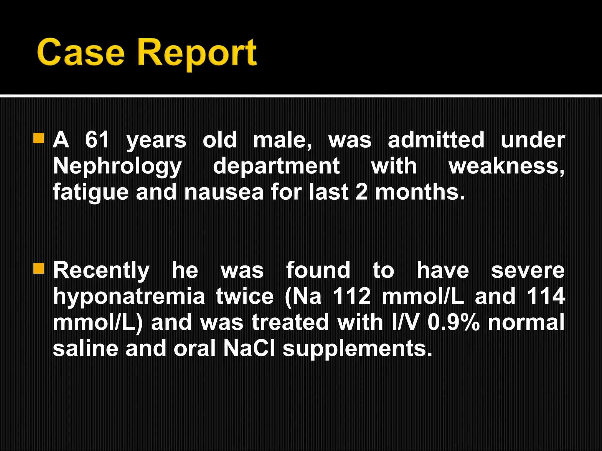
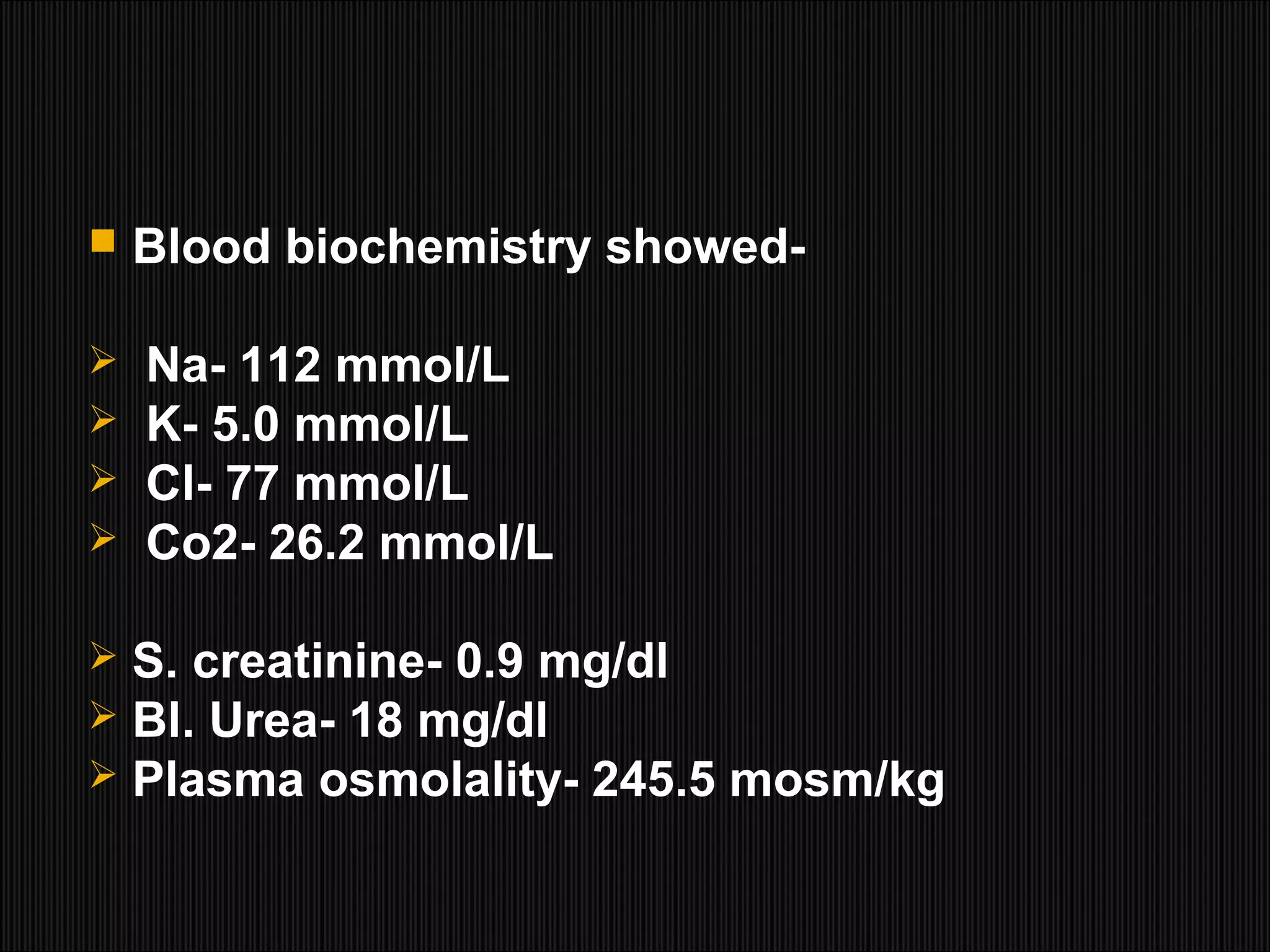
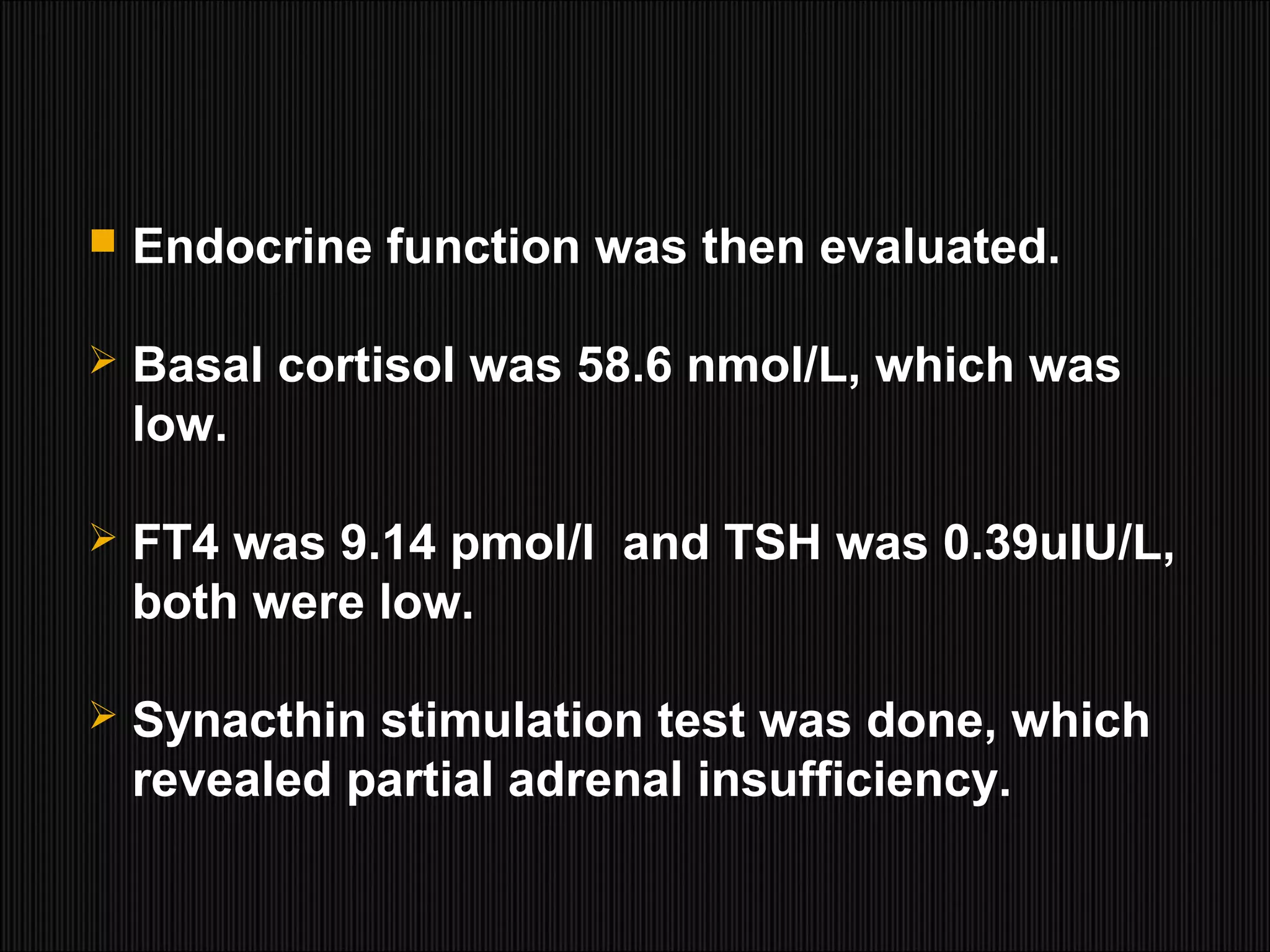
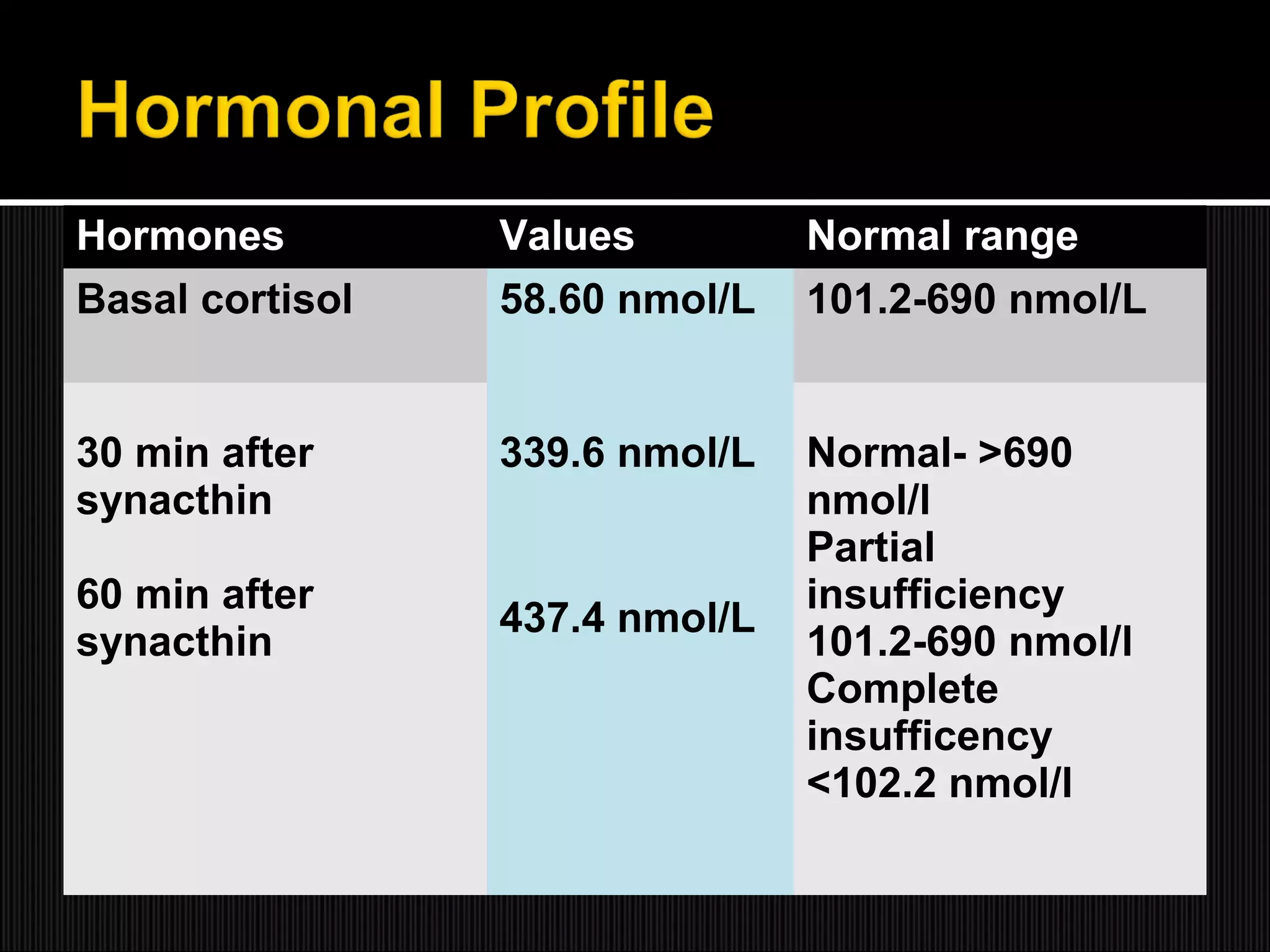
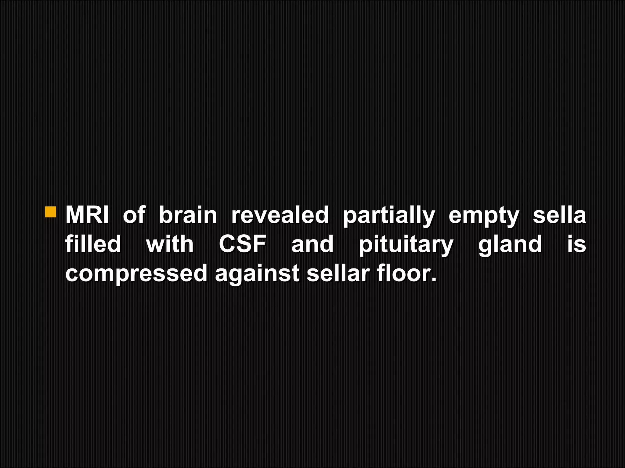
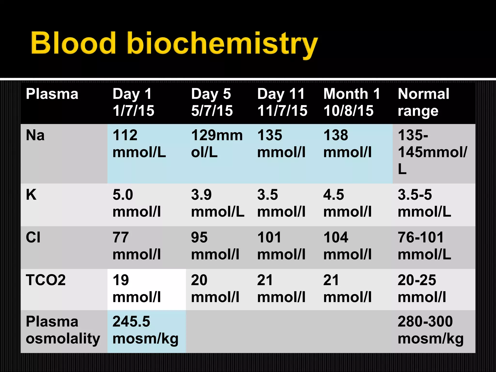
Presented by-
Dr. Jheelam Biswas
Resident Medical Officer
Nephrology Unit I
 Hyponatremia is the most commonHyponatremia is the most common
electrolyte abnormality in hospitalizedelectrolyte abnormality in hospitalized
patients on admission.patients on admission.
 Mild cases are often asymptomatic, butMild cases are often asymptomatic, but
severe hyponatremia may cause dramaticsevere hyponatremia may cause dramatic
symptoms and a high mortality¹ dependingsymptoms and a high mortality¹ depending
on the speed of development and theon the speed of development and the
underlying disease.underlying disease.
1.Andersen RJ, Chung HM, Kluge R & Schrier RW. Hyponatremia: a prospective analysis of its
epidemology and the pathogenic role of vasopressin. Anral’s of Internal Medicine 1985;102:164-
168
 The empty sella is characterized by an
intrasellar herniation of the suprasellar
subarachnoid space within the sella turcica
is often associated with some degree of
flattening of the pituitary gland and can
cause secondary adrenal insufficiency.²
2.Kaufman B. The “empty” sella turcica--a manifestation of the intrasellar subarachnoid space.
Radiology. 1968;90:931-941.
 Severe hyponatremia can, however, also
occur in patients with secondary adrenal
insufficiency in the absence of hypovolumia
and dehydration, and it can be rapidly
corrected by hydrocortisone substitution.³
3. Bethuse JE & Nelson DH. Hyponatremia in hypopitutarism. New England Journal of Medicine.
1965;272: 771-776
 In this report, we described a case with
unrecognized empty sella presented with
recurrent hyponatremia successfully treated
with hydrocortisone supplementation.
 A 61 years old male, was admitted under
Nephrology department with weakness,
fatigue and nausea for last 2 months.
 Recently he was found to have severe
hyponatremia twice (Na 112 mmol/L and 114
mmol/L) and was treated with I/V 0.9% normal
saline and oral NaCl supplements.
 There was no history of taking diuretics or
antipsychotic drugs, nor was there any
history of pituitary surgery.
 He appeared lethargic, his higher psychicHe appeared lethargic, his higher psychic
function was normal.function was normal.
 On admission, his pulse rate was 64 bpm,On admission, his pulse rate was 64 bpm,
BP 120/80 mmHg with no postural drop,BP 120/80 mmHg with no postural drop,
body temp 98° F.body temp 98° F.
 His skin turgor and JVP were normal,His skin turgor and JVP were normal,
indicating normal extracellular fluid volume.indicating normal extracellular fluid volume.
 He didn’t have any Cushing’s appearance,
loss of axillary or pubic hair,
hyperpigmentation and generalized
myxedema.
 Neurological examination exhibited neither
diplopia nor any visual field defects.
Fundoscopic examination revealed normal
findings.
 Blood biochemistry showed-
 Na- 112 mmol/L
 K- 5.0 mmol/L
 Cl- 77 mmol/L
 Co2- 26.2 mmol/L
 S. creatinine- 0.9 mg/dl
 Bl. Urea- 18 mg/dl
 Plasma osmolality- 245.5 mosm/kg
 Urinary electrolytes revealed-
 Na- 51 mmol/L (20-110 mmol/l)
 Cl- 72 mmol/L (55- 153 mmol/l)
 K- 19 mmol/L (12- 62 mmol/l)
 Urine osmolality- 373.14 mosm/kg
(50-1100 mosm/kg)
 Chest radiography, and abdominal
ultrasonography were all nonremarkable.
 Intravenous one liter of 0.9% normal salineIntravenous one liter of 0.9% normal saline
was administered per day along with high Nawas administered per day along with high Na
diet to correct hyponatremia initially, butdiet to correct hyponatremia initially, but
plasma sodium level only raised to 118plasma sodium level only raised to 118
mmol/L .mmol/L .
 Endocrine function was then evaluated.
 Basal cortisol was 58.6 nmol/L, which was
low.
 FT4 was 9.14 pmol/l and TSH was 0.39uIU/L,
both were low.
 Synacthin stimulation test was done, which
revealed partial adrenal insufficiency.
 ACTH, LH and S. testosterone levels were
also low.
 HGH were in lower level of normal.
Hormones Values Normal range
Basal cortisol 58.60 nmol/L 101.2-690 nmol/L
30 min after
synacthin
60 min after
synacthin
339.6 nmol/L
437.4 nmol/L
Normal- >690
nmol/l
Partial
insufficiency
101.2-690 nmol/l
Complete
insufficency
<102.2 nmol/l
Hormones Values Normal range
Basal GH 0.18 ng/ml 0- 4 ng/ml
ACTH 8.32 pg/ml 8.3- 57.8 pg/ml
LH 0.49 mIU/ml 0.57-12.07 mIU/L
Testesterone 0.36 ng/ml 2.8-14 ng/ml
TSH 0.39 uIU/ml 0.4- 5.01 uIU/ml
FT4 9.14 pmol/L 9.1- 23.18 pmol/L
 MRI of brain revealed partially empty sellaMRI of brain revealed partially empty sella
filled with CSF and pituitary gland isfilled with CSF and pituitary gland is
compressed against sellar floor.compressed against sellar floor.
 Inj. Hydrocortisone 100mg I/V was started 8
hrly.
 One day after starting I/V steroid S. Na level
reached 129 mmol/L. After 3 days S. Na level
became normal.
 T. Levothyroxine 50 ugm was also given.
After 9 days oral hydrocortisone was started
instead of I/V hydrocortisone.
 Oral calcium supplementation was also
given.
 With the treatment of oral hydrocortisone 30
mg daily his S. Na level was maintained
within the normal limit and his symptoms
subsided.
 After one month his S. Na level remained
completely normal without requiring sodium
supplementation.
Plasma Day 1
1/7/15
Day 5
5/7/15
Day 11
11/7/15
Month 1
10/8/15
Normal
range
Na 112
mmol/L
129mm
ol/L
135
mmol/l
138
mmol/l
135-
145mmol/
L
K 5.0
mmol/l
3.9
mmol/L
3.5
mmol/l
4.5
mmol/l
3.5-5
mmol/L
Cl 77
mmol/l
95
mmol/l
101
mmol/l
104
mmol/l
76-101
mmol/L
TCO2 19
mmol/l
20
mmol/l
21
mmol/l
21
mmol/l
20-25
mmol/l
Plasma
osmolality
245.5
mosm/kg
280-300
mosm/kg
Plasma Day 1
1/7/15
Normal range
FBS 5.0 mmol/l 3.5- 5.5 mmol/l
Hb 13.3 g/dl 12-16 g/dl
WBC count 7,400 c/cmm 7000-11,000 c/cmm
S creatinine 0.9 mg/dl (0.5-1 mg/dl)
S. Urea 18 mg/dl 10-50 mg/dl
S. Albumin 37.8 mg/dl (43-50 mg/dL)
AST 33 IU/L Upto 37
ALT 23 IU/L Upto 40
S. billirubin 0.5 mg/dl o.4-1.2 mg/dl
Urine electrolytes Day 1
1/7/15
Normal range
Na 51 mmol/l 20-110 mmol/l
K 19 mmol/l 55- 153 mmol/l
Cl 72 mmol/l 12- 62 mmol/l
Urine
osmolality
373.14 mosm/kg 50-1100
mosm/kg
 In this patient with recurrent euvolemic
hyponatremia low level of cortisol and ACTH
and parital respond to synacthin confirm the
presence of secondary adrenal insufficiency.
 The pituitary MRI helped us to search for
underline structure abnormalities of pituitary
inducing secondary adrenal insufficiency.
 The most reasonable explanation for
development of primary empty sella in a
patient who has either a transient elevation
or constant elevation of intracranial
pressure, and who has incomplete
diaphragm sella that allows the
subarachnoid space to be forced into the
sella by hydrostatic pressure and pulsatile
movement of CSF. ¹
1. Bragagnia G, Bianconcini G, Mazzali F. 43 Essay of Primary Empty Sella Syndrome, A Case
Series. Ann Hal Med Int. 1995;10: 138-142
 Secondary empty sella may be caused by
pituitary adenomas with spontaneous
necrosis, infection, autoimmune, trauma,
radiotherapy, drugs, and surgery.²
2. De Marinis L, Bonadonna S, Bianchi A, Maira G, Giustina A. Primary empty sella. J Clin
Endocrinol Metab. 2005;90:5471-5477.
 There are are wide variation in the reported
prevalence of endocrine abnormalities of
primary empty sella.
 Ghatnatti et al¹ noted endocrine dysfuction
on 50% of primary empty sella patients while
De Mariris et al² found endocrine
abnormalities in 19%.
1. De Marinis L, Bonadonna S, Bianchi A, Maira G, Giustina A. Primary empty sella. J Clin
Endocrinol Metab. 2005;90:5471-5477.
2. Ghatnattiv, Sarma D, and Saikia U. Empty Sella Syndrome- Beyond Being an Incidental Finding.
Indian Journal of Endocrinology & Metabolism. 2012;16: 5321-5323
 It has been hypothesized that compression
of pituitary gland due to progressive
hypotrophy or an increase in intra-sellar
pressure requires is heterogenous
endocrine abnormalities ranging from global
or partial hypopituitarism to various isolated
adrenocorticotropics deficiency.
 Partial hypopituitarism has been described in
5% of patients. ¹´²
 Hyponatremia can be a severe complication
of hypopituitarism in empty sella syndrome.³
1.Cannavo S, Curto L, Venturino M, Squadrito S, Almoto B, Narbone MC, Rao R, Trimarchi F.
Abnormalities of hypothalamic-pituitary-thyroid axis in patients with primary empty sella. J
Endocrinol Invest. 2002;25:236-239.
2.Nakagawa H, Nagasaka A, Koie K, Yuasa T, Sakabe Y, Kato O, Suzuki T, Hattori K, Katada K.
Isolated adrenocorticotropin defi ciency associated with an empty sella. J Clin Endocrinol Metab.
1982;55:795-797.
3. Petridis AK, Nabavi A, Doukas A, Buhl R, Mehdorn HM. Severe hyponatraemia in the setting of
hypopituitarism associated with empty sella and herniation of the optic chiasm and gyrus rectus. J
Clin Neurosci. 2009;16:723-724.
 ADH is known to play a role in the
pathogenic mechanism.¹
 However the cause of ADH secretion in
hyponatremia associated with
hypopituitarism is related to adrenocortical
insufficiency. ²
1.Oelkers W. Hyponatremia and inappropriate secretion of vasopressin (antidiuretic hormone) in
patients with hypopituitarism. N Engl J Med 1989;321:492-6.
2. Wakui H, Nishinari T, Nishimura S, Endo Y, Nakamoto Y, Miura AB. Inappropriate secretion of
antidiuretic hormone in isolated adrenocorticotropin deficiency. Am J Med Sci 1991;301:319-21.
 The glucocorticoid deficiency is not an
osmotic but a physiological stimulus for
ADH secretion. ¹´²
 Glucocorticoids have been shown to
reverse the impaired water diuresis of this
disorder by increasing the renal excretion of
solutefree water.
1. Oelkers W. Hyponatremia and inappropriate secretion of vasopressin (antidiuretic hormone) in
patients with hypopituitarism. N Engl J Med 1989;321:492-6.
2. . Gross PA, Ketteler M, Hausmann C, Ritz E. The charted and uncharted waters of
hyponatremia. Kidney Int Suppl 1987;21:S67-75.
 Ahmed et al. suggested that glucocorticoids
promote normal water diuresis by inhibiting
the secretion of ADH from the
neurohypophysis.¹
 Once secondary adrenal insufficiency is
diagnosed hyponatremia could be rapidly
corrected by hydrocortisone replacement (15
to 25 mg of hydrocortisone).²
1. Ahmed ABJ, George BC, Gonzalez-Auvert C, Dingman JF. Increased plasma arginine vasopressin
in clinical adrenocortical insufficiency and its inhibition by glucocorticoids. J Clin Invest
1967;46:111-23.
2. Bornstein SR. Predisposing factors for adrenal insufficiency. N Engl J Med. 2009;360:2328-2339.
 Hyponatremia is the most common
electrolyte disorder.
 Complete differential diagnosis including
endocrinology, laboratory and imaging
techniques are necessary to obtain a correct
diagnosis and treatment.
 Department of Endocrinology
 Department of Radiology
Case report: Empty sella syndrome

Case report: Empty sella syndrome

  • 1.
    Presented by- Dr. JheelamBiswas Resident Medical Officer Nephrology Unit I
  • 2.
     Hyponatremia isthe most commonHyponatremia is the most common electrolyte abnormality in hospitalizedelectrolyte abnormality in hospitalized patients on admission.patients on admission.  Mild cases are often asymptomatic, butMild cases are often asymptomatic, but severe hyponatremia may cause dramaticsevere hyponatremia may cause dramatic symptoms and a high mortality¹ dependingsymptoms and a high mortality¹ depending on the speed of development and theon the speed of development and the underlying disease.underlying disease. 1.Andersen RJ, Chung HM, Kluge R & Schrier RW. Hyponatremia: a prospective analysis of its epidemology and the pathogenic role of vasopressin. Anral’s of Internal Medicine 1985;102:164- 168
  • 3.
     The emptysella is characterized by an intrasellar herniation of the suprasellar subarachnoid space within the sella turcica is often associated with some degree of flattening of the pituitary gland and can cause secondary adrenal insufficiency.² 2.Kaufman B. The “empty” sella turcica--a manifestation of the intrasellar subarachnoid space. Radiology. 1968;90:931-941.
  • 4.
     Severe hyponatremiacan, however, also occur in patients with secondary adrenal insufficiency in the absence of hypovolumia and dehydration, and it can be rapidly corrected by hydrocortisone substitution.³ 3. Bethuse JE & Nelson DH. Hyponatremia in hypopitutarism. New England Journal of Medicine. 1965;272: 771-776
  • 5.
     In thisreport, we described a case with unrecognized empty sella presented with recurrent hyponatremia successfully treated with hydrocortisone supplementation.
  • 6.
     A 61years old male, was admitted under Nephrology department with weakness, fatigue and nausea for last 2 months.  Recently he was found to have severe hyponatremia twice (Na 112 mmol/L and 114 mmol/L) and was treated with I/V 0.9% normal saline and oral NaCl supplements.
  • 7.
     There wasno history of taking diuretics or antipsychotic drugs, nor was there any history of pituitary surgery.
  • 8.
     He appearedlethargic, his higher psychicHe appeared lethargic, his higher psychic function was normal.function was normal.  On admission, his pulse rate was 64 bpm,On admission, his pulse rate was 64 bpm, BP 120/80 mmHg with no postural drop,BP 120/80 mmHg with no postural drop, body temp 98° F.body temp 98° F.  His skin turgor and JVP were normal,His skin turgor and JVP were normal, indicating normal extracellular fluid volume.indicating normal extracellular fluid volume.
  • 9.
     He didn’thave any Cushing’s appearance, loss of axillary or pubic hair, hyperpigmentation and generalized myxedema.  Neurological examination exhibited neither diplopia nor any visual field defects. Fundoscopic examination revealed normal findings.
  • 10.
     Blood biochemistryshowed-  Na- 112 mmol/L  K- 5.0 mmol/L  Cl- 77 mmol/L  Co2- 26.2 mmol/L  S. creatinine- 0.9 mg/dl  Bl. Urea- 18 mg/dl  Plasma osmolality- 245.5 mosm/kg
  • 11.
     Urinary electrolytesrevealed-  Na- 51 mmol/L (20-110 mmol/l)  Cl- 72 mmol/L (55- 153 mmol/l)  K- 19 mmol/L (12- 62 mmol/l)  Urine osmolality- 373.14 mosm/kg (50-1100 mosm/kg)
  • 12.
     Chest radiography,and abdominal ultrasonography were all nonremarkable.  Intravenous one liter of 0.9% normal salineIntravenous one liter of 0.9% normal saline was administered per day along with high Nawas administered per day along with high Na diet to correct hyponatremia initially, butdiet to correct hyponatremia initially, but plasma sodium level only raised to 118plasma sodium level only raised to 118 mmol/L .mmol/L .
  • 13.
     Endocrine functionwas then evaluated.  Basal cortisol was 58.6 nmol/L, which was low.  FT4 was 9.14 pmol/l and TSH was 0.39uIU/L, both were low.  Synacthin stimulation test was done, which revealed partial adrenal insufficiency.
  • 14.
     ACTH, LHand S. testosterone levels were also low.  HGH were in lower level of normal.
  • 15.
    Hormones Values Normalrange Basal cortisol 58.60 nmol/L 101.2-690 nmol/L 30 min after synacthin 60 min after synacthin 339.6 nmol/L 437.4 nmol/L Normal- >690 nmol/l Partial insufficiency 101.2-690 nmol/l Complete insufficency <102.2 nmol/l
  • 16.
    Hormones Values Normalrange Basal GH 0.18 ng/ml 0- 4 ng/ml ACTH 8.32 pg/ml 8.3- 57.8 pg/ml LH 0.49 mIU/ml 0.57-12.07 mIU/L Testesterone 0.36 ng/ml 2.8-14 ng/ml TSH 0.39 uIU/ml 0.4- 5.01 uIU/ml FT4 9.14 pmol/L 9.1- 23.18 pmol/L
  • 18.
     MRI ofbrain revealed partially empty sellaMRI of brain revealed partially empty sella filled with CSF and pituitary gland isfilled with CSF and pituitary gland is compressed against sellar floor.compressed against sellar floor.
  • 19.
     Inj. Hydrocortisone100mg I/V was started 8 hrly.  One day after starting I/V steroid S. Na level reached 129 mmol/L. After 3 days S. Na level became normal.
  • 20.
     T. Levothyroxine50 ugm was also given. After 9 days oral hydrocortisone was started instead of I/V hydrocortisone.  Oral calcium supplementation was also given.
  • 21.
     With thetreatment of oral hydrocortisone 30 mg daily his S. Na level was maintained within the normal limit and his symptoms subsided.  After one month his S. Na level remained completely normal without requiring sodium supplementation.
  • 22.
    Plasma Day 1 1/7/15 Day5 5/7/15 Day 11 11/7/15 Month 1 10/8/15 Normal range Na 112 mmol/L 129mm ol/L 135 mmol/l 138 mmol/l 135- 145mmol/ L K 5.0 mmol/l 3.9 mmol/L 3.5 mmol/l 4.5 mmol/l 3.5-5 mmol/L Cl 77 mmol/l 95 mmol/l 101 mmol/l 104 mmol/l 76-101 mmol/L TCO2 19 mmol/l 20 mmol/l 21 mmol/l 21 mmol/l 20-25 mmol/l Plasma osmolality 245.5 mosm/kg 280-300 mosm/kg
  • 23.
    Plasma Day 1 1/7/15 Normalrange FBS 5.0 mmol/l 3.5- 5.5 mmol/l Hb 13.3 g/dl 12-16 g/dl WBC count 7,400 c/cmm 7000-11,000 c/cmm S creatinine 0.9 mg/dl (0.5-1 mg/dl) S. Urea 18 mg/dl 10-50 mg/dl S. Albumin 37.8 mg/dl (43-50 mg/dL) AST 33 IU/L Upto 37 ALT 23 IU/L Upto 40 S. billirubin 0.5 mg/dl o.4-1.2 mg/dl
  • 24.
    Urine electrolytes Day1 1/7/15 Normal range Na 51 mmol/l 20-110 mmol/l K 19 mmol/l 55- 153 mmol/l Cl 72 mmol/l 12- 62 mmol/l Urine osmolality 373.14 mosm/kg 50-1100 mosm/kg
  • 25.
     In thispatient with recurrent euvolemic hyponatremia low level of cortisol and ACTH and parital respond to synacthin confirm the presence of secondary adrenal insufficiency.  The pituitary MRI helped us to search for underline structure abnormalities of pituitary inducing secondary adrenal insufficiency.
  • 26.
     The mostreasonable explanation for development of primary empty sella in a patient who has either a transient elevation or constant elevation of intracranial pressure, and who has incomplete diaphragm sella that allows the subarachnoid space to be forced into the sella by hydrostatic pressure and pulsatile movement of CSF. ¹ 1. Bragagnia G, Bianconcini G, Mazzali F. 43 Essay of Primary Empty Sella Syndrome, A Case Series. Ann Hal Med Int. 1995;10: 138-142
  • 27.
     Secondary emptysella may be caused by pituitary adenomas with spontaneous necrosis, infection, autoimmune, trauma, radiotherapy, drugs, and surgery.² 2. De Marinis L, Bonadonna S, Bianchi A, Maira G, Giustina A. Primary empty sella. J Clin Endocrinol Metab. 2005;90:5471-5477.
  • 28.
     There areare wide variation in the reported prevalence of endocrine abnormalities of primary empty sella.  Ghatnatti et al¹ noted endocrine dysfuction on 50% of primary empty sella patients while De Mariris et al² found endocrine abnormalities in 19%. 1. De Marinis L, Bonadonna S, Bianchi A, Maira G, Giustina A. Primary empty sella. J Clin Endocrinol Metab. 2005;90:5471-5477. 2. Ghatnattiv, Sarma D, and Saikia U. Empty Sella Syndrome- Beyond Being an Incidental Finding. Indian Journal of Endocrinology & Metabolism. 2012;16: 5321-5323
  • 29.
     It hasbeen hypothesized that compression of pituitary gland due to progressive hypotrophy or an increase in intra-sellar pressure requires is heterogenous endocrine abnormalities ranging from global or partial hypopituitarism to various isolated adrenocorticotropics deficiency.
  • 30.
     Partial hypopituitarismhas been described in 5% of patients. ¹´²  Hyponatremia can be a severe complication of hypopituitarism in empty sella syndrome.³ 1.Cannavo S, Curto L, Venturino M, Squadrito S, Almoto B, Narbone MC, Rao R, Trimarchi F. Abnormalities of hypothalamic-pituitary-thyroid axis in patients with primary empty sella. J Endocrinol Invest. 2002;25:236-239. 2.Nakagawa H, Nagasaka A, Koie K, Yuasa T, Sakabe Y, Kato O, Suzuki T, Hattori K, Katada K. Isolated adrenocorticotropin defi ciency associated with an empty sella. J Clin Endocrinol Metab. 1982;55:795-797. 3. Petridis AK, Nabavi A, Doukas A, Buhl R, Mehdorn HM. Severe hyponatraemia in the setting of hypopituitarism associated with empty sella and herniation of the optic chiasm and gyrus rectus. J Clin Neurosci. 2009;16:723-724.
  • 31.
     ADH isknown to play a role in the pathogenic mechanism.¹  However the cause of ADH secretion in hyponatremia associated with hypopituitarism is related to adrenocortical insufficiency. ² 1.Oelkers W. Hyponatremia and inappropriate secretion of vasopressin (antidiuretic hormone) in patients with hypopituitarism. N Engl J Med 1989;321:492-6. 2. Wakui H, Nishinari T, Nishimura S, Endo Y, Nakamoto Y, Miura AB. Inappropriate secretion of antidiuretic hormone in isolated adrenocorticotropin deficiency. Am J Med Sci 1991;301:319-21.
  • 32.
     The glucocorticoiddeficiency is not an osmotic but a physiological stimulus for ADH secretion. ¹´²  Glucocorticoids have been shown to reverse the impaired water diuresis of this disorder by increasing the renal excretion of solutefree water. 1. Oelkers W. Hyponatremia and inappropriate secretion of vasopressin (antidiuretic hormone) in patients with hypopituitarism. N Engl J Med 1989;321:492-6. 2. . Gross PA, Ketteler M, Hausmann C, Ritz E. The charted and uncharted waters of hyponatremia. Kidney Int Suppl 1987;21:S67-75.
  • 33.
     Ahmed etal. suggested that glucocorticoids promote normal water diuresis by inhibiting the secretion of ADH from the neurohypophysis.¹  Once secondary adrenal insufficiency is diagnosed hyponatremia could be rapidly corrected by hydrocortisone replacement (15 to 25 mg of hydrocortisone).² 1. Ahmed ABJ, George BC, Gonzalez-Auvert C, Dingman JF. Increased plasma arginine vasopressin in clinical adrenocortical insufficiency and its inhibition by glucocorticoids. J Clin Invest 1967;46:111-23. 2. Bornstein SR. Predisposing factors for adrenal insufficiency. N Engl J Med. 2009;360:2328-2339.
  • 34.
     Hyponatremia isthe most common electrolyte disorder.  Complete differential diagnosis including endocrinology, laboratory and imaging techniques are necessary to obtain a correct diagnosis and treatment.
  • 35.
     Department ofEndocrinology  Department of Radiology