The Endocrine System
•Pituitary gland
• Thyroid gland
• Parathyroid glands
• Endocrine Pancreas
• Adrenal glands
• Pineal gland
this lecture's
main focus
3.
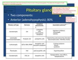
Pituitary gland
• Twocomponents:
• Anterior (adenohypophysis); 80%
Pituitary cell type Hormone
Cytoplasm
characteristic
s
Associated syndrome
Somatotrophs GH
acidophilic
cells
(eosinophilic
cytoplasm)
Gigantism (children)
Acromegaly
(adults)
Lactotrophs
(mammosomatotrophs) Prolactin, GH acidophilic cells
Galactorrhea and amenorrhea (females)
Sexual dysfunction, infertility (males)
Corticotrophs ACTH, MSH, POMC
basophilic cells
(basophil cytoplasm)
Cushing syndrome
Nelson
syndrome
Thyrotrophs TSH pale basophilic cells Hyperthyroidism
Gonadotrophs FSH, LH basophilic cells
Hypogonadism, mass effects,
hypopituitarism
The anterior pituitary is the main producer of hormones in the pituitary gland. The table lists the types of cells
located in it and their properties.
From what germ layer does the
adenohypophysis arise? What
embryological structure formed from this
germ layer is the precursor to the
adenohypophysis?
Associated
syndrome due to
hyperproduction
of the hormone
tcl12
---------------------------------
-----------
ectoderm; Rathke's
pouch
4.
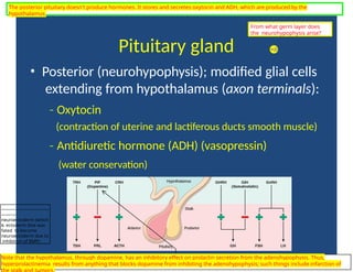
Pituitary gland
• Posterior(neurohypophysis); modified glial cells
extending from hypothalamus (axon terminals):
‐ Oxytocin
(contraction of uterine and lactiferous ducts smooth muscle)
‐ Antidiuretic hormone (ADH) (vasopressin)
(water conservation)
The posterior pituitary doesn't produce hormones. It stores and secretes oxytocin and ADH, which are produced by the
hypothalamus.
Note that the hypothalamus, through dopamine, has an inhibitory effect on prolactin secretion from the adenohypophysis. Thus,
hyperprolactinemia results from anything that blocks dopamine from inhibiting the adenohypophysis; such things include infarction of
the stalk and tumors.
From what germ layer does
the neurohypophysis arise?
---------------------------------
-----------
neuroectoderm (which
is ectoderm that was
fated to become
neuroectoderm due to
inhibition of BMP)
5.
Clinical manifestations ofpituitary
gland disease
• Hyperpituitarism (excess secretion of trophic
hormones)
‐ Adenomas, hyperplasia, carcinoma of
anterior pituitary, etc
• Hypopituitarism (deficiency of trophic
hormones)
‐ Ischemic injury, surgery, radiation,
inflammation
• Local mass effects
‐ sella turcica abnormalities
most common cause of
hyperpituitarism is adenoma
Pituitary gland diseases of various etiologies can either present as too much trophic hormone (hyper-) or too little (hypo-).
What hormones do the
neurohypophysis secrete?
Where are these hormones
produced?
tcl12
--------------------------------------------
oxytocin and ADH; the
hypothalamus
6.
Case 1
• 35year old female presents with amenorrhea,
galactorrhea, visual complaints and headache. CT
scan discloses a 2 cm mass in the anterior
pituitary.
• What’s the most likely cause?
remo
Pve
it
thi
us te
ix
tt-
abox
rt
yo re
ave
dal t
ehe
nan
oswe
mr. Th
ae
s
(tic
Pky
rn
oote
le
axp
cou
tn
id
ns u
opon
mthe
aan
)swer.
• What could explain the “visual disturbances”?
remoMve thais stexst-beox ftofreevecatl thoe afnswaerd.
Tehensticokymnoteaexpcouondms upponrthee asnsswienr. g
decussating fibers of optic chiasm
(bitemporal hemianopsia)
tcl12
--------------------------------------------
Remember from the previous slide that a
d
e
n
o
m
a is the most likely cause of
hyperpituitarism. Prolactinomas are the most common adenomas of
the anterior pituitary.
tcl12
--------------------------------------------
In bitemporal hemianopsia, you lose the outer half of
your left and right visual fields. In this situation, this is
due to compression of the fibers that cross (i.e.
decussate) in the optic chiasm. Remember that these
fibers project from the nasal retina, which receives
information from the peripheral halves of your visual
field.
7.
visual information comingfrom the part of
the visual field where this text box is
located would not be relayed if a pituitary
adenoma impinged upon the decussating
fibers of the optic chiasm
Notice the
proximity of the
pituitary to the
optic chiasm. It
should be no
surprise that a
pituitary
adenoma could
push on the
optic chiasm and
cause
bitemporal
hemianopsia.
The anatomy of the brain shows how mass effect
from a pituitary adenoma can cause visual field
deficits.
the pituitary
would be right
about there
8.
Pituitary Adenomas
• Mostcommon cause of hyperpituitarism
• Functional or non functional
‐
• Affects adults (35 60)
‐
• Microadenomas: < 1 cm
• Macroadenomas: > 1 cm
• Gross appearance: soft, well circumscribed,
‐ and
confined to sella turcica
• Microscopically: monotonous population of
polygonal cells lacking significant reticulin
framework.
The non-functional adenomas
(i.e. no hypersecretory
symptoms like galactorrhea)
tend to get caught when big
and cause compressive
symptoms, like the bitemporal
hemianopsia previously
described. The functional
adenomas tend to get caught
when they are small since
their hypersecretory
symptoms are pronounced
even when the tumor is not
very large.
9.
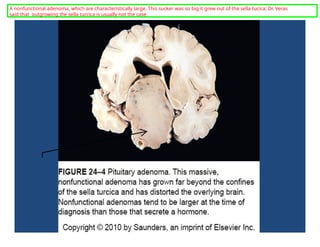
A nonfunctional adenoma,which are characteristically large. This sucker was so big it grew out of the sella tucica; Dr. Veras
said that outgrowing the sella turcica is usually not the case.
10.
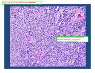
This is themicroscopic appearance of a pituitary
adenoma.
Note that the sheets of cells all look the
same (in her words: "monotonous
population of polygonal cells).
11.
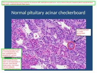
Normal pituitary acinarcheckerboard
This is normal pituitary tissue noted for its diverse cells, well-demarcated acini, and a robust reticulin network (which would be best
seen with a special reticulin fiber stain).
Note the diversity of cells
in normal pituitary. You
have acidophils and
basophils of various
staining intensity.
note the example
of a well-
demarcated
acinus
Look at how the acini
seem to be separated. A
reticulin stain would
show a very nice intact
reticulin network
surrounding each acinus.
12.
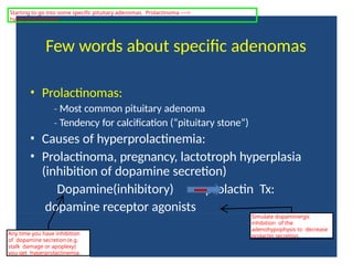
Few words aboutspecific adenomas
• Prolactinomas:
‐ Most common pituitary adenoma
‐ Tendency for calcification (“pituitary stone”)
• Causes of hyperprolactinemia:
• Prolactinoma, pregnancy, lactotroph hyperplasia
(inhibition of dopamine secretion)
Dopamine(inhibitory) prolactin Tx:
dopamine receptor agonists
Any time you have inhibition
of dopamine secretion (e.g.
stalk damage or apoplexy)
you get hyperprolactinemia.
Simulate dopaminergic
inhibition of the
adenohypophysis to decrease
prolactin secretion.
Starting to go into some specific pituitary adenomas. Prolactinoma ---->
hyperprolactinemia
13.
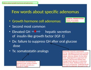
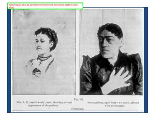
Few words aboutspecific adenomas
• Growth hormone cell adenomas:
• Second most common
• Elevated GH hepatic secretion
of insulin like
‐ growth factor (IGF 1)
‐
• Dx: failure to suppress GH after oral glucose
dose
• Tx: somatostatin analogs
CLINICAL PRESENTATION:
Adults: acromegaly
Children: gigantism
Remember that GH is stimulated
during hypoglycemia because it causes
hepatic gluconeogenesis and reduced
hepatic glucose uptake. On the other
hand, hyperglycemia suppresses GH
secretion because we already have
enough blood glucose. In a pituitary
adenoma, these feedback loops are
lost so an oral glucose challenge would
not yield the expected decrease in GH.
Somatostatin secreted by the
periventricular nucleus inhibits
GH secretion and acts as a
counter to GHRH
Talking about growth hormone cell adenomas
Few words aboutspecific adenomas
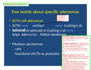
• ACTH cell adenomas:
• ACTH cortisol
(adrenal)
Cushing’s dz
• Removal of adrenals in Cushing’s sd
large adenomas Nelson syndrome
• Pituitary carcinomas:
‐ rare
‐ functional (ACTH or prolactin)
Cushing's disease is pituitary in origin,
and is defined as excess ACTH secretion
from the pituitary.
Cushing's syndrome is simply a result
of excess glucocorticoid whether it is
pituitary or adrenal in origin.
Thus, Cushing's disease is one cause
of Cushing's syndrome but not vice
versa.
Nelson syndrome: Removal of the
adrenal glands leads to no more cortisol
to inhibit ACTH production by negative
feedback.
Thus, ACTH cell adenomas grow
unchecked. This presents as muscle
weakness due to excess ACTH and skin
hyperpigmentation due to excess
melanocyte stimulating hormone,
released from
the adrenal
cortex, hence,
CORTisol
Talking about ACTH cell adenomas
16.
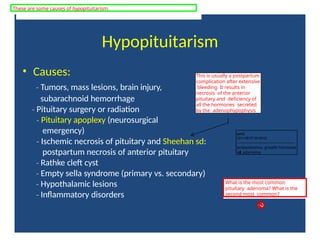
Hypopituitarism
• Causes:
‐ Tumors,mass lesions, brain injury,
subarachnoid hemorrhage
‐ Pituitary surgery or radiation
‐ Pituitary apoplexy (neurosurgical
emergency)
‐ Ischemic necrosis of pituitary and Sheehan sd:
postpartum necrosis of anterior pituitary
‐ Rathke cleft cyst
‐ Empty sella syndrome (primary vs. secondary)
‐ Hypothalamic lesions
‐ Inflammatory disorders
This is usually a postpartum
complication after extensive
bleeding. It results in
necrosis of the anterior
pituitary and deficiency of
all the hormones secreted
by the adenophypophysis
What is the most common
pituitary adenoma? What is the
second most common?
These are some causes of hypopituitarism.
tcl12
2011-06-07 03:00:02
--------------------------------------------
prolactinoma; growth hormone
cell adenoma
17.
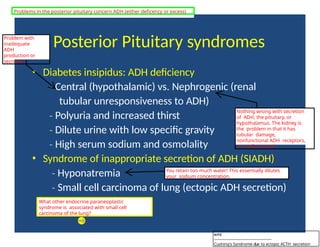
Posterior Pituitary syndromes
•Diabetes insipidus: ADH deficiency
‐ Central (hypothalamic) vs. Nephrogenic (renal
tubular unresponsiveness to ADH)
‐ Polyuria and increased thirst
‐ Dilute urine with low specific gravity
‐ High serum sodium and osmolality
• Syndrome of inappropriate secretion of ADH (SIADH)
‐ Hyponatremia
‐ Small cell carcinoma of lung (ectopic ADH secretion)
Nothing wrong with secretion
of ADH, the pituitary, or
hypothalamus. The kidney is
the problem in that it has
tubular damage,
nonfunctional ADH receptors,
or nonfunctional
You retain too much water! This essentially dilutes
your sodium concentration.
What other endocrine paraneoplastic
syndrome is associated with small cell
carcinoma of the lung?
Problems in the posterior pituitary concern ADH (either deficency or excess)
Problem with
inadequate
ADH
production or
secretion
tcl12
--------------------------------------------
Cushing's Syndrome due to ectopic ACTH secretion
18.
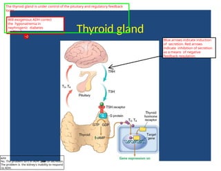
Thyroid gland
Will exogenousADH correct
the hyponatremia in
nephrogenic diabetes
mellitus?
Blue arrows indicate induction
of secretion. Red arrows
indicate inhibition of secretion
as a means of negative
feedback regulation.
The thyroid gland is under control of the pituitary and regulatory feedback
loops.
tcl12
No. The problem isn't in ADH p
r
o
d
u
c
t
i
o
n or secretion.
The problem is the kidney's inability to respond
to ADH.
19.
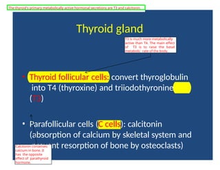
Thyroid gland
• Thyroidfollicular cells: convert thyroglobulin
into T4 (thyroxine) and triiodothyronine
(T3)
• Parafollicular cells (C cells): calcitonin
(absorption of calcium by skeletal system and
prevent resorption of bone by osteoclasts)
T3 is much more metabolically
active than T4. The main effect
of T3 is to raise the basal
metabolic rate of the body.
Calcitonin conserves
calcium in bone. It
has the opposite
effect of parathyroid
hormone.
The thyroid's primary metabolically active hormonal secretions are T3 and calcitonin.
20.
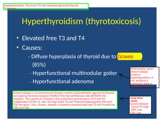
Hyperthyroidism (thyrotoxicosis)
• Elevatedfree T3 and T4
• Causes:
‐ Diffuse hyperplasia of thyroid due to Graves
(85%)
‐ Hyperfunctional multinodular goiter
‐ Hyperfunctional adenoma
Multinodular goiter
doesn't always
produce
hyperthyroidism. It
can produce a
euthyroid state or
hypothyroidism.
Grave's disease is an autoimmune disease in which autoantibodies against the thyroid
stimulating hormone receptor (TSHR) of the thyroid follicular cells ACTIVATE the
receptor. This causes an increase in the production and secretion of T3 and T4
independent of TSH. In fact, the high levels T3 and T4 would downregulate TRH and
TSH secretion. Thus, Grave's disease is noted for paradoxically high T3 and T4 with low
TRH and TSH.
Long story
short...
Grave's Disease:
T3 and T4: high
TRH: Low
TSH: Low
Hyperthyroidism: Too much T3, the metabolically active thyroid
hormone.
21.
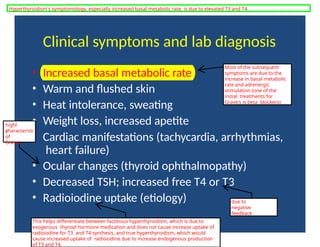
Clinical symptoms andlab diagnosis
• Increased basal metabolic rate
• Warm and flushed skin
• Heat intolerance, sweating
• Weight loss, increased apetite
• Cardiac manifestations (tachycardia, arrhythmias,
heart failure)
• Ocular changes (thyroid ophthalmopathy)
• Decreased TSH; increased free T4 or T3
• Radioiodine uptake (etiology)
Most of the subsequent
symptoms are due to the
increase in basal metabolic
rate and adrenergic
stimulation (one of the
initial treatments for
Grave's is beta blockers)
highl
y
characteristi
c
of
Graves
due to
negative
feedback
This helps differentiate between factitious hyperthyroidism, which is due to
exogenous thyroid hormone medication and does not cause increase uptake of
radioiodine for T3 and T4 synthesis, and true hyperthyroidism, which would
cause increased uptake of radioiodine due to increase endogenous production
of T3 and T4.
Hyperthyroidism's symptomology, especially increased basal metabolic rate, is due to elevated T3 and T4.
22.
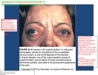
The increased basal
metabolicrate and
subsequent sympathetic
overdrive causes the
wide- eyed staring gaze.
Remember, that
sympathetic drive is fight-
or-flight, in which you'd
want your eyes to be wide
open to get a handle on
your surroundings.
Buildup of
glycosaminoglcans (GAG)
in retro-ocular muscles
and connective tissue
cause the protuberant
eyeballs.
What are the
concentrations of
T3 and T4, TRH,
and TSH in Grave's
disease relative to
physiologic levels?
Hyperthyroidism is noted for very protuberant and wide
eyes.
--------------------------------------------
T3 and T4: high TRH: low
TSH: low
23.
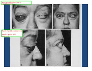
Panels A andB: pre treatment
Panels C and D: post
treatment
More ophthalmopathy due to
hyperthyroidism.
24.
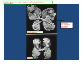
edematous
and engorged
due toGAG
deposition
Dissected eye and retro-ocular muscles with ophthalmopathy due to
hyperthyroidism.
Normal
hyperthyroidism
25.
Hypothyroidism
• Decreased levelsof thyroid hormone
• Common in the population (0.3%)
• F:M = 10:1
• Primary:
‐ Autoimmune thyroiditis (Hashimoto)
‐ Iodine deficiency
‐ Drugs (lithium, etc)
‐ Dyshormonogenetic goiter
• Secondary:
‐ TSH or TRH deficiency
This is rare nowadays,
but you see this in
people far away from
the ocean and with less
access to iodine
enriched salt. ENDEMIC
Hypothyroidism: too little thyroid
hormone
26.
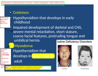
• Cretinism:
• Hypothyroidismthat develops in early
childhood
• Impaired development of skeletal and CNS,
severe mental retardation, short stature,
coarse facial features, protruding tongue and
umbilical hernia
• Myxedema:
• Hypothyroidism that
develops in older child or
adult
Reversibility
is
dependent
on
timing
of
treatment.
NOTE: Pretibial myxedema is different and occurs in
Grave's disease, which is a hyperthyroid disease.
Presentations of hypothyroidism: Cretinism = children. Myxedema = older children and
adults
a specific
form
of
cutaneous
and dermal
edema due
to
deposition
of
connective
tissue
components
like GAGs
27.
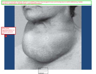
An extreme exampleof goiter, which is a swelling of the thyroid gland most commonly due to iodine deficiency. Goiter
can be a presentation of both hypo- and hyperthyroidism.
What does
hypothyroidism in
children present
as?
tcl12
-----------------------
---------------------
cretinism
28.
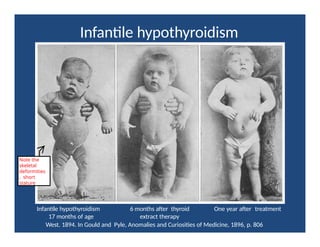
Infantile hypothyroidism
Infantile hypothyroidism6 months after thyroid One year after treatment
17 months of age extract therapy
West. 1894. In Gould and Pyle, Anomalies and Curiosities of Medicine, 1896, p. 806
Note the
skeletal
deformities
, short
stature
29.
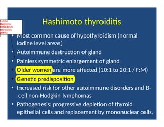
Hashimoto thyroiditis
• Mostcommon cause of hypothyroidism (normal
iodine level areas)
• Autoimmune destruction of gland
• Painless symmetric enlargement of gland
• Older women are more affected (10:1 to 20:1 / F:M)
• Genetic predisposition
• Increased risk for other autoimmune disorders and B‐
cell non Hodgkin
‐ lymphomas
• Pathogenesis: progressive depletion of thyroid
epithelial cells and replacement by mononuclear cells.
helpful
for
diagnosis
because
other kinds
of
thyroiditis
are
painfu
l
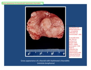
Gross appearance ofa thyroid with Hashimoto’s thyroiditis
(mimicks lymphoma)
Very fleshy and
whitish because it
is completely
infiltrated by
mononuclear cells.
It actually looks
like the cut
surface of a
lymph node,
which makes
sense given that
there is
infiltration by
mononuclear
inflammatory cells
that are forming
germinal centers.
Gross appearance of a thyroid with Hashimoto's thyroiditis.
32.
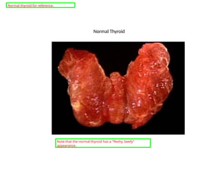
Normal Thyroid
Note thatthe normal thyroid has a "fleshy, beefy"
appearance.
Normal thyroid for reference.
33.
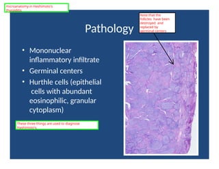
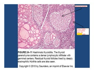
Pathology
• Mononuclear
inflammatory infiltrate
•Germinal centers
• Hurthle cells (epithelial
cells with abundant
eosinophilic, granular
cytoplasm)
microanatomy in Hashimoto's
thyroiditis
Note that the
follicles have been
destroyed and
replaced by
germinal centers
These three things are used to diagnose
Hashimoto's.
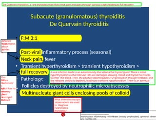
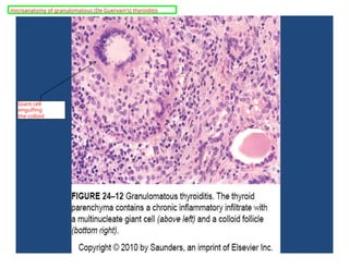
Subacute (granulomatous) thyroiditis
DeQuervain thyroiditis
• F:M 3:1
• Post viral
‐ inflammatory process (seasonal)
• Neck pain, fever
• Transient hyperthyroidism > transient hypothyroidism >
full recovery
• Pathology:
Follicles destroyed by neutrophilic microabscesses
• Multinucleate giant cells enclosing pools of colloid
different
from
Hashimoto's
,
which
is
painles
s
A viral infection leads to an autoimmunity that attacks the thyroid gland. There is initial
hyperthyroidism as the follicular cells are damaged, allowing colloid and thyroid hormones
to enter the blood. Then, the pituitary downregulates TSH production through feedback, and
the released colloid is depleted, leading to transient hypothyroidism. There is an eventual
full recovery.
What three microscopic
observations are used
to diagnose
Hashimoto's
thyroiditis?
This
differs
fro
m
Hashimoto'
s
which has no •
full
recovery
since
the
gland
is
destroyed
and
fibrosed
.
De Quervain thyroiditis: a rare thyroiditis that elicits neck pain and goes through various stages leading to full recovery
tcl12
--------------------------------------------
mononuclear inflammatory cell infiltrates (mostly lymphocytes), germinal centers
and Hurthle cells
Subacute lymphocytic (painless)thyroiditis
• Uncommon cause of hypothyroidism
• Commonly “postpartum thyroiditis”
• Mild gland enlargement and hyperthyroidism
• Pathology:
• Lymphocytic infiltration and germinal centers
• No Hurthle cells or fibrosis
tends to be
transient
It goes through the same transient hyperthyroidism by the
same mechanism as in De Quervain's thyroiditis.
Yet another kind of thyroiditis
38.
Graves Disease
• Mostcommon cause of endogenous hyperthyroidism
• Triad: hyperthyroidism; infiltrative ophthalmopathy
(exophthalmos); localized, infiltrative dermopathy
(pretibial myxedema)
• F:M > 7:1
• Genetic susceptibility (HLA B8
‐ and –DR3)
• Autoimmune disorder:
• Autoantibodies to TSH receptor:
• LATS (long acting
‐ thyroid stimulator); IgG Ab
• Thyroid growth stimulating
‐ Ig (TGI)
• TSH binding
‐ inhibitor Igs (TBII)
85-90%
this is different from the
myxedema associated with
adult hypothyroidism
very high genetic
susceptibility
Instead of destroying the gland like in
Hashimoto's thyroiditis, the autoantibodies
stimulate the TSH receptor. This causes the
gland to grow and keep secreting thyroid
hormones.
Memorize
LATS! It is the
IgG Ab that
targets
the TSH-
R to
cause the
gland to
grow
and
secrete
thyroid
hormones.
Details about Grave's Disease
39.
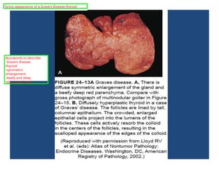
Gross appearance ofa Grave's Disease thyroid
Buzzwords to describe
Grave's disease
thyroid:
-symmetric
enlargement
-beefy and deep
red parenchyma
40.
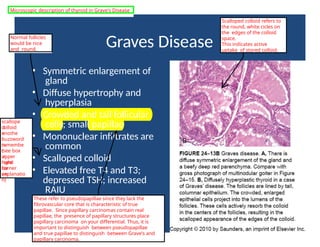
Graves Disease
• Symmetricenlargement of
gland
• Diffuse hypertrophy and
hyperplasia
• Crowded and tall follicular
cells; small papillae
• Mononuclear infiltrates are
common
• Scalloped colloid
• Elevated free T4 and T3;
depressed TSH; increased
RAIU
Normal follicles
would be nice
and round.
These refer to pseudopapillae since they lack the
fibrovascular core that is characteristic of true
papillae. Since papillary carcinomas contain real
papillae, the presence of papillary structures place
papillary carcinoma on your differential. Thus, it is
important to distinguish between pseudopapillae
and true papillae to distinguish between Grave's and
papillary carcinoma.
Scalloped colloid refers to
the round, white cicles on
the edges of the colloid
space.
This indicates active
uptake of stored colloid.
scallope
d
colloid
=
anothe
r
buzzword
to
remembe
r
(see box
in
upper
right
hand
corner
for
an
explanatio
n)
Microscopic description of thyroid in Grave's Disease
41.
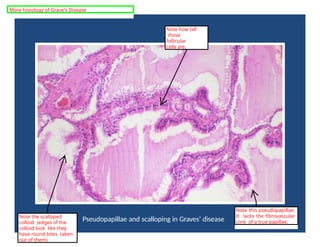
Pseudopapillae and scallopingin Graves’ disease
Note this pseudopapillae.
It lacks the fibrovascular
core of a true papillae.
Note the scalloped
colloid (edges of the
colloid look like they
have round bites taken
out of them)
Note how tall
those
follicular
cells are.
More histology of Grave's Disease
42.
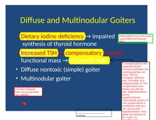
Diffuse and MultinodularGoiters
• Dietary iodine deficiency → impaired
synthesis of thyroid hormone
(goiter)
• Increased TSH → compensatory in gland
functional mass → euthyroid state
• Diffuse nontoxic (simple) goiter
• Multinodular goiter
To compensate for the
impaired thyroid
hormone synthesis, the
pituitary pumps out
more TSH via
increased synthetic
rate. This leads to a
euthyroid state. If this
compensation is in
excess, you end up
with hyperthyroidism
due to
hyperfunctional,
multinodular goiters. If
the compensation is
insufficient, then you
can end up with
hypothyroidism. Thus,
goiter can present as
eu-, hypo-, or
Iodine deficiency is the usual
cause (90% according to
Wiki).
In Grave's disease,
how do you describe
the colloid?
--------------------------------------------
scalloped
43.
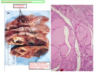
multinodularity
on cut section
roundand encapsulated
follicular adenoma. It is not
uncommon to see multiple
diseases in a thyroid.
Gross appearance of a multinodular goiter.
44.
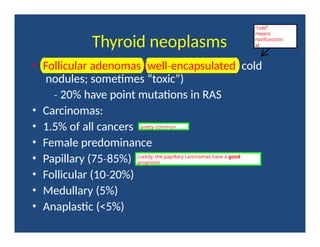
Thyroid neoplasms
• Follicularadenomas (well encapsulated
‐ ; cold
nodules; sometimes “toxic”)
‐ 20% have point mutations in RAS
• Carcinomas:
• 1.5% of all cancers
• Female predominance
• Papillary (75 85%)
‐
• Follicular (10 20%)
‐
• Medullary (5%)
• Anaplastic (<5%)
pretty common
"cold"
means
nonfunction
al
Luckily, the papillary carcinomas have a good
prognosis
45.
This emphasizes thatfollicular
adenomas are
encapsulated. It is
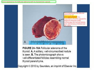
important to find the capsule for
diagnosis because under
microscopy, the normal follicles
and adenomatous follicles look
the same.
Gross appearance of a follicular adenoma with its characteristic capsule.
46.
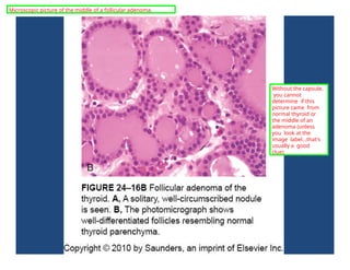
Without the capsule,
youcannot
determine if this
picture came from
normal thyroid or
the middle of an
adenoma (unless
you look at the
image label...that's
usually a good
clue).
Microscopic picture of the middle of a follicular adenoma.
47.
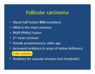
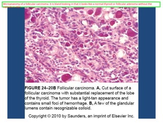
Follicular carcinoma
• Abouthalf harbor RAS mutations
• NRAS is the most common
• PAX8 PPARγ1
‐ fusion
• 2nd most common
• Female predominance; older age
• Increased incidence in areas of iodine deficiency
• Cold nodules
• Tendency for vascular invasion (not lymphatic)
48.
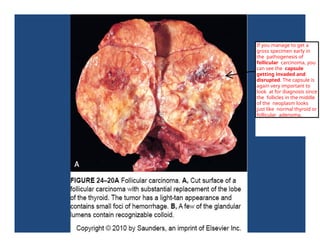
If you manageto get a
gross specimen early in
the pathogenesis of
follicular carcinoma, you
can see the capsule
getting invaded and
disrupted. The capsule is
again very important to
look at for diagnosis since
the follicles in the middle
of the neoplasm looks
just like normal thyroid or
follicular adenoma.
49.
Microanatomy of afollicular carcinoma. It is bland looking in that it looks like a normal thyroid or follicular adenoma without the
capsule in view.
50.
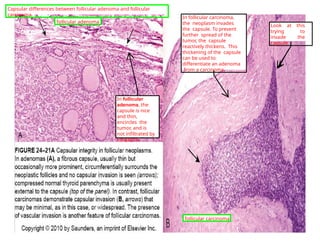
In follicular
adenoma, the
capsuleis nice
and thin,
encircles the
tumor, and is
not infiltrated by
neoplasm.
In follicular carcinoma,
the neoplasm invades
the capsule. To prevent
further spread of the
tumor, the capsule
reactively thickens. This
thickening of the capsule
can be used to
differentiate an adenoma
from a carcinoma.
Look at this
trying to
invade the
capsule
follicular adenoma
follicular carcinoma
Capsular differences between follicular adenoma and follicular
carcinoma.
51.
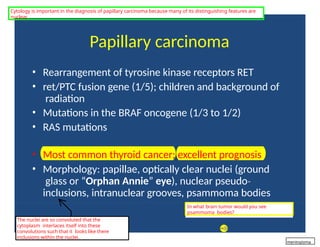
Papillary carcinoma
• Rearrangementof tyrosine kinase receptors RET
• ret/PTC fusion gene (1/5); children and background of
radiation
• Mutations in the BRAF oncogene (1/3 to 1/2)
• RAS mutations
• Most common thyroid cancer; excellent prognosis
• Morphology: papillae, optically clear nuclei (ground
glass or “Orphan Annie” eye), nuclear pseudo‐
inclusions, intranuclear grooves, psammoma bodies
The nuclei are so convoluted that the
cytoplasm interlaces itself into these
convolutions such that it looks like there
inclusions within the nuclei.
In what brain tumor would you see
psammoma bodies?
Cytology is important in the diagnosis of papillary carcinoma because many of its distinguishing features are
nuclear.
meningioma
52.
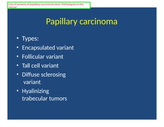
Papillary carcinoma
• Types:
•Encapsulated variant
• Follicular variant
• Tall cell variant
• Diffuse sclerosing
variant
• Hyalinizing
trabecular tumors
Lots of variants of papillary carcinomas exist. Pathologists to the
rescue!
53.
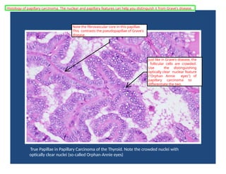
True Papillae inPapillary Carcinoma of the Thyroid. Note the crowded nuclei with
optically clear nuclei (so called
‐ Orphan Annie
‐ eyes)
Note the fibrovascular core in this papillae.
This contrasts the pseudopapillae of Grave's
disease.
Just like in Grave's disease, the
follicular cells are crowded.
Use the distinguishing
optically clear nuclear feature
("Orphan Annie eyes") of
papillary carcinoma to
differentiate the two.
Histology of papillary carcinoma. The nuclear and papillary features can help you distinguish it from Grave's disease.
54.
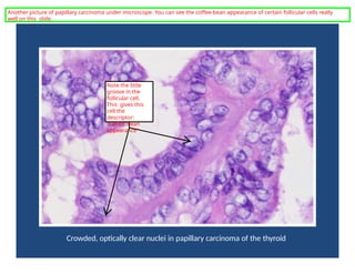
Crowded, optically clearnuclei in papillary carcinoma of the thyroid
Note the little
groove in the
follicular cell.
This gives this
cell the
descriptor:
"coffee bean
appearance"
Another picture of papillary carcinoma under microscope. You can see the coffee-bean appearance of certain follicular cells really
well on this slide.
55.
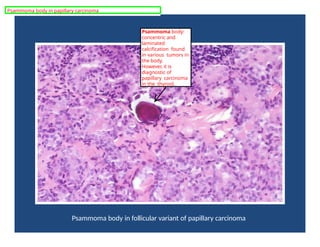
Psammoma body infollicular variant of papillary carcinoma
Psammoma body:
concentric and
laminated
calcification found
in various tumors in
the body.
However, it is
diagnostic of
papillary carcinoma
in the thyroid.
Psammoma body in papillary carcinoma
56.
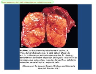
Medullary Carcinoma
• ParafollicularC cells (neuroendocrine neoplasm)
→ calcitonin secretion
• Sporadic: 80% of cases
• Familial forms: MEN 2
‐ (RET protooncogene point
mutation)
• Solitary of multiple nodules
• Polygonal or spindle shaped
‐ cells
• Nests, trabeculae or follicles
• Amyloid deposits
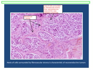
Nests of cellssurrounded by fibrovascular stroma is characteristic of neuroendocrine tumors
nests of cells surrounded
by fibrovascular stroma:
think neuroendocrine
tumor (in this case
medullary carcinoma)
fibrovascula
r stroma
59.
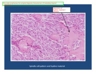
Spindle cell patternand hyaline material
deposition of
hyaline material =
amyloid
This slide emphasizes the amyloid deposits characteristic of medullary thyroid
carcinomas.
60.
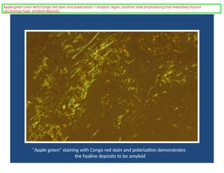
"Apple green" stainingwith Congo red stain and polarization demonstrates
the hyaline deposits to be amyloid
Apple-green color with Congo red stain and polarization = amyloid. Again, another slide emphasizing that medullary thyroid
carcinomas have amyloid deposits.
61.
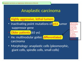
Anaplastic carcinoma
• Highlyaggressive, lethal tumors
• Inactivating point mutations of p53 tumor
suppressor gene
• Older patients (65 yo)
• Hx: multinodular goiter, differentiated
carcinoma
• Morphology: anaplastic cells (pleomorphic,
giant cells, spindle cells, small cells)
The formerly
differentiated
carcinoma
could have
progressed
due to
accumulation
of more
mutations,
leading to
anaplastic
carcinoma.
Anaplastic carcinoma: an aggressive, lethal cancer of the old.
62.
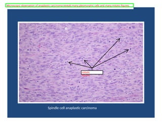
Spindle cell anaplasticcarcinoma
Microscopic observation of anaplastic carcinoma reveals many pleomorphic cells and many mitotic figures.
mitotic
figures
63.
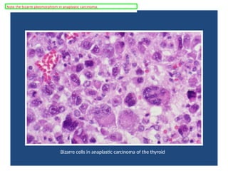
Bizarre cells inanaplastic carcinoma of the thyroid
Note the bizarre pleomorphism in anaplastic carcinoma.
64.
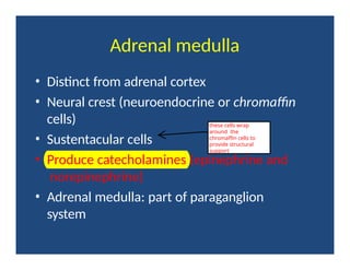
Adrenal medulla
• Distinctfrom adrenal cortex
• Neural crest (neuroendocrine or chromaffin
cells)
• Sustentacular cells
• Produce catecholamines (epinephrine and
norepinephrine)
• Adrenal medulla: part of paraganglion
system
these cells wrap
around the
chromaffin cells to
provide structural
support
65.
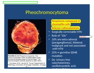
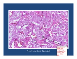
Pheochromocytoma
• Neoplasms composedof
chromaffin cells
• Release cathecolamines
• Surgically correctable HTN
• Rule of “10s”
• 10% are extra adrenal
‐
(paragangliomas), bilateral,
malignant and not associated
with HTN
• 25%→ germline SDHB
mutation
• Dx: Urinary free
catecholamines,
vanillylmandelic acid,
metanephrines
10% each
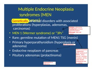
Multiple Endocrine Neoplasia
syndromes(MEN
syndromes)
• Genetically inherited disorders with associated
proliferations (hyperplasias, adenomas,
carcinomas)
• MEN 1 (Wermer syndrome) or “3Ps”
• Rare; germline mutation of MEN1 TSG (menin)
• Primary hyperparathyroidism (hyperplasia or
adenoma)
• Endocrine neoplasm of pancreas
• Pituitary adenomas (prolactinoma)
Pituitary: adenomas
Parathyroid: primary
hyperthyroidism
Pancreas: Islet cell
tumors
islet cell tumors
hyperplasia or
adenoma
Menin is a tumor
suppressor
gene product of
MEN1. Thus,
loss-of- function
mutation of this
gene would
predispose one
to cancer.
68.
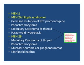
• MEN 2
•MEN 2A (Sipple syndrome)
• Germline mutation of RET protooncogene
• Pheochromocytoma
• Medullary Carcinoma of thyroid
• Parathyroid hyperplasia
• MEN 2B
• Medullary Carcinoma of thryoid
• Pheochromocytoma
• Mucosal neuromas or ganglioneuromas
• Marfanoid habitus
shared; then
remember what
is different to
differentiate
between MEN2A
and MEN2B