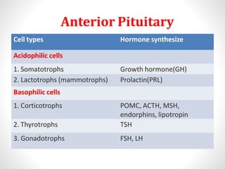
10. Hormone Features of deficiency
GH Children: Growth retardation
Adults: Decrease in muscle bulk
Hypoglycemic tendency
ACTH Weight loss, hypotension, hypoglycemia, decrease skin
pigmentation, hyponatremia, nausea, vomiting
Prolactin Lactation failure
Gonadotropins Children: delayed puberty
Female: oligomenorrhoea, infertility, atrophy of breast &
genitalia, loss of libido
Male: Impotence, azoospermia, testicular atrophy, loss of
libido
Both sexes : loss of libido, loss of body hair
TSH Weight gain, cold intolerance, fatique, hair loss, constipation,
dry skin, hoarseness, bradycardia, fatigue
ADH Thirst, polyuria
13. Septo-Optic Dysplasia
Hypothalamic dysfunction and hypopituitarism
■may result from dysgenesis of the septum pellucidum or corpus
callosum
Affected children have mutations in the HESX1 gene
These children exhibit variable combinations of:
■cleft palate
■syndactyly
■ear deformities
■optic atrophy
■micropenis
■anosmia
Pituitary dysfunction
■Diabetes insipidus
■GH deficiency and short stature
■Occasionally TSH deficiency
14. Prader Willi Syndrome
• Result from deletion of patternal copy of SRNPN and
NECDIN gene (chr 15q).
• Clinical feature:- Hypogonadotropic Hypogonadism,
Hyperphagia, obesity, muscle hypotonia , mental
retardation, T2DM.
15. Kallman Syndrome
• Defective hypothalamic gonadotropin-releasing hormone
(GnRH) synthesis
• Associated with anosmia or hyposmia due to olfactory bulb
agenesis or hypoplasia
• May also be associated with: color blindness,optic atrophy,
nerve deafness, cleft palate, renal abnormalities,
cryptorchidism
• GnRH deficiency prevents progression
through puberty
• Characterized by
✓low LH and FSH levels
✓low concentrations of sex steroids
16. Kallman Syndrome
• Males patients
✓Delayed puberty and hypogonadism(small testis), including
micropenis
✓Long-term treatment:
• human chorionic gonadotropin (hCG) or testosterone
• Female patients
✓Primary amenorrhea and failure of secondary sexual
development
✓Long-term treatment:
• cyclic estrogen and progestin
• Repetitive GnRH administration restores normal fertility
• Fertility may also be restored by the administration of
gonadotropins or by using a portable infusion pump to deliver
subcutaneous, pulsatile GnRH
17. Laurence-Moon-Bardet-Biedl Syndrome
• Rare autosomal recessive disorder
• Clinical feature:- Retinal degenration, Renal
abnormality, Retardation(Mental), obesity
other: hexadactyly/ brachydactyly/ syndactyly
• Central diabetes insipidus may or may not be associated
• GnRH deficiency occurs in 75% of males and half of
affected females
• Retinal degeneration begins in early childhood
– most patients are blind by age 30
18. Pituitary adenoma(Non functioning)
• Consist of 10% of all intracranial
neoplasm.
• 25-35% are nonfunctioning .
• Most of them arise from gonadotropin cells &
monoclonal in origin.
• Most of them are macroadenoma.
• Usually discovered because of space-
occupying effects, or inadvertently.
19. Management of nonfunctioning pituitary adenoma
Non functioning pit. adenoma
MRI differential diagnosis
Assess pituitary function
Nonfunctioning
pituitary
adenoma
Macroadenoma
Transspenoidal surgery
Follow up: MRI at 1,2,5 year
Reassess if symptomatic
Microadenoma
Observe
MRI and hormone testing
every 6 month for 2yr,
annually thereafter, hormone
replacement if required
21. Pituitary Apoplexy
• Risk factors:
✓DM, HTN
✓Sickle cell anemia
✓Anticoagulant use:- Warfarin, Heprin
• Usually resolve completely
• Transient or permanent hypopituitarism is possible
✓undiagnosed acute adrenal insufficiency
• Diagnose with CT/MRI
• Differentiate from leaking aneurysm
• Treatment:
✓If visual defects and altered consciousness then Surgical –
Transsphenoidal decompression
• Medical therapy – if symptoms are mild
✓Corticosteroids
22. Sheehan’s Syndrome
• Infarction of pituitary after substantial blood loss
during childbirth.
• Incidence: 3.6%
• No correlation between severity of hemorrhage and
symptoms
• Severe: recognized month to years postpartum
period
• Lethargy, anorexia, weight loss, unable to breast
feeding
23. Sheehan’s Syndrome
• Typically long interval between obstetric event and
diagnosis( months to years)
• Of total patients:
✓50% permanent amenorrhea
✓The rest had scanty-rare menses
✓Mostly lactation was poor to absent
•
•
Dx: MRI empty sella turcica
T/t: Hormone replacement &
Corticosteroids
24. Lymphocytic Hypophysitis
• Deficiency of one or more anterior pituitary hormones (Diabetes
Insipidus): ACTH deficiency is the most common.
Diagnosis:
MRI - may be indistinguishable from pituitary adenoma
Treatment:
✓Non compressive symptoms→ Hormone support + Steroid
✓Compressive symptoms→ Surgery(TSS)
25. Pituitary Trauma
•The pituitary may be partially or totally damaged by birth
trauma, cranial hemorrhage, fetal asphyxia, or breech
delivery.
• Head trauma can lead to direct pituitary damage by
1) Sella turcica fracture,
2) Pituitary stalk section
3) Trauma-induced vasospasm, or ischemic infarction
The most common traumatic cause of compromised
pituitary function in the adult is iatrogenic neurosurgical
trauma.
26. • Hypopituitarism following head trauma usually
appears within a year after the insult.
• Virtually all patients with subsequent
pituitary failure have a history of loss of
consciousness following trauma.
• 50% has documented skull fracture.
• 30% of these patients have demonstrable signs
of hypothalamic or post pituitary hemorrhage
(or both) or ant lobe infarction on MRI.
27. • Diabetes insipidus is the most common endocrine
disorder, encountered in about 30% of these
patients, later on other hormone deficiency may
also occurs.
• 75% of patients have evidence of hypopituitarism, and
the degree of pituitary failure correlates with severity
of head trauma.
28.
29. Radiation Injury (Cranial Irradiation)
• Children and adolescents, are more susceptible.
• Up to two-thirds of patients ultimately develop
hormone insufficiency after a median dose of 50
Gy (5000 rad) .
• The development of hypopituitarism occurs over 5–
15 years and usually reflects hypothalamic damage
rather than primary destruction of pituitary cells.
• GH deficiency is most common, followed by
gonadotropin and ACTH deficiency.
• Replacement therapy to be instituted when
appropriate .
30. Empty Sella Syndrome
• An empty sellacan developas a consequenceof a primary
congenitalweaknessof the diaphragm.
• Damageto the sellardiaphragmcan lead to arachnoid
herniationintothe sella.
• Oftenan incidental MRIfinding.
31. Empty Sella Syndrome
• Usually have normal pituitary function implying that
the surrounding rim of pituitary tissue is fully
functional
• Hypopituitarism may develop insidiously when > 90% of
tissue is compressed.
• Rarely, functional pituitary adenomas may arise within
the rim of pituitary tissue, and these are not always
visible on MRI.
• Aetiology:-
1.Congenital
2.Primary hypophysitis
3.Pituitary Adenoma (silent infarction)
4.Other: Trauma, Surgery, Radiation
34. Clinical Presentation
• Can present with features of deficiency of one or more
anterior pituitary hormones.
• Up to 50% of patients with primary empty sella have
associated benign intracranial hypertension
• Clinical presentation depends on:
✓Age at onset
✓Hormone affected, extent
✓Speed of onset
✓Duration of the deficiency
35.
36. Diagnosis of GH Deficiency
Test Blood sample timing Interpretation
Insulin tolerance test: -30,0,30,60,120 min for Glu˂40mg/dl
Regular insulin(.05-.15U/kg glucose & GH GH˃3µg/L-Normal
I.V.)
Clonidine stimulation test: 0,30,60,90,120,150,180 min GH >7µg/L- Normal
Clonidine 150 µg/m2 orally
GHRH test: 1µg/kg I.V. 0,15,30,45,60,120 min GH˃3µg/L-Normal
L-Arginine test:30gm I.V. over 0,30,60,120 min GH˃3µg/L-Normal
30min
L-Dopa test:500mg PO 0,30,60,120 min GH˃3µg/L-Normal
38. Test Time of Sample collection Result(normal response)
ITT: regular insulin
(0.05-0.15U/Kg I.V.)
-30,0,30,60,90 min for
glucose and cortisol
1. Glucose<40mg/dl
2. Cortisol↑ by >7ug/dl or
to>20ug/dl
CRH Test: 1ug/kg CRH I.V. at
8am
0,15,30,60,90,120 min for
ACTH and Cortisol
1. ACTH↑ 2 to 4 fold
2. Cortisol level>20-25 ug/dl
Metyrapone test:
Metyrapone
30mg/kg at midnight
Plasma 11-deoxycortisol,
ACTH, cortisol at 8 am
1. Cortisol< 4g/dl
2. 11-deoxycortisol>7.5ug/dl
3. ACTH> 75pg/ml
Std ACTH stimulation test :
ACTH(1-24 cosyntropin)
0.25mg IM or IV
0,30,60min for cortisol and
aldosterone
1. Cortisol >21g/dl
2.Aldosterone >4ng/dl above
baseline
Low dose ACTH test:
ACTH 1ug IV
0,30,60 min for cortisol Cortisol >21g/dl
3 day ACTH stimulation test:
0.25mg ACTH I.V. 8 hrly
Cortisol >21g/dl
•ACTH Reserve