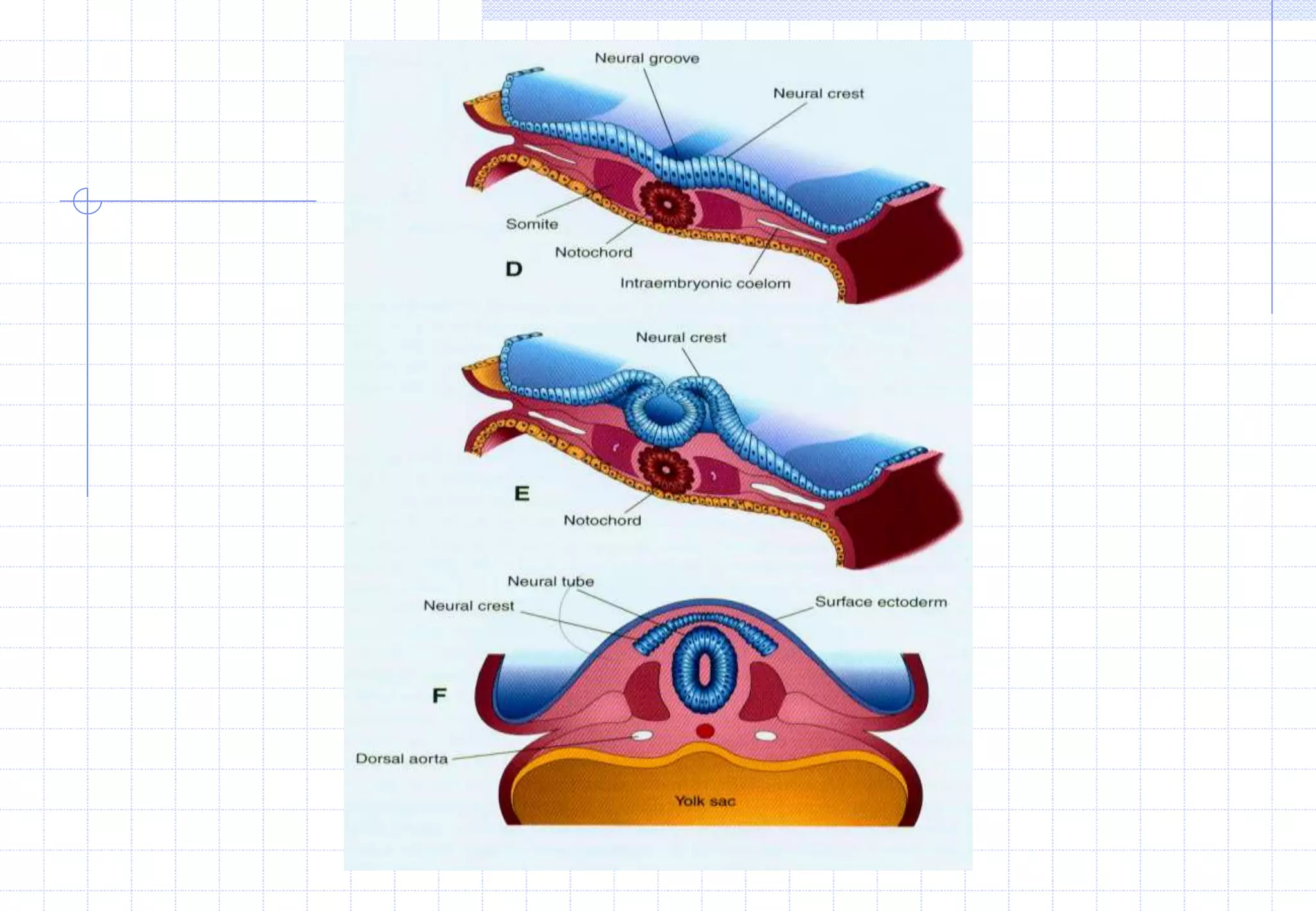
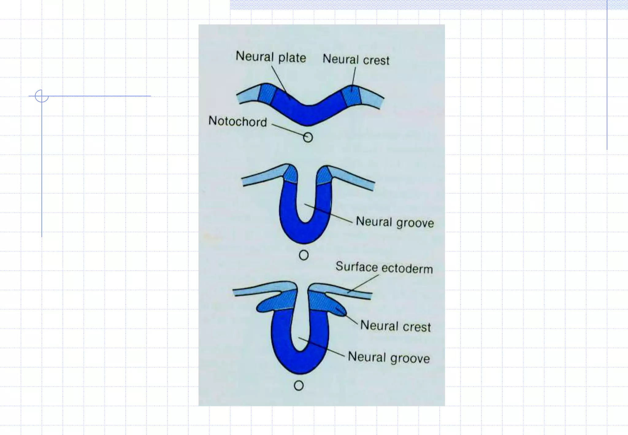
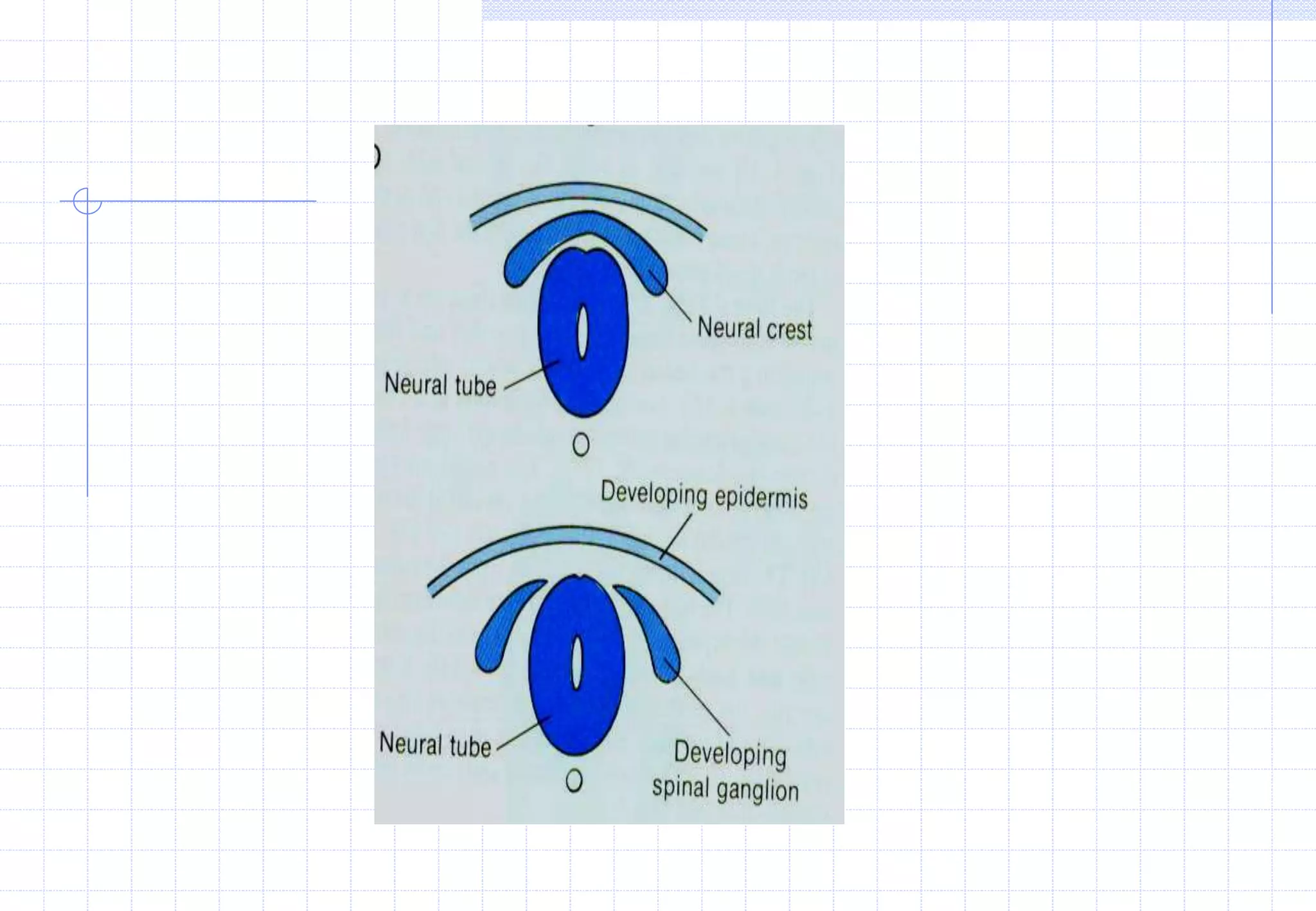
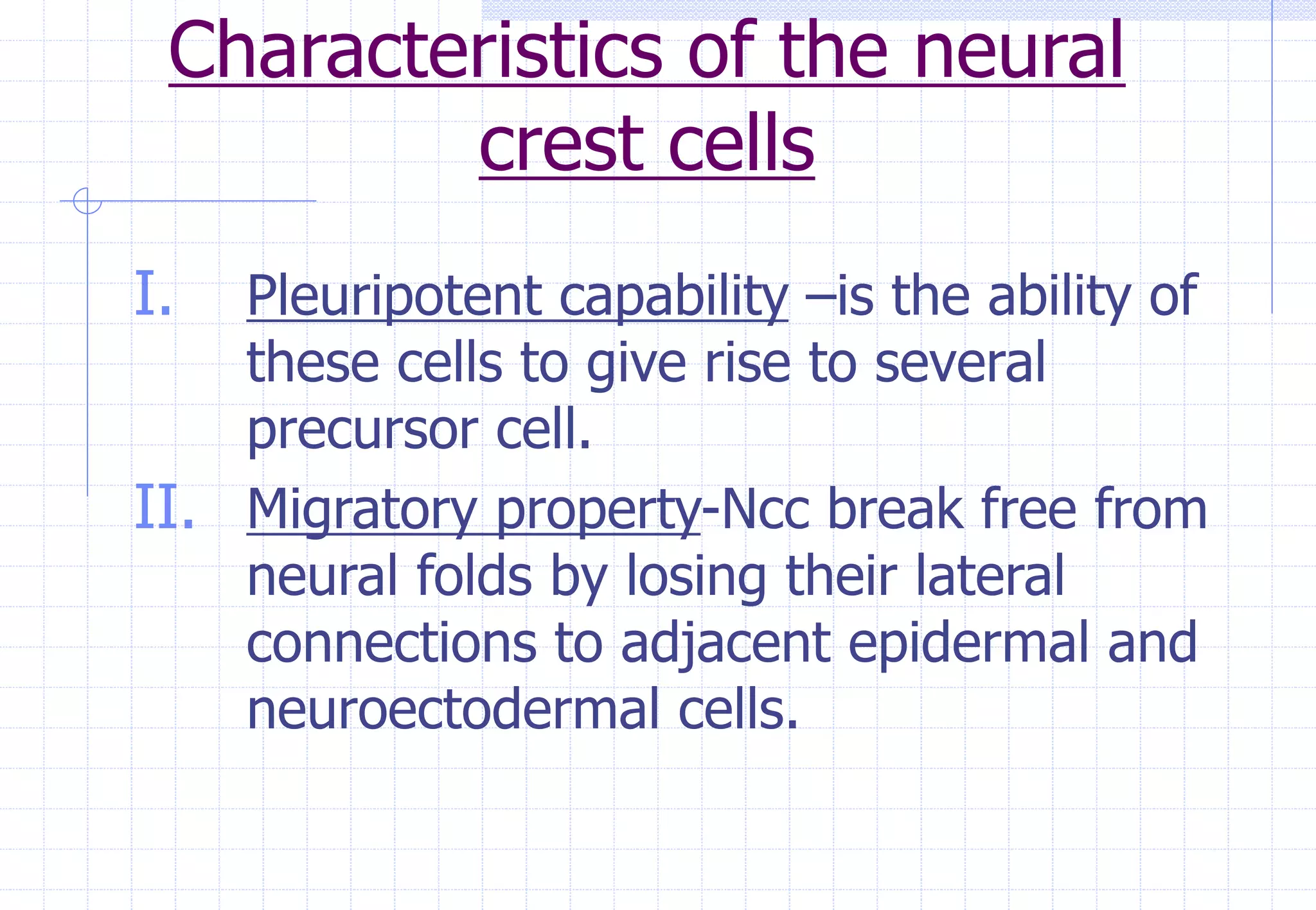
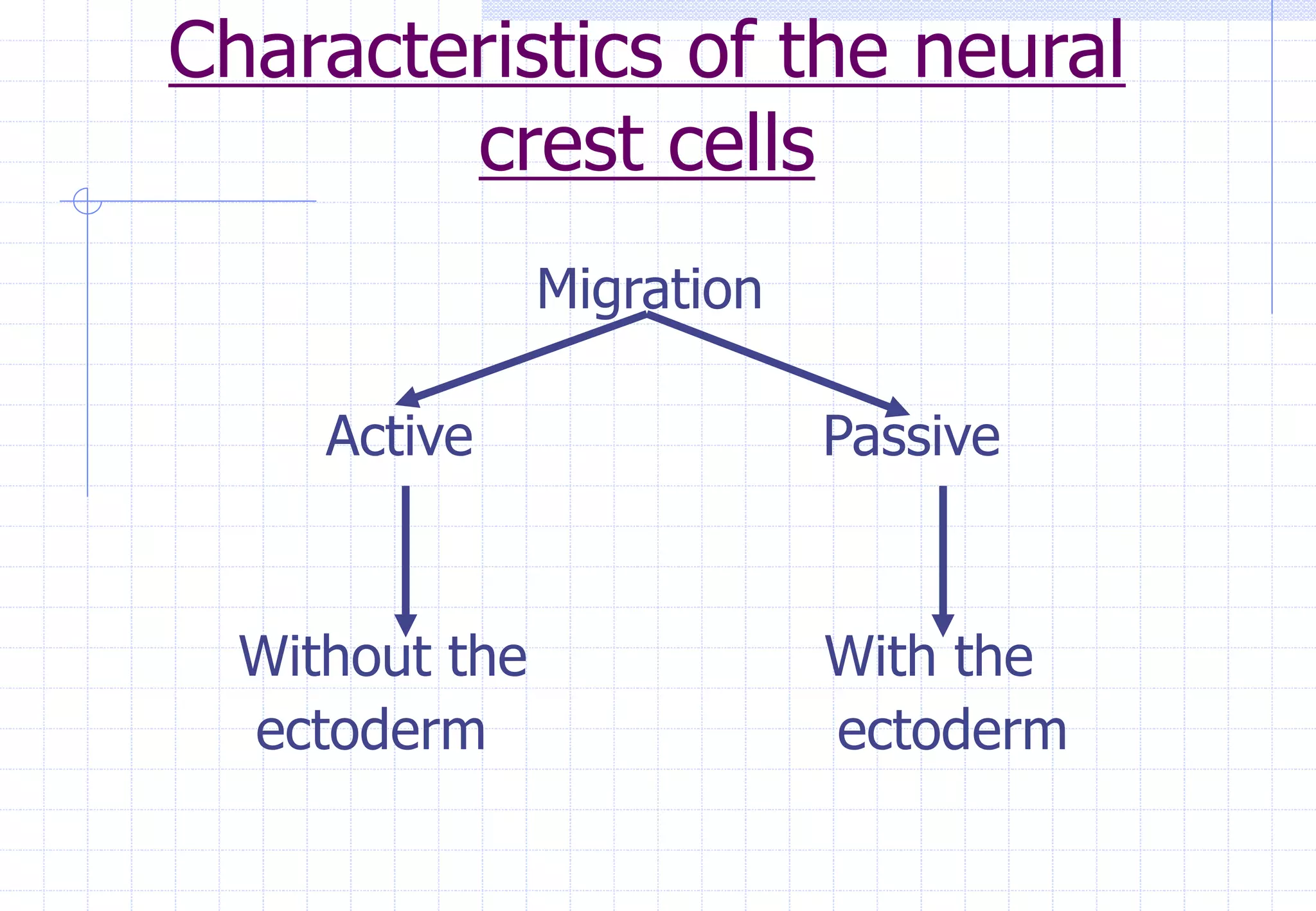
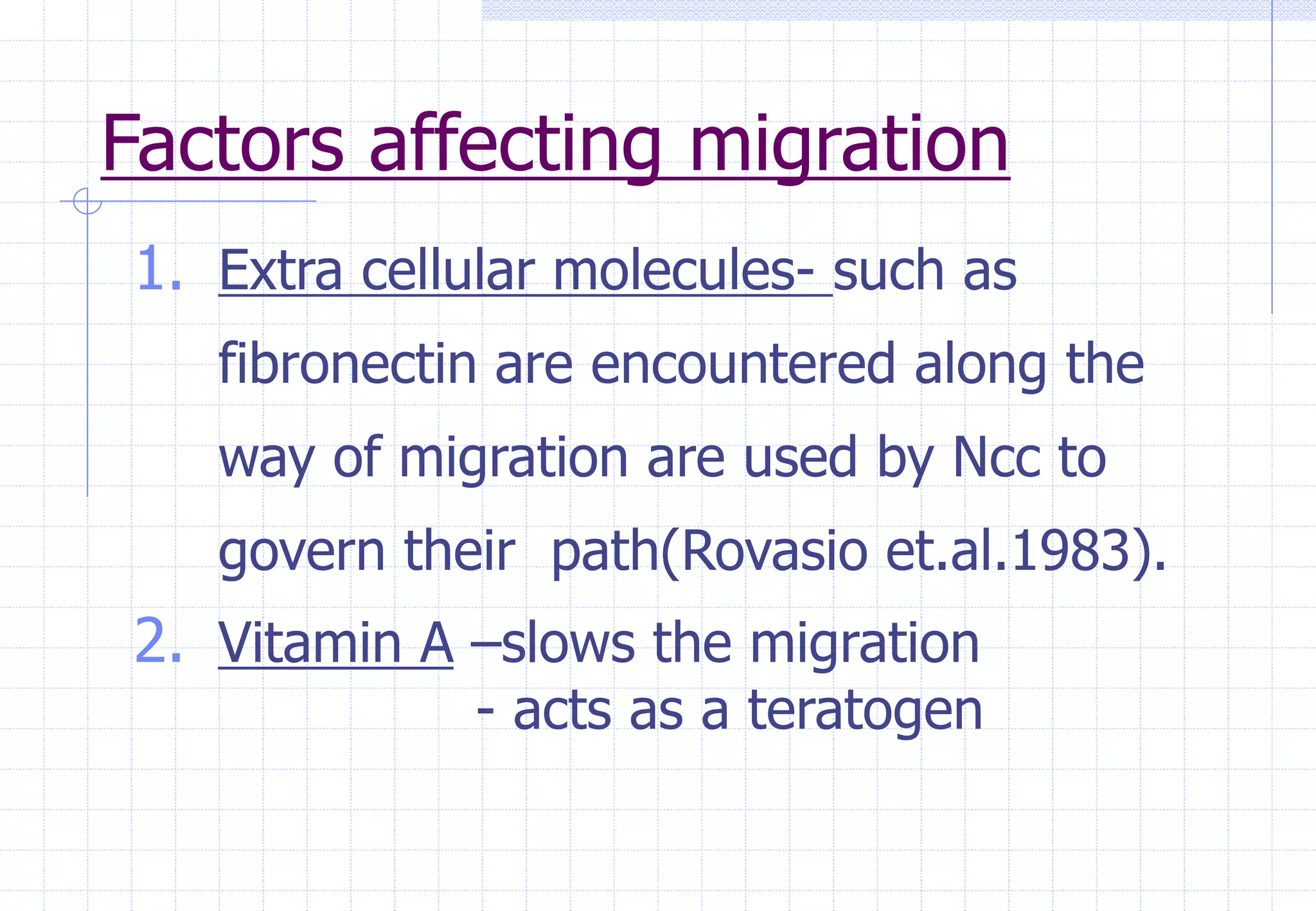
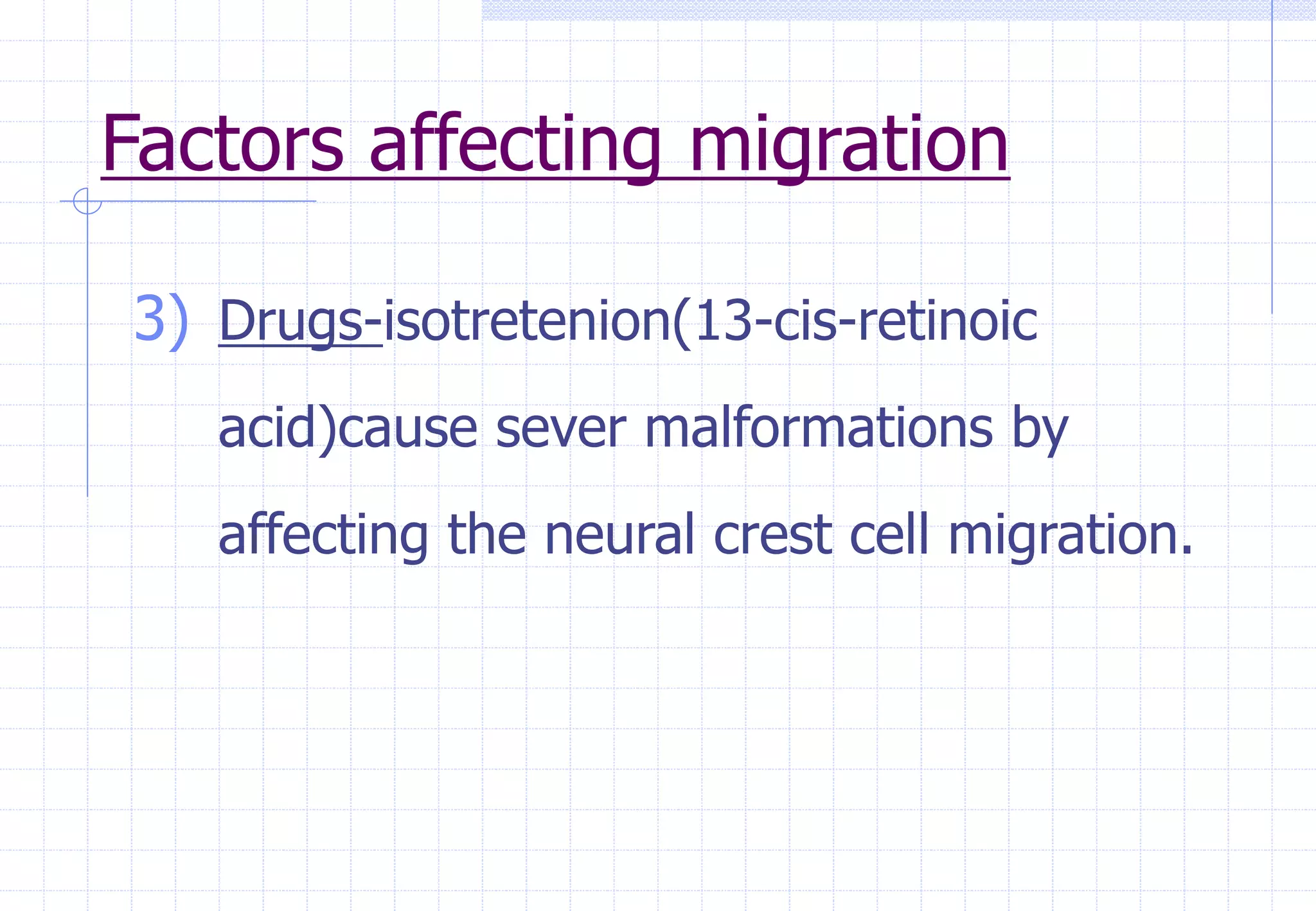
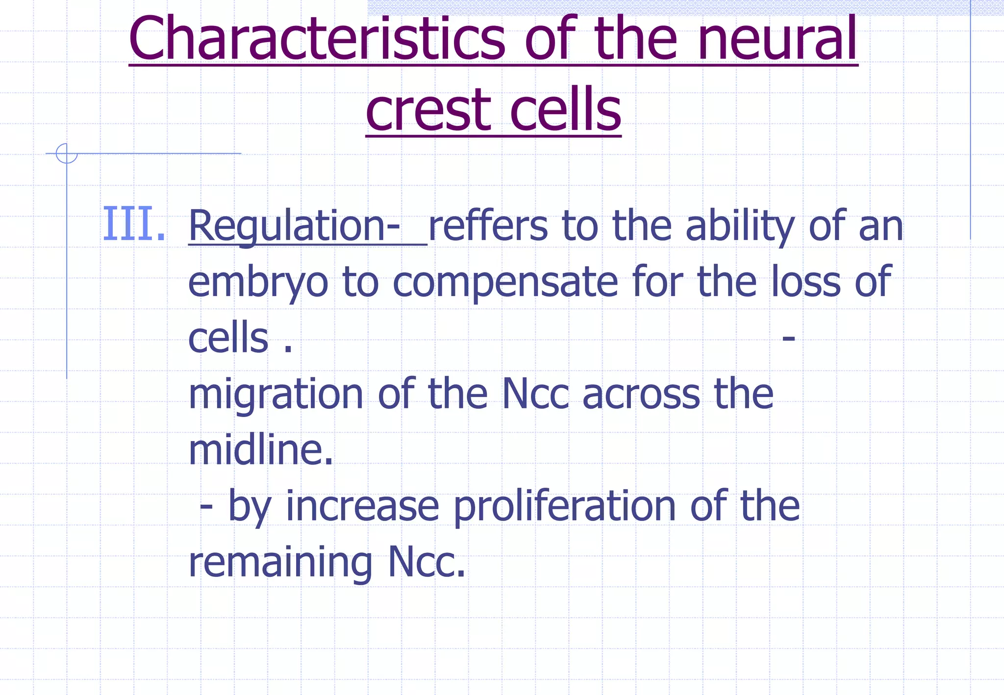
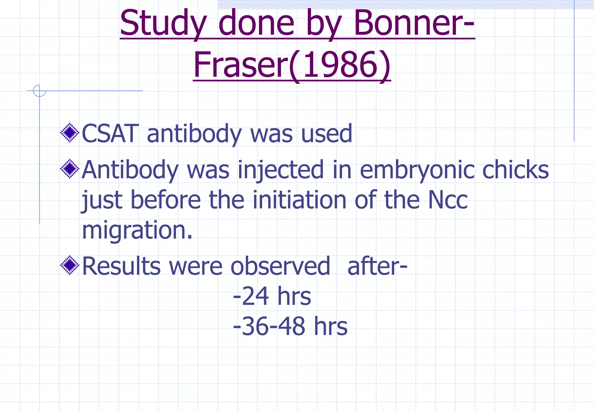
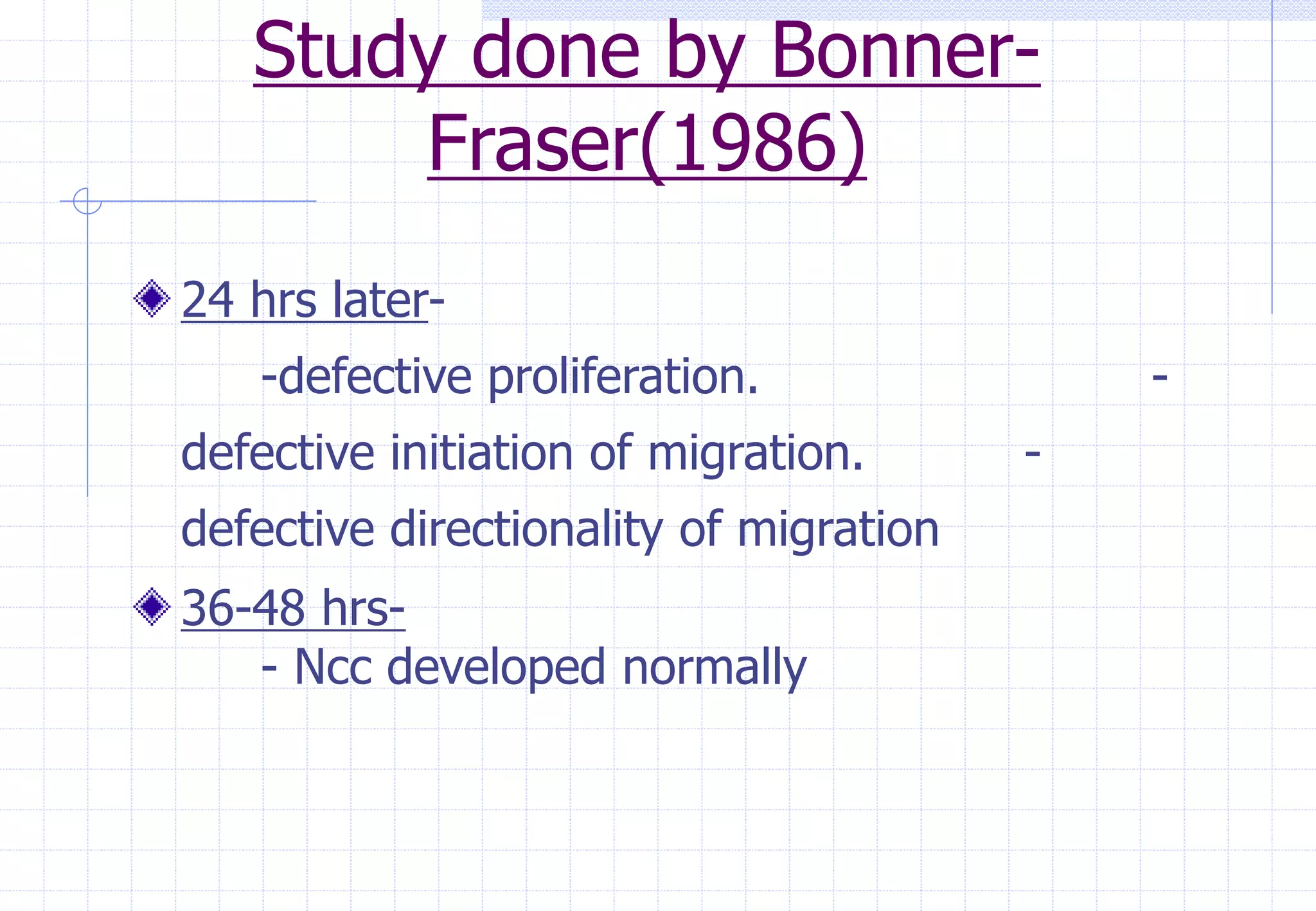
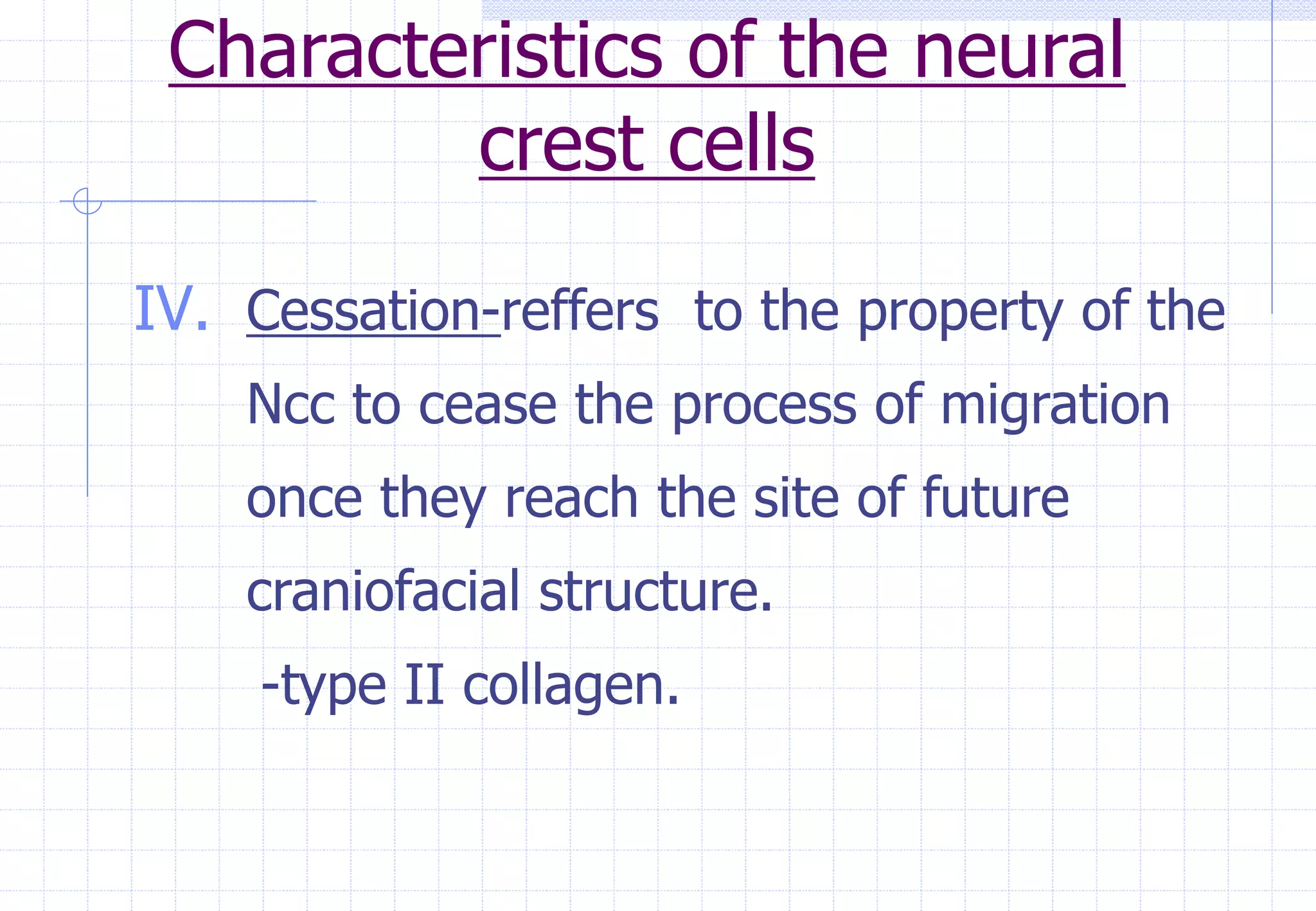
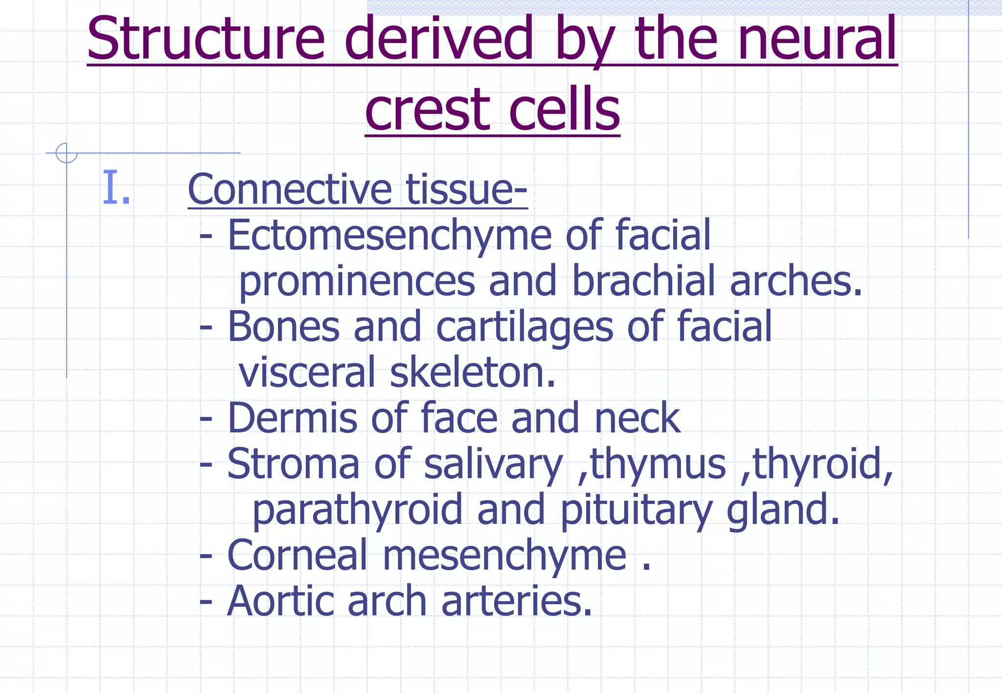
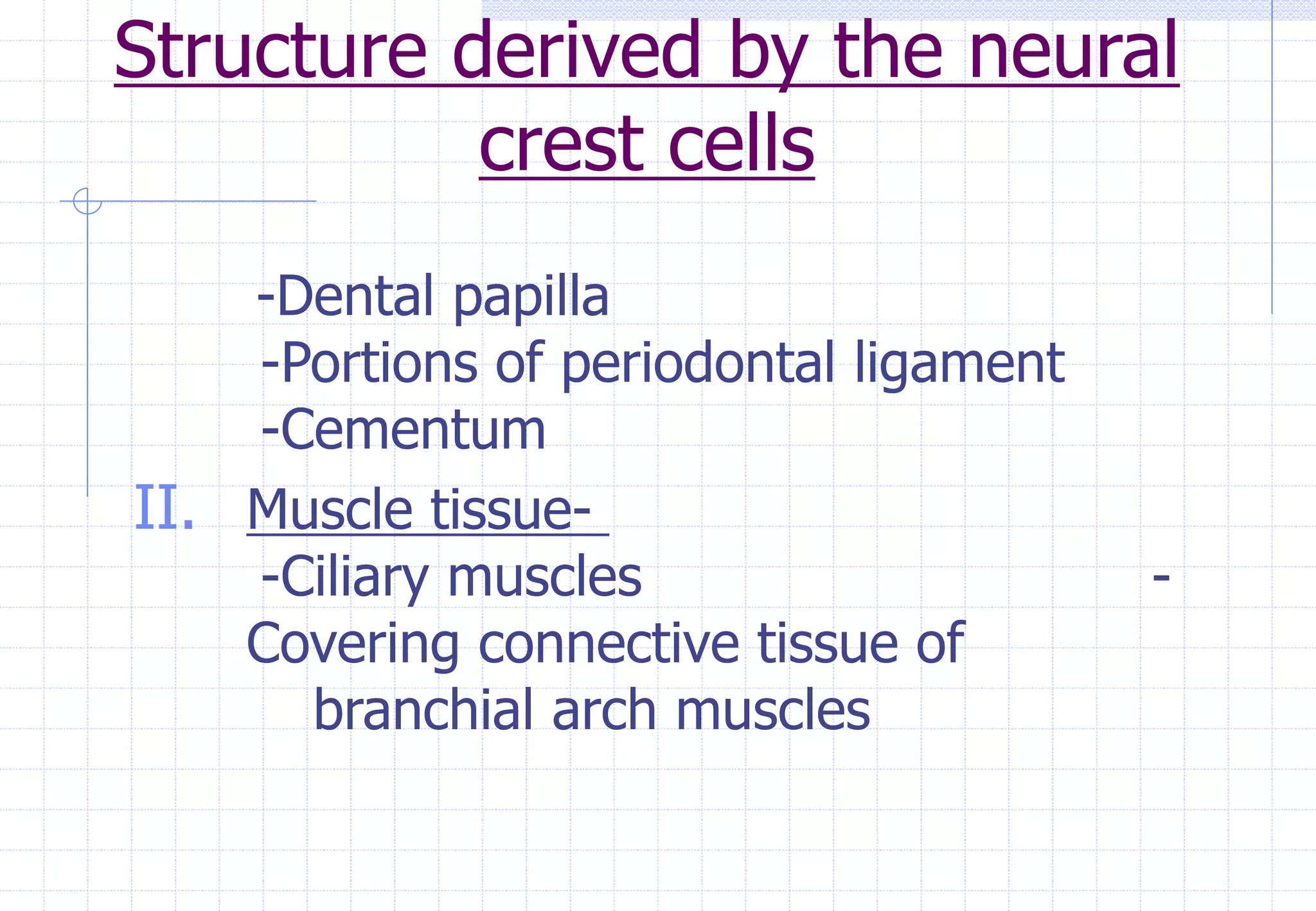
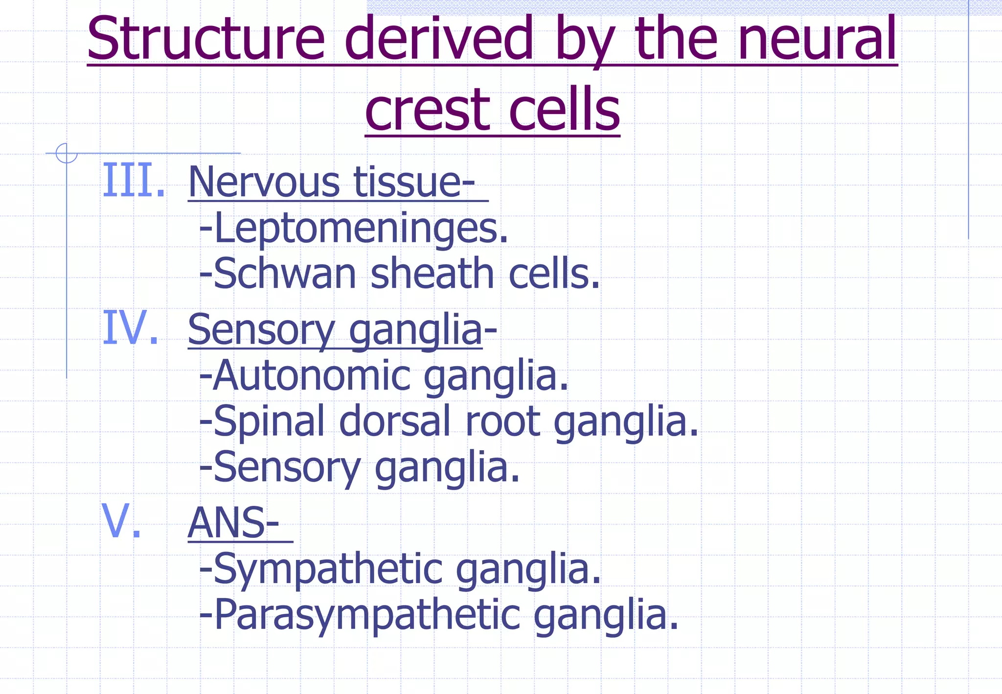
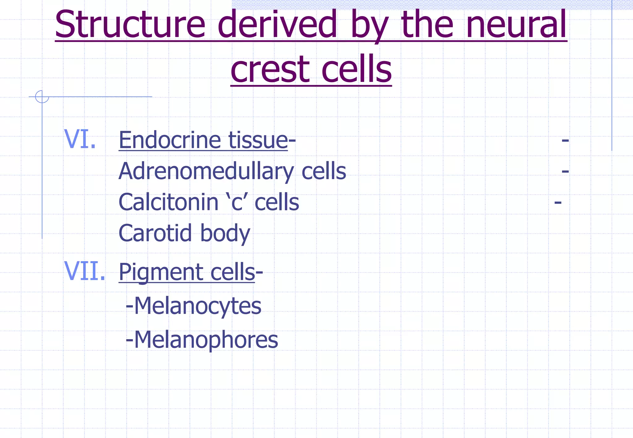
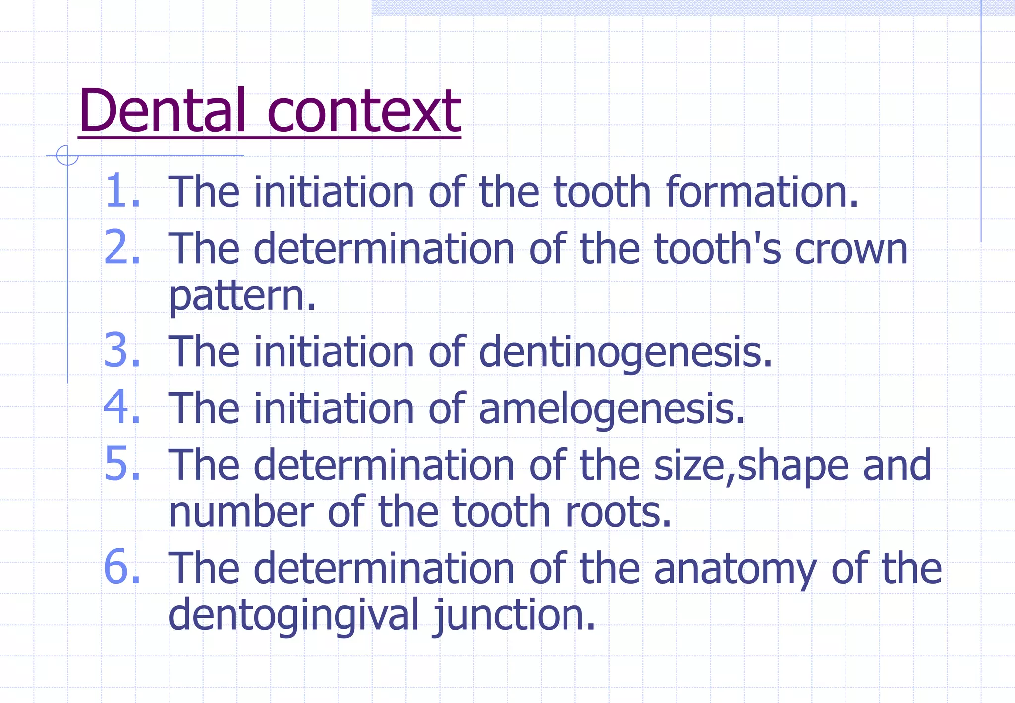
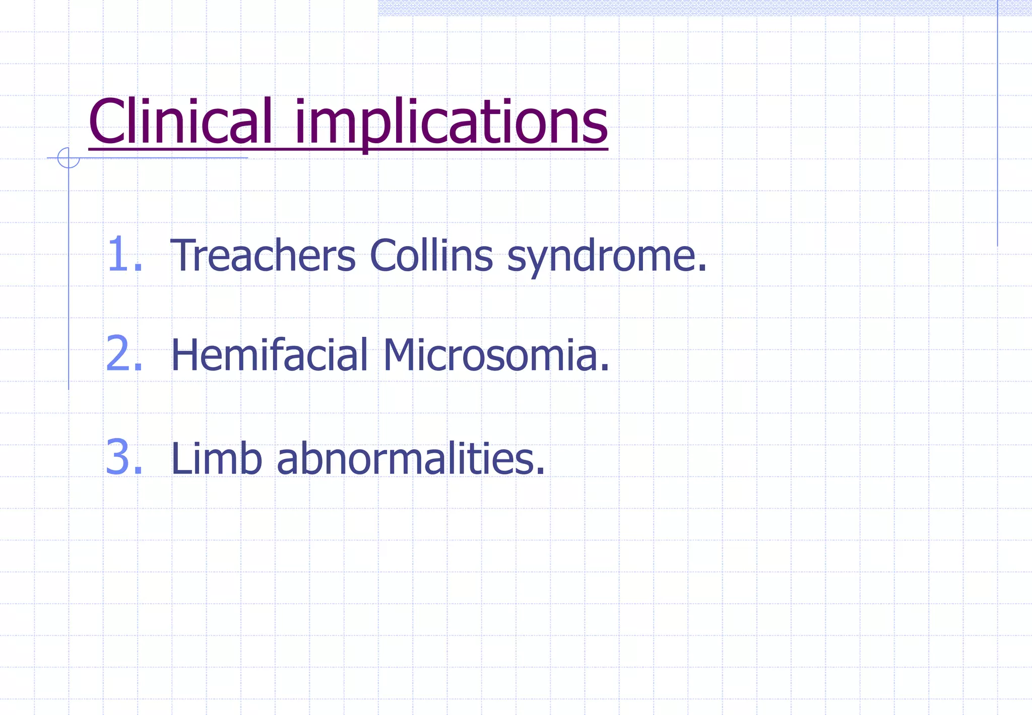
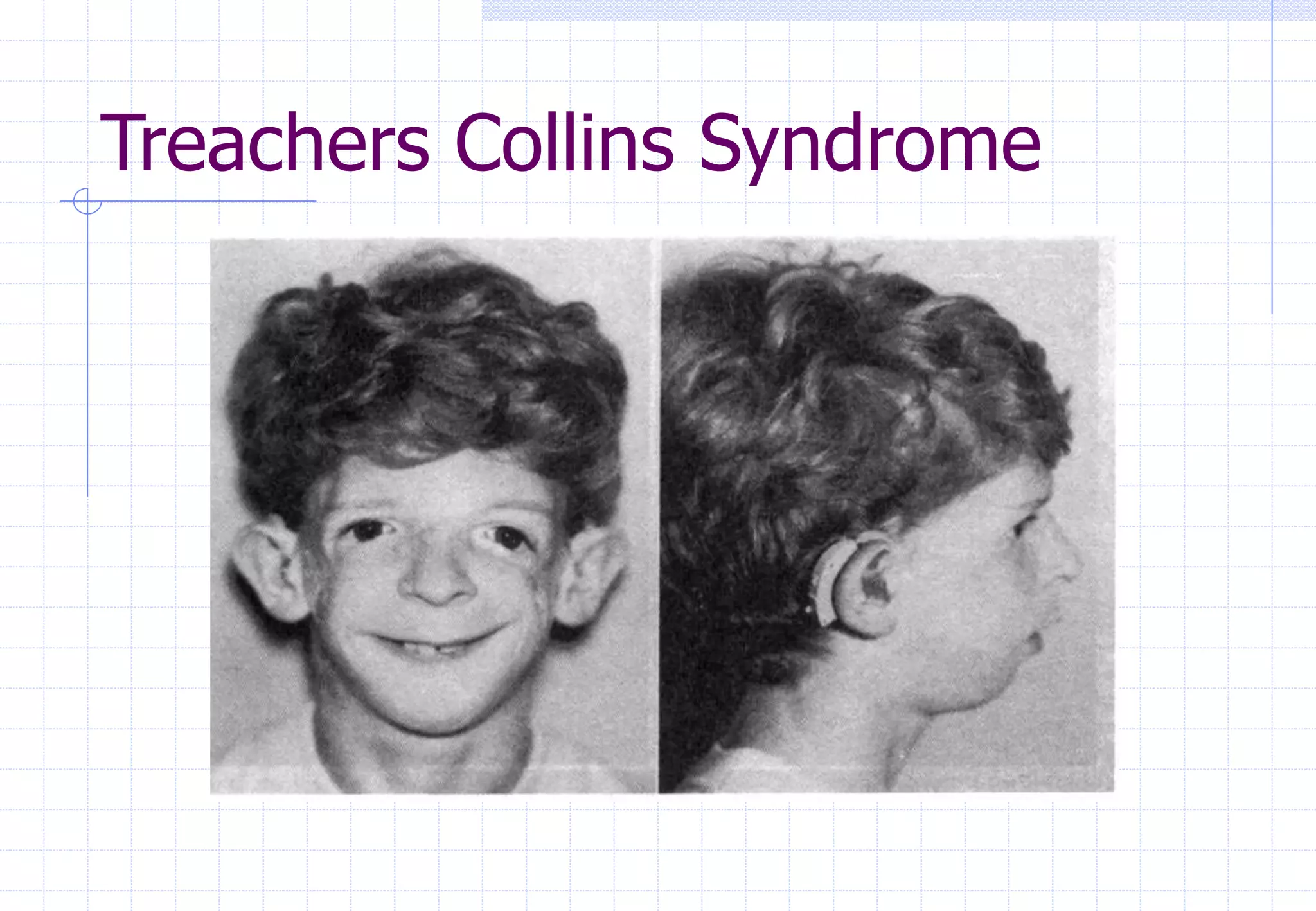
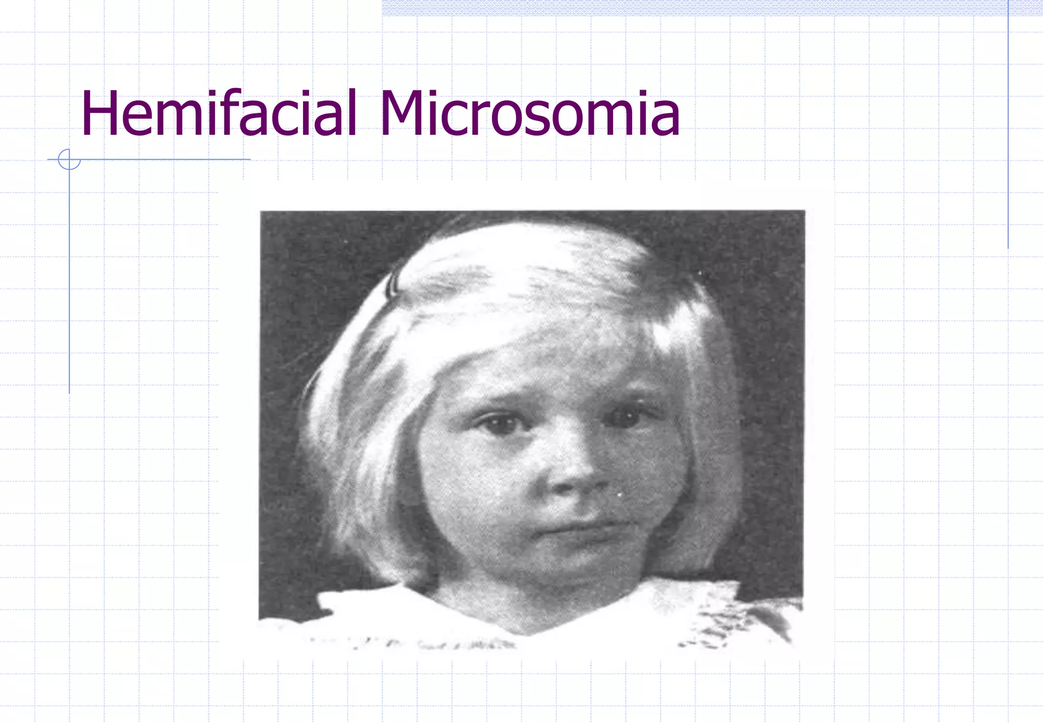
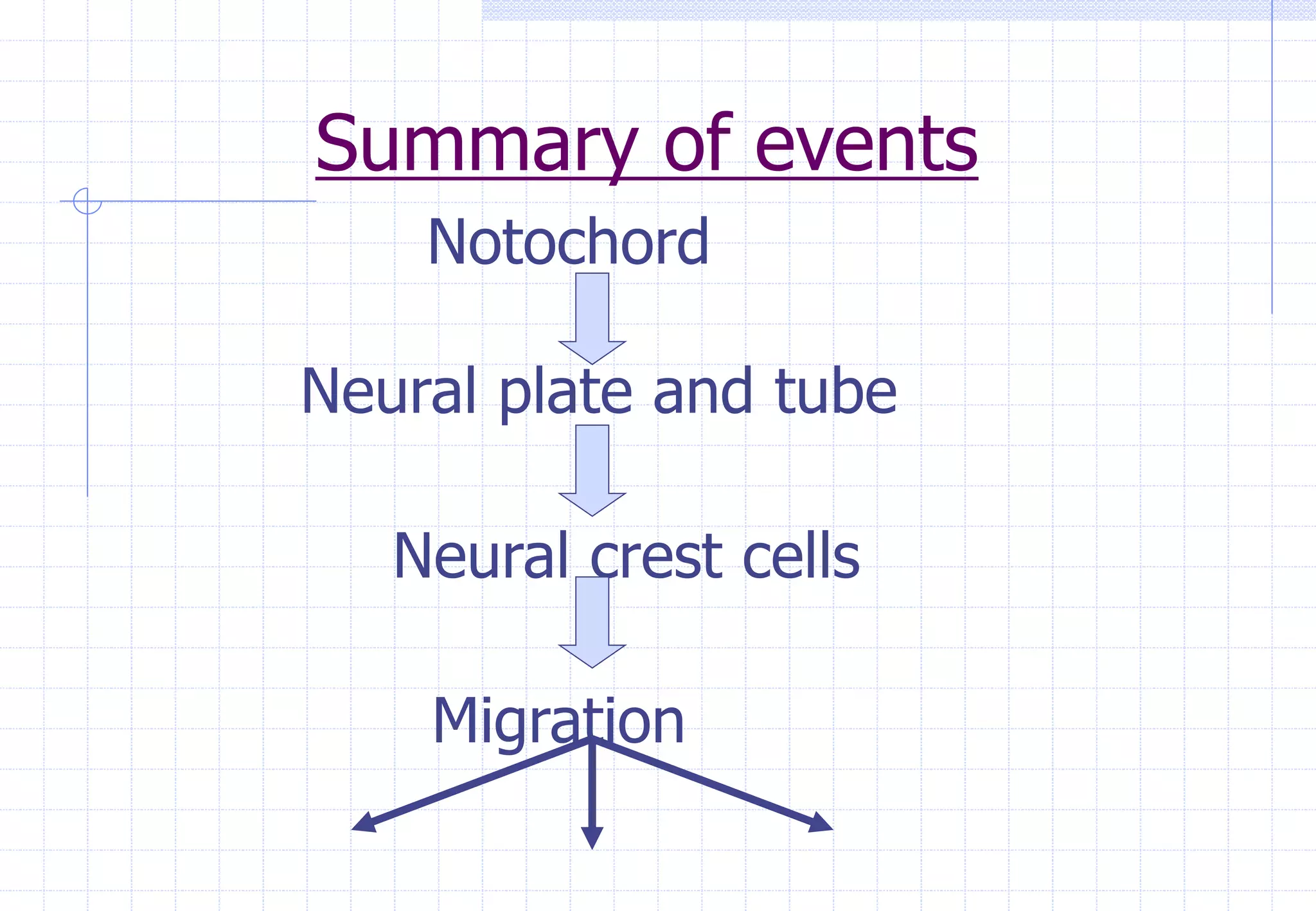
Neural Crest Cells
Dr.Akash Kencha
Dept of Orthodontics and Dentofacial
Orthopedics
S.B. Patil institute for Dental Sciences
and Research
“He who sees things grow from the beginning
will have the finest view of them”
-Aristotle
Fertilization
It is sequence of events that begins with
contact between a sperm and a oocyte
and finally ends with the fusion of nuclei
of the sperm and ovum and the
intermingling of maternal and paternal
chromosomes.
Zygote
Cleavage
It is a process by which zygote
undergoes a series of rapid mitotic
division.
First the zygote divides into two cells
known as Blastomeres
Zygote showing the process of
cleavage
Time: The division starts 30 hours after
fertilization.
Site: This occurs as the zygote passes
along the uterine tube towards the uterus.
Important point:
There is increase in the number of cell
without an increase in the cytoplasmic
mass as a result of which cells
progressively become smaller in size.
Morula
This is so called when the number of
cells becomes 16
Time: 3-4 days after fertilizations
Site: Uterus
Morula
BLASTOCYST
Space appear between the central
Blastomeres of Morula. This space gets
filled with the fluid from the uterine cavity.
Trophoblast Inner cell mass
Blastocyst
BLASTOCYST
Following this fluid – filled spaces fuse to
form a single, large blastocyst cavity.
Blastocyst
Implantation
Time: After six days
The blastocyst attaches itself to the
endometrial epithelium, adjacent t the
inner cell mass
Embryonic pole
Implantation
As this occurs the trophoblast start
proliferating rapidly
cytotrophoblast syncytiotrophoblast
Implantation of the blastocyst
Implantation
Finger like processes of
syncytiotrophoblast extend through the
endometrial epithelium
Time:by the end of the first week
blastocyst superficially implants in the
endometrium.
Also a flattened layer of cells called
hypoblast appear on the surface of the
inner cell mass.
Completion of implantation
Time: the process of implantation is
completed by the end of second week.
As the blastocyst slowly embeds itself in
the endometrium trophoblast goes on
differentiating into the two cell layer.
Formation of the amniotic
cavity
As the implantation is progressing small
cavity appears at the embryonic pole
between embryoblast and trophoblast.
This space forms the primodium of the
amniotic cavity.
Developing amniotic cavity
Formation of Bilaminar plate
At the same time morphologic changes
occur in the embryoblast resulting in the
formation of circular,bilaminar plate of
the cells called embryonic disc
epiblast hypoblast
Bilaminar plate
Formation of the primary yolk
sac
Few cells from the hypoblast migrate to
form a membrane called exocoelomic
membrane
This membrane surrounds the blastocyst
cavity called exocoelomic cavity.
Primary yolk cell
Primary yolk sac
Formation of the secondary
yolk cell
Some cells from the hypoblast gives rise
to a layer of loosely arranged tissue
called-extra embryonic mesoderm.
Extra embryonic coelom
yolk sac
Development of the secondary
yolk sac
Development of chorionic sac
Extra embryonic coelom splits the extra
embryonic mesoderm into two layers
Extraembryonic
Somatic mesoderm
Extraembryonic
Splanchic mesoderm
Development of chorionic sac
Extra embryonic Somatic mesoderm
+
Two layers of trophoblast
Chorion
Chorionic sac
Prochodral plate
Time:around the 14th day
few hypoblastic cells change into
columnar shape and form a
thickened ,circular area
Prochodral plate .
Prochodral plate
The third week
Gastrulation.
Primitive streak.
The notochordal process.
Notochord.
Neuralation.
The neural crest cells.
Gastrulation
It is process by which the bilaminar
embryonic disc is converted into a
trilaminar embryonic disc resulting in the
formation of the three germ layers and
the primitive streak.
Primitive streak
It is a thickened linear band of epiblast
which appears caudally in the median
plane and on the dorsal aspect of the
embryonic disc.
Primitive node
Primitive pit
Primitive node
Germ layers
Mesoblast/Mesenchyme--mesoderm
Primitive streak cell
displace hypoblast -- endoderm
Epiblast -- ectoderm
Developing germ layers
Notochordal process
Some mesenchymal cells migrate cranially
from the primitive node to form a median
cellular cord known as notochordal
process
notochordal canal
The notochordal process
prochordal plate
Notochordal canal
cloacal membrane
Formation of the notochord
It is a cellular rod that develops by
transformation of notochordal process
- defines the primitive axis of embryo
- gives rigidity
- future site of vertebral column.
Formation of the notochord
1. As notochordal process
elongates ,notochordal canal extends
cranially from primitive node to
prochordal plate .
2. Floor of the notochordal process fuses
with the underlying embryonic
endoderm.
3. Fused region gradually undergo
degeneration creating an opening.
Developing notochord
Formation of the notochord
4. The opening rapidly becomes confluent
and notochordal canal disappears .
5. Beginning at cranial end ,notochordal
plate infolds to form notochord.
6. The notochord finally detached from the
embryonic endoderm.
Neuralation
It is process involved in the formation of
the neural plate and neural folds and the
closure of these folds to form the neural
tube.
Neurla
Neural plate
As the notochord develops ,embryonic
ectoderm over it thickens to form the
neural plate-neuroectoderm.
Around the 18th day
Neural tube
The neural crest
Some neuroectodermal cells lying along
the crest of each neural fold lose their
epithelial affinities and are called neural
crest cells.
These form a irregular flattened mass
called neural crest .
Characteristics of the neural
crest cells
I. Pleuripotent capability –is the ability of
these cells to give rise to several
precursor cell.
II. Migratory property-Ncc break free from
neural folds by losing their lateral
connections to adjacent epidermal and
neuroectodermal cells.
Characteristics of the neural
crest cells
Migration
Active Passive
Without the With the
ectoderm ectoderm
Factors affecting migration
1. Extra cellular molecules- such as
fibronectin are encountered along the
way of migration are used by Ncc to
govern their path(Rovasio et.al.1983).
2. Vitamin A –slows the migration
- acts as a teratogen
Factors affecting migration
3) Drugs-isotretenion(13-cis-retinoic
acid)cause sever malformations by
affecting the neural crest cell migration.
Characteristics of the neural
crest cells
III. Regulation- reffers to the ability of an
embryo to compensate for the loss of
cells . -
migration of the Ncc across the
midline.
- by increase proliferation of the
remaining Ncc.
Study done by Bonner-
Fraser(1986)
CSAT antibody was used
Antibody was injected in embryonic chicks
just before the initiation of the Ncc
migration.
Results were observed after-
-24 hrs
-36-48 hrs
Study done by Bonner-
Fraser(1986)
24 hrs later-
-defective proliferation. -
defective initiation of migration. -
defective directionality of migration
36-48 hrs-
- Ncc developed normally
Characteristics of the neural
crest cells
IV. Cessation-reffers to the property of the
Ncc to cease the process of migration
once they reach the site of future
craniofacial structure.
-type II collagen.
Structure derived by the neural
crest cells
I. Connective tissue-
- Ectomesenchyme of facial
prominences and brachial arches.
- Bones and cartilages of facial
visceral skeleton.
- Dermis of face and neck
- Stroma of salivary ,thymus ,thyroid,
parathyroid and pituitary gland.
- Corneal mesenchyme .
- Aortic arch arteries.
Structure derived by the neural
crest cells
-Dental papilla
-Portions of periodontal ligament
-Cementum
II. Muscle tissue-
-Ciliary muscles -
Covering connective tissue of
branchial arch muscles
Structure derived by the neural
crest cells
III. Nervous tissue-
-Leptomeninges.
-Schwan sheath cells.
IV. Sensory ganglia-
-Autonomic ganglia.
-Spinal dorsal root ganglia.
-Sensory ganglia.
V. ANS-
-Sympathetic ganglia.
-Parasympathetic ganglia.
Structure derived by the neural
crest cells
VI. Endocrine tissue- -
Adrenomedullary cells -
Calcitonin ‘c’ cells -
Carotid body
VII. Pigment cells-
-Melanocytes
-Melanophores
Dental context
1. The initiation of the tooth formation.
2. The determination of the tooth's crown
pattern.
3. The initiation of dentinogenesis.
4. The initiation of amelogenesis.
5. The determination of the size,shape and
number of the tooth roots.
6. The determination of the anatomy of the
dentogingival junction.
Clinical implications
1. Treachers Collins syndrome.
2. Hemifacial Microsomia.
3. Limb abnormalities.
Treachers Collins Syndrome
Hemifacial Microsomia
Summary of events
Zygote
Morula
Blastula
Implantation
Amniotic cavity
Summary of events
Bilaminar embryonic disc
Yolk sac
Chorionic sac
Germ layers
Primitive streak
Summary of events
Notochord
Neural plate and tube
Neural crest cells
Migration
References
Contemporary Orthodontics-
William R Proffit
The Developing Human -
Moore and Persaud
Craniofacial Morphogenesis and
Dysmorphogenesis-
Katherine and Alphonse
Craniofacial Embryology-
G.H.Sperber
Oral histology-
Tencate

Neural Crest Cells - 1.ppt

  • 1.
    Neural Crest Cells Dr.AkashKencha Dept of Orthodontics and Dentofacial Orthopedics S.B. Patil institute for Dental Sciences and Research
  • 2.
    “He who seesthings grow from the beginning will have the finest view of them” -Aristotle
  • 3.
    Fertilization It is sequenceof events that begins with contact between a sperm and a oocyte and finally ends with the fusion of nuclei of the sperm and ovum and the intermingling of maternal and paternal chromosomes. Zygote
  • 4.
    Cleavage It is aprocess by which zygote undergoes a series of rapid mitotic division. First the zygote divides into two cells known as Blastomeres
  • 5.
    Zygote showing theprocess of cleavage
  • 6.
    Time: The divisionstarts 30 hours after fertilization. Site: This occurs as the zygote passes along the uterine tube towards the uterus.
  • 8.
    Important point: There isincrease in the number of cell without an increase in the cytoplasmic mass as a result of which cells progressively become smaller in size.
  • 9.
    Morula This is socalled when the number of cells becomes 16 Time: 3-4 days after fertilizations Site: Uterus
  • 10.
  • 11.
    BLASTOCYST Space appear betweenthe central Blastomeres of Morula. This space gets filled with the fluid from the uterine cavity. Trophoblast Inner cell mass
  • 12.
  • 13.
    BLASTOCYST Following this fluid– filled spaces fuse to form a single, large blastocyst cavity. Blastocyst
  • 14.
    Implantation Time: After sixdays The blastocyst attaches itself to the endometrial epithelium, adjacent t the inner cell mass Embryonic pole
  • 15.
    Implantation As this occursthe trophoblast start proliferating rapidly cytotrophoblast syncytiotrophoblast
  • 16.
  • 17.
    Implantation Finger like processesof syncytiotrophoblast extend through the endometrial epithelium Time:by the end of the first week blastocyst superficially implants in the endometrium. Also a flattened layer of cells called hypoblast appear on the surface of the inner cell mass.
  • 18.
    Completion of implantation Time:the process of implantation is completed by the end of second week. As the blastocyst slowly embeds itself in the endometrium trophoblast goes on differentiating into the two cell layer.
  • 19.
    Formation of theamniotic cavity As the implantation is progressing small cavity appears at the embryonic pole between embryoblast and trophoblast. This space forms the primodium of the amniotic cavity.
  • 20.
  • 21.
    Formation of Bilaminarplate At the same time morphologic changes occur in the embryoblast resulting in the formation of circular,bilaminar plate of the cells called embryonic disc epiblast hypoblast
  • 22.
  • 23.
    Formation of theprimary yolk sac Few cells from the hypoblast migrate to form a membrane called exocoelomic membrane This membrane surrounds the blastocyst cavity called exocoelomic cavity. Primary yolk cell
  • 24.
  • 25.
    Formation of thesecondary yolk cell Some cells from the hypoblast gives rise to a layer of loosely arranged tissue called-extra embryonic mesoderm. Extra embryonic coelom yolk sac
  • 26.
    Development of thesecondary yolk sac
  • 27.
    Development of chorionicsac Extra embryonic coelom splits the extra embryonic mesoderm into two layers Extraembryonic Somatic mesoderm Extraembryonic Splanchic mesoderm
  • 28.
    Development of chorionicsac Extra embryonic Somatic mesoderm + Two layers of trophoblast Chorion
  • 29.
  • 30.
    Prochodral plate Time:around the14th day few hypoblastic cells change into columnar shape and form a thickened ,circular area Prochodral plate .
  • 31.
  • 32.
    The third week Gastrulation. Primitivestreak. The notochordal process. Notochord. Neuralation. The neural crest cells.
  • 33.
    Gastrulation It is processby which the bilaminar embryonic disc is converted into a trilaminar embryonic disc resulting in the formation of the three germ layers and the primitive streak.
  • 34.
    Primitive streak It isa thickened linear band of epiblast which appears caudally in the median plane and on the dorsal aspect of the embryonic disc. Primitive node Primitive pit
  • 35.
  • 36.
    Germ layers Mesoblast/Mesenchyme--mesoderm Primitive streakcell displace hypoblast -- endoderm Epiblast -- ectoderm
  • 37.
  • 38.
    Notochordal process Some mesenchymalcells migrate cranially from the primitive node to form a median cellular cord known as notochordal process notochordal canal
  • 39.
    The notochordal process prochordalplate Notochordal canal cloacal membrane
  • 40.
    Formation of thenotochord It is a cellular rod that develops by transformation of notochordal process - defines the primitive axis of embryo - gives rigidity - future site of vertebral column.
  • 41.
    Formation of thenotochord 1. As notochordal process elongates ,notochordal canal extends cranially from primitive node to prochordal plate . 2. Floor of the notochordal process fuses with the underlying embryonic endoderm. 3. Fused region gradually undergo degeneration creating an opening.
  • 42.
  • 43.
    Formation of thenotochord 4. The opening rapidly becomes confluent and notochordal canal disappears . 5. Beginning at cranial end ,notochordal plate infolds to form notochord. 6. The notochord finally detached from the embryonic endoderm.
  • 45.
    Neuralation It is processinvolved in the formation of the neural plate and neural folds and the closure of these folds to form the neural tube. Neurla
  • 46.
    Neural plate As thenotochord develops ,embryonic ectoderm over it thickens to form the neural plate-neuroectoderm. Around the 18th day Neural tube
  • 48.
    The neural crest Someneuroectodermal cells lying along the crest of each neural fold lose their epithelial affinities and are called neural crest cells. These form a irregular flattened mass called neural crest .
  • 51.
    Characteristics of theneural crest cells I. Pleuripotent capability –is the ability of these cells to give rise to several precursor cell. II. Migratory property-Ncc break free from neural folds by losing their lateral connections to adjacent epidermal and neuroectodermal cells.
  • 52.
    Characteristics of theneural crest cells Migration Active Passive Without the With the ectoderm ectoderm
  • 53.
    Factors affecting migration 1.Extra cellular molecules- such as fibronectin are encountered along the way of migration are used by Ncc to govern their path(Rovasio et.al.1983). 2. Vitamin A –slows the migration - acts as a teratogen
  • 54.
    Factors affecting migration 3)Drugs-isotretenion(13-cis-retinoic acid)cause sever malformations by affecting the neural crest cell migration.
  • 55.
    Characteristics of theneural crest cells III. Regulation- reffers to the ability of an embryo to compensate for the loss of cells . - migration of the Ncc across the midline. - by increase proliferation of the remaining Ncc.
  • 56.
    Study done byBonner- Fraser(1986) CSAT antibody was used Antibody was injected in embryonic chicks just before the initiation of the Ncc migration. Results were observed after- -24 hrs -36-48 hrs
  • 57.
    Study done byBonner- Fraser(1986) 24 hrs later- -defective proliferation. - defective initiation of migration. - defective directionality of migration 36-48 hrs- - Ncc developed normally
  • 58.
    Characteristics of theneural crest cells IV. Cessation-reffers to the property of the Ncc to cease the process of migration once they reach the site of future craniofacial structure. -type II collagen.
  • 59.
    Structure derived bythe neural crest cells I. Connective tissue- - Ectomesenchyme of facial prominences and brachial arches. - Bones and cartilages of facial visceral skeleton. - Dermis of face and neck - Stroma of salivary ,thymus ,thyroid, parathyroid and pituitary gland. - Corneal mesenchyme . - Aortic arch arteries.
  • 60.
    Structure derived bythe neural crest cells -Dental papilla -Portions of periodontal ligament -Cementum II. Muscle tissue- -Ciliary muscles - Covering connective tissue of branchial arch muscles
  • 61.
    Structure derived bythe neural crest cells III. Nervous tissue- -Leptomeninges. -Schwan sheath cells. IV. Sensory ganglia- -Autonomic ganglia. -Spinal dorsal root ganglia. -Sensory ganglia. V. ANS- -Sympathetic ganglia. -Parasympathetic ganglia.
  • 62.
    Structure derived bythe neural crest cells VI. Endocrine tissue- - Adrenomedullary cells - Calcitonin ‘c’ cells - Carotid body VII. Pigment cells- -Melanocytes -Melanophores
  • 63.
    Dental context 1. Theinitiation of the tooth formation. 2. The determination of the tooth's crown pattern. 3. The initiation of dentinogenesis. 4. The initiation of amelogenesis. 5. The determination of the size,shape and number of the tooth roots. 6. The determination of the anatomy of the dentogingival junction.
  • 64.
    Clinical implications 1. TreachersCollins syndrome. 2. Hemifacial Microsomia. 3. Limb abnormalities.
  • 65.
  • 66.
  • 67.
  • 68.
    Summary of events Bilaminarembryonic disc Yolk sac Chorionic sac Germ layers Primitive streak
  • 69.
    Summary of events Notochord Neuralplate and tube Neural crest cells Migration
  • 70.
    References Contemporary Orthodontics- William RProffit The Developing Human - Moore and Persaud Craniofacial Morphogenesis and Dysmorphogenesis- Katherine and Alphonse Craniofacial Embryology- G.H.Sperber Oral histology- Tencate