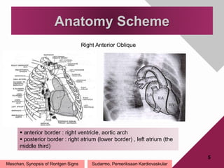
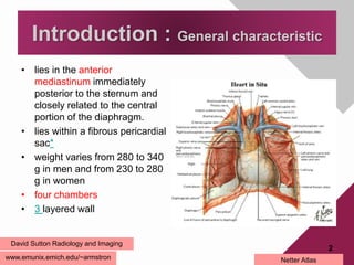
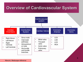
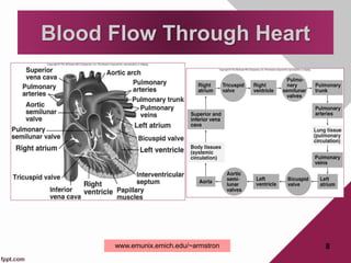
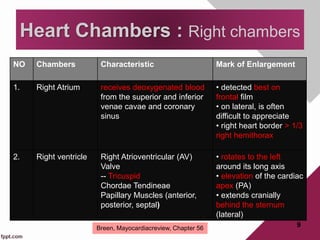
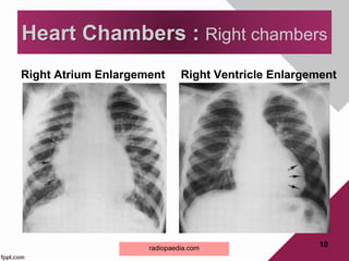
Anatomy of The Heart
Text Book Reading David Sutton Radiology and Imaging
7th ed vol.1 Section 1, page 265-271
THE NORMAL HEART: ANATOMY AND TECHNIQUES OF
EXAMINATION
Oleh: Reni Indrastuti
Pembimbing : dr. Edy Moeljono, Sp.Rad (K) RA
Introduction : General characteristic
• lies in the anterior
mediastinum immediately
posterior to the sternum and
closely related to the central
portion of the diaphragm.
• lies within a fibrous pericardial
sac*
• weight varies from 280 to 340
g in men and from 230 to 280
g in women
• four chambers
• 3 layered wall
www.emunix.emich.edu/~armstron
2
David Sutton Radiology and Imaging
Netter Atlas
Cardiovascular
system
Cardiac
Chambers
Conduction
System
Cardiac Valves
Coronary
Arteries
Coronary
Veins
3
Overview of Cardiovascular System
• Right Atrium
• Left Atrium
• Right
Ventricle
• Left Ventricle
• Sinus node
• Internodal
pathways
• Atrioventricul
ar node
• His bundle
and bundle
branches
• Mitral valve
• Tricuspid
valve
• Aortic valve
• Pulmonary
valve
• LCA
• LAD
• LCX
• RCA
Mancini, Medscape reference
Anatomy Scheme
4
Meschan, Synopsis of Rontgen Signs
 right border : SVC, right atrium, IVC
 left border : aortic arch, left pulmonary artery, left atrial
appendage, left ventricle
Frontal Projection
Sudarmo, Pemeriksaan Kardiovaskular
5
Anatomy Scheme
Meschan, Synopsis of Rontgen Signs
Right Anterior Oblique
 anterior border : right ventricle, aortic arch
 posterior border : right atrium (lower border) , left atrium (the
middle third)
Sudarmo, Pemeriksaan Kardiovaskular
Blood Flow Through Heart
8www.emunix.emich.edu/~armstron
NO Chambers Characteristic Mark of Enlargement
1. Right Atrium receives deoxygenated blood
from the superior and inferior
venae cavae and coronary
sinus
• detected best on
frontal film
• on lateral, is often
difficult to appreciate
• right heart border > 1/3
right hemithorax
2. Right ventricle Right Atrioventricular (AV)
Valve
-- Tricuspid
Chordae Tendineae
Papillary Muscles (anterior,
posterior, septal)
• rotates to the left
around its long axis
• elevation of the cardiac
apex (PA)
• extends cranially
behind the sternum
(lateral)
9
Heart Chambers : Right chambers
Breen, Mayocardiacreview, Chapter 56
Right Atrium Enlargement Right Ventricle Enlargement
10
Heart Chambers : Right chambers
radiopaedia.com
NO Chambers Characteristics Marks of Enlargement
1. Left Atrium  receives blood from the four
pulmonary veins
--superior/inferior, right/left
• walls are slightly thicker than
right atrium and smooth except
for auricles whichhave pectinate
muscles
• PA : double density (
double contour) of right
heart border
• bulging left atrial
appendage
• Upward and posterior
displacement of left main
bronchus ---- widening
carinal angle
2. Left Ventricle • performs more work than the
right ventricle
⇒ wall is twice as thick as that
of the right ventricle
Left AV Valve (Mitral Valve)
--bicuspid (anterior & posterior
papillary muscle)
•PA : cardiac apex may be
displaced to the extent
that it projects below the
diaphragm
11
Heart Chambers : Left chambers
Breen, Mayocardiacreview, Chapter 56
Left Atrium Enlargement Left Ventricle Enlargement
12
Heart Chambers : Left chambers
radiopaedia.com
• Introduction
• Overview of Cardiovascular System
• Anatomy Scheme
• Blood Flow Through Heart
• Heart Chambers
Resume
13
QUIZ
A 75 years old woman had clinical
presentation : Shortness of
breath and poor exercise
tolerance. The CXR shows
beside.
1. Please describe this picture!
2. What is the most likely
diagnosis of this patient?
14
Terimakasih dan Mohon Asupan
Pericardial cavity
• which is continuous with the central tendon of the diaphragm and which
extends to the root of the aorta and the pulmonary artery.
16
Pericardial Fluid
• secreted by pericardial membranes
• acts as lubricant, reducing friction
between the opposing surfaces as the
heart beats
3 Layers of Heart Wall
17
18
• Left main coronary artery : The left main coronary artery (LCA) originates from the ostium of the
left sinus of Valsalva. The LCA, which courses between the left atrial appendage and the
pulmonary artery, typically is 1-2 cm in length. When it reaches the left AV groove, the LCA
bifurcates into the left anterior descending (LAD) and the LCX branches. The LCA supplies most
of the left atrium, left ventricle, interventricular septum, and AV bundles. The LCA arises from the
left aortic sinus and courses between the left auricle and the pulmonary trunk to reach the
coronary groove
• Left anterior descending artery : After originating from the left main artery, the LAD artery runs
along the anterior interventricular sulcus and supplies the apical portion of both ventricles. The
LAD artery is mostly epicardial but can be intramuscular in places. An important identifying
characteristic of the LAD artery during angiography is the identification of 4-6 perpendicular septal
branches. These branches, approximately 7.5 cm in length, supply the interventricular septum.
The first branch of the LAD artery is termed the ramus intermedius. In fewer than 1% of patients,
the LCA is absent, and the LAD and LCX arteries originate from the aorta via 2 separate ostia. As
the LAD artery passes along the anterior interventricular groove toward the apex, it turns sharply
to anastomose with the posterior interventricular branch of the RCA. As the LAD artery courses
anteriorly along the ventricular septum, it sends off diagonal branches to the lateral wall of the left
ventricle. Congenital LAD artery variations may include its duplication as 2 parallel arteries (4%
incidence) and length variations (premature or delayed distal termination).
• Left circumflex artery : The LCA gives off the LCX artery at a right angle
near the base of the left atrial appendage. The LCX artery courses in the
coronary groove around the left border of the heart to the posterior surface
of the heart to anastomose to the end of the RCA. In the AV groove, the
LCX artery lies close to the annulus of the mitral valve. The atrial circumflex
artery, the first branch off the LCX artery, supplies the left atrium. The LCX
artery gives off an obtuse marginal (OM) branch at the left border of the
heart near the base of the left atrial appendage to supply the posterolateral
surface of the left ventricle. The color contrast between the yellow-orange
OM and the adjacent red-brown myocardium may be the most reliable way
to identify this artery intraoperatively. In patients with a left-dominant heart,
the LCX artery supplies the PDA. Many variations in the origin and length of
the LCX artery are noted. In fewer than 40% of patients, the sinus node
artery may originate from the LCX artery.
• Right coronary artery
19
Coronary Circulation
20
Coronary Circulation
• rt. & lt. coronary arteries originate at
base of ascending aorta
Right Coronary Artery
--marginal branch
--posterior interventricular branch
Left Coronary Artery
--circumflex branch
--anterior interventricular branch
• Great cardiac vein
• Coronary sinus
21
posterior cardiac vein
middle cardiac vein
anterior cardiac vein
small cardiac vein
Anatomy frontal projection
22
23
Heart Measurement
RVH
24
A 39-year-old woman presents with the
complaint of dyspnea on exertion and
paroxysmal nocturnal dyspnea. The
onset of symptoms began insidiously
about 1 year ago. The symptoms seem
to have worsened over the past several
weeks. She describes a chronic cough
with occasional blood-streaked sputum.
She denies chest pain, fever, weight
loss or other symptoms.
The patient's medical history is significant
for childhood rheumatic fever. She does
not consume alcohol, nicotine or illicit
drugs.
• Ppt radiologi jantung prof arif = slideshare
• Sudarmo
• Meschan
• Learningradiology
• Medscape
• Fundamental
• Sutton
• slideshare
25
• Gambar skema jantung dr berbagai
proyeksi
• Batas jantung
• SCV overview
• Hallmark pembesaran chamber
• Measurement
• Skema blood flow
26
27

the normal heart

  • 1.
    Anatomy of TheHeart Text Book Reading David Sutton Radiology and Imaging 7th ed vol.1 Section 1, page 265-271 THE NORMAL HEART: ANATOMY AND TECHNIQUES OF EXAMINATION Oleh: Reni Indrastuti Pembimbing : dr. Edy Moeljono, Sp.Rad (K) RA
  • 2.
    Introduction : Generalcharacteristic • lies in the anterior mediastinum immediately posterior to the sternum and closely related to the central portion of the diaphragm. • lies within a fibrous pericardial sac* • weight varies from 280 to 340 g in men and from 230 to 280 g in women • four chambers • 3 layered wall www.emunix.emich.edu/~armstron 2 David Sutton Radiology and Imaging Netter Atlas
  • 3.
    Cardiovascular system Cardiac Chambers Conduction System Cardiac Valves Coronary Arteries Coronary Veins 3 Overview ofCardiovascular System • Right Atrium • Left Atrium • Right Ventricle • Left Ventricle • Sinus node • Internodal pathways • Atrioventricul ar node • His bundle and bundle branches • Mitral valve • Tricuspid valve • Aortic valve • Pulmonary valve • LCA • LAD • LCX • RCA Mancini, Medscape reference
  • 4.
    Anatomy Scheme 4 Meschan, Synopsisof Rontgen Signs  right border : SVC, right atrium, IVC  left border : aortic arch, left pulmonary artery, left atrial appendage, left ventricle Frontal Projection Sudarmo, Pemeriksaan Kardiovaskular
  • 5.
    5 Anatomy Scheme Meschan, Synopsisof Rontgen Signs Right Anterior Oblique  anterior border : right ventricle, aortic arch  posterior border : right atrium (lower border) , left atrium (the middle third) Sudarmo, Pemeriksaan Kardiovaskular
  • 6.
    Blood Flow ThroughHeart 8www.emunix.emich.edu/~armstron
  • 7.
    NO Chambers CharacteristicMark of Enlargement 1. Right Atrium receives deoxygenated blood from the superior and inferior venae cavae and coronary sinus • detected best on frontal film • on lateral, is often difficult to appreciate • right heart border > 1/3 right hemithorax 2. Right ventricle Right Atrioventricular (AV) Valve -- Tricuspid Chordae Tendineae Papillary Muscles (anterior, posterior, septal) • rotates to the left around its long axis • elevation of the cardiac apex (PA) • extends cranially behind the sternum (lateral) 9 Heart Chambers : Right chambers Breen, Mayocardiacreview, Chapter 56
  • 8.
    Right Atrium EnlargementRight Ventricle Enlargement 10 Heart Chambers : Right chambers radiopaedia.com
  • 9.
    NO Chambers CharacteristicsMarks of Enlargement 1. Left Atrium  receives blood from the four pulmonary veins --superior/inferior, right/left • walls are slightly thicker than right atrium and smooth except for auricles whichhave pectinate muscles • PA : double density ( double contour) of right heart border • bulging left atrial appendage • Upward and posterior displacement of left main bronchus ---- widening carinal angle 2. Left Ventricle • performs more work than the right ventricle ⇒ wall is twice as thick as that of the right ventricle Left AV Valve (Mitral Valve) --bicuspid (anterior & posterior papillary muscle) •PA : cardiac apex may be displaced to the extent that it projects below the diaphragm 11 Heart Chambers : Left chambers Breen, Mayocardiacreview, Chapter 56
  • 10.
    Left Atrium EnlargementLeft Ventricle Enlargement 12 Heart Chambers : Left chambers radiopaedia.com
  • 11.
    • Introduction • Overviewof Cardiovascular System • Anatomy Scheme • Blood Flow Through Heart • Heart Chambers Resume 13
  • 12.
    QUIZ A 75 yearsold woman had clinical presentation : Shortness of breath and poor exercise tolerance. The CXR shows beside. 1. Please describe this picture! 2. What is the most likely diagnosis of this patient? 14
  • 13.
  • 14.
    Pericardial cavity • whichis continuous with the central tendon of the diaphragm and which extends to the root of the aorta and the pulmonary artery. 16 Pericardial Fluid • secreted by pericardial membranes • acts as lubricant, reducing friction between the opposing surfaces as the heart beats
  • 15.
    3 Layers ofHeart Wall 17
  • 16.
    18 • Left maincoronary artery : The left main coronary artery (LCA) originates from the ostium of the left sinus of Valsalva. The LCA, which courses between the left atrial appendage and the pulmonary artery, typically is 1-2 cm in length. When it reaches the left AV groove, the LCA bifurcates into the left anterior descending (LAD) and the LCX branches. The LCA supplies most of the left atrium, left ventricle, interventricular septum, and AV bundles. The LCA arises from the left aortic sinus and courses between the left auricle and the pulmonary trunk to reach the coronary groove • Left anterior descending artery : After originating from the left main artery, the LAD artery runs along the anterior interventricular sulcus and supplies the apical portion of both ventricles. The LAD artery is mostly epicardial but can be intramuscular in places. An important identifying characteristic of the LAD artery during angiography is the identification of 4-6 perpendicular septal branches. These branches, approximately 7.5 cm in length, supply the interventricular septum. The first branch of the LAD artery is termed the ramus intermedius. In fewer than 1% of patients, the LCA is absent, and the LAD and LCX arteries originate from the aorta via 2 separate ostia. As the LAD artery passes along the anterior interventricular groove toward the apex, it turns sharply to anastomose with the posterior interventricular branch of the RCA. As the LAD artery courses anteriorly along the ventricular septum, it sends off diagonal branches to the lateral wall of the left ventricle. Congenital LAD artery variations may include its duplication as 2 parallel arteries (4% incidence) and length variations (premature or delayed distal termination).
  • 17.
    • Left circumflexartery : The LCA gives off the LCX artery at a right angle near the base of the left atrial appendage. The LCX artery courses in the coronary groove around the left border of the heart to the posterior surface of the heart to anastomose to the end of the RCA. In the AV groove, the LCX artery lies close to the annulus of the mitral valve. The atrial circumflex artery, the first branch off the LCX artery, supplies the left atrium. The LCX artery gives off an obtuse marginal (OM) branch at the left border of the heart near the base of the left atrial appendage to supply the posterolateral surface of the left ventricle. The color contrast between the yellow-orange OM and the adjacent red-brown myocardium may be the most reliable way to identify this artery intraoperatively. In patients with a left-dominant heart, the LCX artery supplies the PDA. Many variations in the origin and length of the LCX artery are noted. In fewer than 40% of patients, the sinus node artery may originate from the LCX artery. • Right coronary artery 19
  • 18.
    Coronary Circulation 20 Coronary Circulation •rt. & lt. coronary arteries originate at base of ascending aorta Right Coronary Artery --marginal branch --posterior interventricular branch Left Coronary Artery --circumflex branch --anterior interventricular branch
  • 19.
    • Great cardiacvein • Coronary sinus 21 posterior cardiac vein middle cardiac vein anterior cardiac vein small cardiac vein
  • 20.
  • 21.
  • 22.
    RVH 24 A 39-year-old womanpresents with the complaint of dyspnea on exertion and paroxysmal nocturnal dyspnea. The onset of symptoms began insidiously about 1 year ago. The symptoms seem to have worsened over the past several weeks. She describes a chronic cough with occasional blood-streaked sputum. She denies chest pain, fever, weight loss or other symptoms. The patient's medical history is significant for childhood rheumatic fever. She does not consume alcohol, nicotine or illicit drugs.
  • 23.
    • Ppt radiologijantung prof arif = slideshare • Sudarmo • Meschan • Learningradiology • Medscape • Fundamental • Sutton • slideshare 25
  • 24.
    • Gambar skemajantung dr berbagai proyeksi • Batas jantung • SCV overview • Hallmark pembesaran chamber • Measurement • Skema blood flow 26
  • 25.