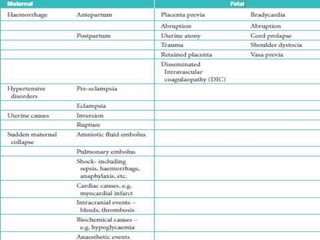
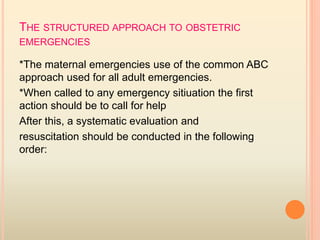
Obstetric emergencies
Done by:
*Safa kadhim
*Rafal mohammed
DEFINITION
An emergency is defined as a
serious situation
or occurrence that happens
unexpectedly
and demands immediate
action.
CLASSIFICATION :
Emergency of pregnancy can be classified
according to the causes into:
*maternal causes
*fetal causes
THE STRUCTURED APPROACH TO OBSTETRIC
EMERGENCIES
*The maternal emergencies use of the common ABC
approach used for all adult emergencies.
*When called to any emergency sitiuation the first
action should be to call for help
After this, a systematic evaluation and
resuscitation should be conducted in the following
order:
1. Airway
2. Breathing and ventilation
3. Circulation with volume replacement and
control of bleeding
4. Disability
5. Environment and exposure
MANAGEMENT OF SPECIFIC OBSTETRIC
EMERGENCIES
*Haemorrhage
Obstetric haemorrhage can occur antenatally or
post-natally, and both can present as obstetric
emergencies.
ANTEPARTUM HAEMORRHAGE
*Antepartum haemorrhage (APH) is any bleeding
occurring in the antenatal period after 20 weeks of
gestation
*The causes of APH are
placental abruption
placenta praevia
Other cause
*When assessing patients presenting with an APH, a
digital examination should not be conducted
until an ultrasound scan has identified the location
of the placenta.
PLACENTA PRAEVIA
*Placenta praevia is defined as a placenta that has
implanted into the lower segment of the uterus.
*It classified into:
-Major: in which the placenta is covering the cervical
os.
-Minor: when the placenta is sited within the lower
segment of the uterus , but doesn’t cover the
internal os.
RISK FACTORS
- older (often multiparous) women
- women with previous uterine surgery
•In women who have a previous cs, there is a risk
that the placenta implants into, and thus invades, into
the previous scar. This is called a ‘morbidly adherent
placenta’ and there are three types:
1. Placenta accreta. Placenta is abnormally
adherent to the uterine wall.
2. Placenta increta. Placenta is abnormally
invading into the uterine wall.
3. Placenta percreta. Placenta is invading through
the uterine wall.
The risk of a morbidly adherent placenta increases with
increasing numbers of previous Caesarean sections
DIAGNOSIS
*The mother will present with painless bleeding,
often recurrent in third trimester , and ultrasound
scans will demonstrate the abnormal location of the
placenta.
*A digital examination is contraindicated as this can
precipitate bleeding
MANAGEMENT
Treatment of placenta praevia depend on many
factors
1- amount of vaginal bleeding
2- whether bleeding has stopped
3- gestational age
4-health of mother and fetus
5-position of placenta
-The patient should be initially resuscitated using the
structured approach of ABC
If the bleeding is relatively minor and the fetus
uncompromised, the patient should be admitted for
observation and not allowed home until at least 24
hours has passed without further bleeding.
Major bleeding will require fluid resuscitation and
delivery of the fetus by Caesearean section by a
senior obstetrician..
The indications for delivery are reaching 37–38
weeks
Management with close observation is indicated in
situations where the fetal gestational age is less
than 36weeks of gestation as long as reassuring
fetal monitoring is present and vaginal bleeding has
resolved or significantly decreased.
If gestational age is less than 34weeks we should
Administer of betamethasone
MODE DELIVERY
1- a vaginal delivery if the placenta is a more than 2
cm away from the cervical os.
2- Caesarean section if the placenta is less than 2
cm away from the cervical os.
PLACENTAL ABRUPTION
A placental abruption is separation of a normally
sited placenta from the uterine wall.
diagnosis
*typically presents as vaginal bleeding associated
with pain.
The pain can be constant ,or as frequent short-
lasting contractions caused by the irritable effect
of blood within the uterus.
MANAGEMENT
1.The patient should be initially resuscitated using
the structured approach of ABC.
2.If the fetus will dead,
vaginal delivery can be accelerated by artificial
rupture of the membranes once the mother is
stable.
3.If the fetus is alive ,delivery without compromising
the mother is urgent and this will usually done be by
cs.
POSTPARTUM HAEMORRHAGE
It is defined as:
• Primary PPH. Loss of 500 mL blood from the
genital tract within 24 hours of delivery;
• Secondary PPH. Loss of 500 mL blood from the
genital tract between 24 hours and 12 weeks post
delivery .
*Post partum haemorrage divided into 2 types:
1.Minor:between 500ml_1000ml
2.Major:greater than 1000ml
-In practice, blood losses between
500 and 1000 mL are relatively common, and can
usually be tolerated well by the woman.
-Thus, it has been suggested that losses over
1000 mL should trigger emergency PPH protocols.
-However, it should be remembered that estimation
of blood loss is inaccurate,and if
a woman demonstrates evidence of cardiovascular
compromise, such as
tachycardia, or if there is continued bleeding, then
protocols should be instituted even if estimated
losses are less than 1000 mL.
CAUSE OF POST PARTUM HEAMORRAGE
DIAGNOSIS
- Early recognition of blood loss and rapid action is vital in
the management of PPH.
- recognition of the maternal signs of cardiovascular
compromise are vital. These include a tachycardia, low
blood pressure, symptoms of nausea, vomiting and
feeling faint, pallor and slow capillary refill (greater than
2 seconds).
- It is important to recognize that young, fit women have
the capacity to tolerate large amounts of blood loss
without demonstrating many clinical symptoms. -The
earliest symptom will be a tachycardia and often blood
pressure does not fall until massive haemorrhage has
occurred (often 1200–1500 mL of blood).
MANAGEMENT
diagnosis and management of PPH occur simultaneously
- The structured ABC approach should be instituted.
- Rapid fluid resuscitation should occur at the same time
as assessing and treating the cause.
- Since uterine atony is the most common cause, the
uterus should be massaged to encourage contraction
and oxytocics given
- Bimanual compression and more potent drugs can also
be used. These include ergometrine, prostaglandin F2α
or misoprostol.
The bladder should be catheterized as an empty bladder
aids uterine contraction
- A vaginal examination should be conducted to expel clots
which will prevent contraction of theuterus and assess for
genital tract trauma.
- Any identified tears will need prompt compression to limit blood
loss followed by repair
- The placenta should be delivered if retained and inspected
- If bleeding continues, the patient should be transferred to
theatre to allow a further thorough examination under
anaesthetic. This will also allow the use of further measures,
including uterine tamponade using uterine balloons,
radiological occlusion of the uterine vessels, laparotomy for
bilateral iliac artery ligation, uterine compression sutures, and,
as a last resort, hysterectomy.
- Massive PPH will require correction of clotting factors using
fresh frozen plasma, platelets and cryoprecipitate.
HYPERTENSIVE DISORDERS
Pre-eclampsia is a disease of pregnancy
characterized by a blood pressure of 140/90 mmHg
or more on two separate occasions after the 20th
week of pregnancy in a previously normotensive
woman. This is accompanied by significant
proteinuria (300 mg in 24 hours).
Eclampsia is the same condition that has proceeded
to the presence of convulsions
- the structured ABC approach should be used.
-Call for help.
-In a woman with severe pre-eclampsia, the airway and breathing are likely
to be secure. However, if a seizure has occurred, these will need
assessment and treatment. Seizures occurring due to eclampsia are
usually short-lasting and self-limiting. However, the patient should be
moved to the side (recovery position) and oxygen applied. Large bore
intravenous cannulae should be sited and blood taken for full blood
count, clotting studies, renal and liver function tests and cross-matching
. -The mother’s condition needs to be stabilized urgently, before considering
delivery in antenatal cases. Stabilizing the mother’s condition will involve
blood pressure control, prevention and treatment of fits and management
of fluid balance. Intravenous magnesium sulphate is used to treat and
prevent fits. An initial loading dose of 4 g is given, followed by an infusion
of 1 g/hour. Magnesium sulphate can also lower blood pressure and
cause some maternal side effects, such as flushing. It is important to
recognize that overdose of magnesium sulphate can cause respiratory
and cardiac depression
This can be reversed using calcium gluconate.
MANAGEMENT
The blood pressure should be reduced to safe levels
 Antihypertensives can be either labetalol (can be given
orally while intravenous access is obtained), oral
nifedipine or intravenous hydralazine
 If given intravenously, a bolus is initially used followed
by an infusion that can be titrated to obtain a safe blood
pressure.
 Maternal observations should be conducted frequently
until the mother has stabilized (every 5–15 minutes
depending on condition) and continuous fetal monitoring
used.
 A gradual reduction of the blood pressure is optimal to
avoid precipitating fetal distress secondary to sudden
drops in maternal blood pressure that reduce uterine
blood flow.
TIMING OF DELIVER
depends upon
1- gestational age
2- the presence of other complicating factors,
3- the severity of the disease
4-the stability of the patient’s condition
-- When it is clear that early delivery is likely, if the
gestation is less than 34 weeks, steroids should be
given to improve lung maturity and decrease
neonatal complications
Delivery is often by Caesarean section, although if
labour is well established, vaginal delivery is
possible. If at all possible, clotting disorders must
be corrected before delivery is attempted.
Postpartum, both fulminating pre-eclampsia and
eclampsia may occur. Management is as for the
antenatal case except that delivery has already
taken place.
*UTERINE CAUSES OF OBSTETRIC EMERGENCY
:-Uterine rupture
Uterine rupture, or a tear in the uterus usually
occur due to a previous uterine injury.It
occurs mainly in association with a previous
Caesarean section .
*other causes or risk factors includes:
-Previous uterine surgery (such as a
surgical evacuation of retained products of
conception
resulting in a perforation).
-Induction and augmentation of labour
-High parity
-Macrosomic fetus
-Placenta percreta
-Fetal version, e.g. breech extraction
-Congenital uterine anomaly, e.g. unicornuate
uterus
DIAGNOSIS
1. The patient may complain of abdominal pain (‘scar
tenderness’,often not masked epidural analgesia)
2. vaginal bleeding.
3.Haematuria may be present if
the uterus has ruptured into the bladder 4.Typically,
contractions stop and decelerationsare present
on the cardiotocography.
5.If the rupture occurs in the late second stage of
labour, it may not be recognized immediately but
usaully the fetus deliver by forceps or ventouse due
to abnormal CTG
In the immediate postnatal peroid,the mother
bleeds internally and shows signs of circulatory
collapse while complaining of abdominal discomfort.
MANAGEMENT
1.Immediate resuscitation of ABC is required
2.Immediate laparotomy to deliver the baby and
repair the uterine is required. Frequently,
the only safe treatment is hysterectomy.
-Uterine inversion
*Uterine inversion is a rare complication
occurring during the third stage of labour.
*It has a reported incidence of between
1:2000 and 1:6000.
*The uterine fundus descends either the
uterine cavity, through
the cervix, and very rarely beyond the
introitus.
CAUSES
-traction on the umbilical cord before the placenta
has separated
-after vaginal deliveries or Caesarean section.
-Associated factors are a fundal placenta, a short
cord and a morbidly adherent placenta.
DIAGNOSIS
*The woman will demonstrate signs of cardiovascular
collapse and shock due to the prolapsed uterus
stretching the cervix causes
vagal stimulation.
*Haemorrhage and the associated syptoms depend
on the severity of blood loss.
*Inverted uterus may be obvious at the introitus.
*On abdominal examination :lack of a palpable uterus
in the abdomen or the feeling of a ‘dimple
’ in the uterine fundus.
MANAGEMENT
1.Resuscitate the patient using the ABC approach.
2.If the placenta is still attached,its very important not
to remove as this will increase the bleeding.
3.Immediately replace the uterus through
the cervix by manual compression.
4.If that fail,hydrostatic pressure can be applied by
pouring warmed saline into the vagina,usaully via
sil cup ventouse.
5. Tocolysis may be helpful to relax the uterine ring.
6.If failed, then do surgical methods to repositon of
uterus.and uterine contraction should maintain with
an oxytotic.
*SUDDEN MATERNAL COLLAPSE
Sudden collapse, as in non-pregnant adults, occurs
due to many reasons. Some will be benign,
such as a vasaovagal attack (simple faint) or an
epileptic fit in a known epileptic, but other causes
are life threatening. The management approach
should be the same,structured ABC approach.
-Pulmonary embolism
common in the puerperium, but it can occur at any
time in the antenatal and post-natal period
*Diagnosis and management
PE can be a cause of sudden cardiorespiratory
collapse. In this situation, diagnosis and management
should occur simultaneously.
Urgent resuscitation using the structured ABC
approach is needed. If PE is suspected,
anticoagulation should be instituted
-Amniotic fluid embolism:
It is a rare cause of maternal collapse specific to
pregnancy, believed to be caused by aminotic fluid
entering the maternal circulation. This cause
cardiorespiratory compromise and sever DIC. In
some cases, there may be an abnormal maternal
reaction to amniotic fluid as the primary event.
It is difficult to diagnose in life, and is typically
diagnosed at postmortem with presence of fetal
cells(squames or hair) in the maternal pulmonary
capillaries.
DIAGNOSIS AND MANAGEMENT
Symptoms occurring just before the collapse may
be helpful in diagnosis, these include:
.breathlessness
• chest pain
• feeling cold
• lightheadedness
• restlessness, distress and panic
• pins and needles in the fingers
• nausea and vomiting
management should be ABC approach.
The prognosis is poor, with around 30 per cent
of patients dying in the first hour and only 10 per
cent surviving overall. Management is supportive,
requiring intensive care and there are no specific
therapies available
*FETAL EMERGENCIES
*The fetus may be severely affected by any of
the preceding maternal emergencies that occur
before delivery.
*Major abnormalities of the fetal heart rate, in
particular prolonged fetal bradycardia, call for
immediate delivery, usually by Caesarean section.
*The two specific causes of fetal emergency:
cord prolapse and shoulder dystocia.
-cord prolapse(Umbilical cord accidents)
*A cord presentation : is defined as the presence of
umbilical cord below the fetal presenting part when
the membranes are intact.
*Cord prolapse :is the presence of the cord below the
presenting part when the membranes are ruptured.
occurs when the fetal presenting part does not fit well
into the maternal pelvis , giving
‘space’ for the cord to prolapse when the membranes
rupture.
Risk factors for cord prolapse
DIAGNOSIS
1.vaginal examination: most commonly, it is
diagnosed by seeing the cord at the introitus , or
feeling it.
2.abnormal fetal heart rate pattern may
suggest it, as compression of the umbilical vein
between the presenting part and the pelvis
3.reduces or stops the flow of oxygenated blood to
the fetus, causing deep variable decelerations then
bradycardia if the situation is not relieved
MANAGEMENT
*Immediate management aims to minimize the
pressure of the fetal presenting part on the cord,
while plans are made to deliver the baby.
*This is achieved by:
- applying pressure vaginally to push the presenting
part out of the pelvis
-or by filling the bladder with 500 mL of saline.
*However, if the cord is beyond the introitus it should
be replaced into the vagina to keep it warmer.
*Emergency Caesarean section is required unless the
cervix is fully dilated
and an assisted vaginal delivery can be safely and
easily performed
-Shoulder dystocia
Shoulder dystocia occurs when a baby’s head passes
through the birth canal and their shoulders become
stuck during labor. This prevents the doctor from
fully delivering the baby and can extend the length
of time for delivery.
RISK FACTORS FOR SHOULDER DYSTOCIA
*diabetes and gestational diabetes
*history of having a baby with large birth weight, or
macrosomia
*history of shoulder dystocia
*labor that’s induced (oxytocin)
* obese
*birth after the due date (post maturity)
*operative vaginal birth, uses forceps or a vacuum.
*pregnant with multiple babies.
*short stature.
COMPLICATIONS OF SHOULDER DYSTONICA
Fetal complications:
-fetal hypoxia lead to loss of O2 into the brain
,which can cause brain damage.
-death
-fetal trauma, in the form of fractures (usually
long bones of the arm,hand,
or clavicle), or brachial plexus
injury.
Mother complications:
-excessive bleeding.
-tearing of a mother’s tissues, such as the
cervix, rectum, uterus, or vagina.
-perineal trauma including 3rd and 4th dgree
tears.
DIAGNOSIS
*Diagnosis is usually obvious when the shoulders
fail to deliver during the next contraction after
delivery of the head.
*It is sometimes preceded by the ‘turtle sign’
which is the head appearing to be pulled back on to
the perineum at delivery.
MANAGEMENT
Shoulder dystocia is managed by a sequence of
manoeuvres designed to facilitate delivery while
minimizing the risk of fetal damage.
The basic principle of all the manoeuvres is to reduce
the anterior _posterior diameter of the
shoulders and to max. space in the maternal pelvis.
DOCTORS USE A MNEMONIC “HELPERR” AS A
GUIDE FOR TREATING SHOULDER DYSTOCIA:
*“H” for help: This involves preparing for the help of
an obstetrician, for anesthesia, and for pediatrics
for subsequent resuscitation of the infant that may
be needed if the methods below fail.
*“E” for episiotomy.
*“L” for legs hyperflexion (McRoberts' maneuver): It
helps to flatten and rotate pelvis, which may help
baby pass through more easily.
*“P” for suprapubic pressure: to encourage baby’s
shoulder to rotate.
*“E” for enter maneuvers: This means helping to
rotate baby’s shoulders to where they can pass
through more easily. Another term for this is internal
rotation.
*“R” for remove the posterior arm from the birth
canal: If one of the baby’s arms pass free from the
birth canal, this makes it easier for baby’s shoulders
to pass through the birth canal.
*“R” for roll the patient: This means asking the
mother to get on her hands and knees. This
movement can help the baby to pass more easily
through the birth canal.
After delivery of the baby, the risks of maternal
morbidity should be remembered: prevent the PPH
and check for vaginal trauma.
Women will require debriefing after the delivery,
and most obstetricians would suggest a CS in the
next pregnancy.
Obestitric emergency

Obestitric emergency

  • 1.
  • 2.
    DEFINITION An emergency isdefined as a serious situation or occurrence that happens unexpectedly and demands immediate action.
  • 3.
    CLASSIFICATION : Emergency ofpregnancy can be classified according to the causes into: *maternal causes *fetal causes
  • 5.
    THE STRUCTURED APPROACHTO OBSTETRIC EMERGENCIES *The maternal emergencies use of the common ABC approach used for all adult emergencies. *When called to any emergency sitiuation the first action should be to call for help After this, a systematic evaluation and resuscitation should be conducted in the following order:
  • 6.
    1. Airway 2. Breathingand ventilation 3. Circulation with volume replacement and control of bleeding 4. Disability 5. Environment and exposure
  • 7.
    MANAGEMENT OF SPECIFICOBSTETRIC EMERGENCIES *Haemorrhage Obstetric haemorrhage can occur antenatally or post-natally, and both can present as obstetric emergencies.
  • 8.
    ANTEPARTUM HAEMORRHAGE *Antepartum haemorrhage(APH) is any bleeding occurring in the antenatal period after 20 weeks of gestation *The causes of APH are placental abruption placenta praevia Other cause *When assessing patients presenting with an APH, a digital examination should not be conducted until an ultrasound scan has identified the location of the placenta.
  • 9.
    PLACENTA PRAEVIA *Placenta praeviais defined as a placenta that has implanted into the lower segment of the uterus. *It classified into: -Major: in which the placenta is covering the cervical os. -Minor: when the placenta is sited within the lower segment of the uterus , but doesn’t cover the internal os.
  • 10.
    RISK FACTORS - older(often multiparous) women - women with previous uterine surgery •In women who have a previous cs, there is a risk that the placenta implants into, and thus invades, into the previous scar. This is called a ‘morbidly adherent placenta’ and there are three types: 1. Placenta accreta. Placenta is abnormally adherent to the uterine wall. 2. Placenta increta. Placenta is abnormally invading into the uterine wall. 3. Placenta percreta. Placenta is invading through the uterine wall. The risk of a morbidly adherent placenta increases with increasing numbers of previous Caesarean sections
  • 11.
    DIAGNOSIS *The mother willpresent with painless bleeding, often recurrent in third trimester , and ultrasound scans will demonstrate the abnormal location of the placenta. *A digital examination is contraindicated as this can precipitate bleeding
  • 12.
    MANAGEMENT Treatment of placentapraevia depend on many factors 1- amount of vaginal bleeding 2- whether bleeding has stopped 3- gestational age 4-health of mother and fetus 5-position of placenta
  • 13.
    -The patient shouldbe initially resuscitated using the structured approach of ABC If the bleeding is relatively minor and the fetus uncompromised, the patient should be admitted for observation and not allowed home until at least 24 hours has passed without further bleeding. Major bleeding will require fluid resuscitation and delivery of the fetus by Caesearean section by a senior obstetrician.. The indications for delivery are reaching 37–38 weeks
  • 14.
    Management with closeobservation is indicated in situations where the fetal gestational age is less than 36weeks of gestation as long as reassuring fetal monitoring is present and vaginal bleeding has resolved or significantly decreased. If gestational age is less than 34weeks we should Administer of betamethasone
  • 15.
    MODE DELIVERY 1- avaginal delivery if the placenta is a more than 2 cm away from the cervical os. 2- Caesarean section if the placenta is less than 2 cm away from the cervical os.
  • 16.
    PLACENTAL ABRUPTION A placentalabruption is separation of a normally sited placenta from the uterine wall. diagnosis *typically presents as vaginal bleeding associated with pain. The pain can be constant ,or as frequent short- lasting contractions caused by the irritable effect of blood within the uterus.
  • 17.
    MANAGEMENT 1.The patient shouldbe initially resuscitated using the structured approach of ABC. 2.If the fetus will dead, vaginal delivery can be accelerated by artificial rupture of the membranes once the mother is stable. 3.If the fetus is alive ,delivery without compromising the mother is urgent and this will usually done be by cs.
  • 18.
    POSTPARTUM HAEMORRHAGE It isdefined as: • Primary PPH. Loss of 500 mL blood from the genital tract within 24 hours of delivery; • Secondary PPH. Loss of 500 mL blood from the genital tract between 24 hours and 12 weeks post delivery . *Post partum haemorrage divided into 2 types: 1.Minor:between 500ml_1000ml 2.Major:greater than 1000ml
  • 19.
    -In practice, bloodlosses between 500 and 1000 mL are relatively common, and can usually be tolerated well by the woman. -Thus, it has been suggested that losses over 1000 mL should trigger emergency PPH protocols. -However, it should be remembered that estimation of blood loss is inaccurate,and if a woman demonstrates evidence of cardiovascular compromise, such as tachycardia, or if there is continued bleeding, then protocols should be instituted even if estimated losses are less than 1000 mL.
  • 20.
    CAUSE OF POSTPARTUM HEAMORRAGE
  • 21.
    DIAGNOSIS - Early recognitionof blood loss and rapid action is vital in the management of PPH. - recognition of the maternal signs of cardiovascular compromise are vital. These include a tachycardia, low blood pressure, symptoms of nausea, vomiting and feeling faint, pallor and slow capillary refill (greater than 2 seconds). - It is important to recognize that young, fit women have the capacity to tolerate large amounts of blood loss without demonstrating many clinical symptoms. -The earliest symptom will be a tachycardia and often blood pressure does not fall until massive haemorrhage has occurred (often 1200–1500 mL of blood).
  • 22.
    MANAGEMENT diagnosis and managementof PPH occur simultaneously - The structured ABC approach should be instituted. - Rapid fluid resuscitation should occur at the same time as assessing and treating the cause. - Since uterine atony is the most common cause, the uterus should be massaged to encourage contraction and oxytocics given - Bimanual compression and more potent drugs can also be used. These include ergometrine, prostaglandin F2α or misoprostol. The bladder should be catheterized as an empty bladder aids uterine contraction
  • 23.
    - A vaginalexamination should be conducted to expel clots which will prevent contraction of theuterus and assess for genital tract trauma. - Any identified tears will need prompt compression to limit blood loss followed by repair - The placenta should be delivered if retained and inspected - If bleeding continues, the patient should be transferred to theatre to allow a further thorough examination under anaesthetic. This will also allow the use of further measures, including uterine tamponade using uterine balloons, radiological occlusion of the uterine vessels, laparotomy for bilateral iliac artery ligation, uterine compression sutures, and, as a last resort, hysterectomy. - Massive PPH will require correction of clotting factors using fresh frozen plasma, platelets and cryoprecipitate.
  • 24.
    HYPERTENSIVE DISORDERS Pre-eclampsia isa disease of pregnancy characterized by a blood pressure of 140/90 mmHg or more on two separate occasions after the 20th week of pregnancy in a previously normotensive woman. This is accompanied by significant proteinuria (300 mg in 24 hours). Eclampsia is the same condition that has proceeded to the presence of convulsions
  • 25.
    - the structuredABC approach should be used. -Call for help. -In a woman with severe pre-eclampsia, the airway and breathing are likely to be secure. However, if a seizure has occurred, these will need assessment and treatment. Seizures occurring due to eclampsia are usually short-lasting and self-limiting. However, the patient should be moved to the side (recovery position) and oxygen applied. Large bore intravenous cannulae should be sited and blood taken for full blood count, clotting studies, renal and liver function tests and cross-matching . -The mother’s condition needs to be stabilized urgently, before considering delivery in antenatal cases. Stabilizing the mother’s condition will involve blood pressure control, prevention and treatment of fits and management of fluid balance. Intravenous magnesium sulphate is used to treat and prevent fits. An initial loading dose of 4 g is given, followed by an infusion of 1 g/hour. Magnesium sulphate can also lower blood pressure and cause some maternal side effects, such as flushing. It is important to recognize that overdose of magnesium sulphate can cause respiratory and cardiac depression This can be reversed using calcium gluconate. MANAGEMENT
  • 26.
    The blood pressureshould be reduced to safe levels  Antihypertensives can be either labetalol (can be given orally while intravenous access is obtained), oral nifedipine or intravenous hydralazine  If given intravenously, a bolus is initially used followed by an infusion that can be titrated to obtain a safe blood pressure.  Maternal observations should be conducted frequently until the mother has stabilized (every 5–15 minutes depending on condition) and continuous fetal monitoring used.  A gradual reduction of the blood pressure is optimal to avoid precipitating fetal distress secondary to sudden drops in maternal blood pressure that reduce uterine blood flow.
  • 27.
    TIMING OF DELIVER dependsupon 1- gestational age 2- the presence of other complicating factors, 3- the severity of the disease 4-the stability of the patient’s condition -- When it is clear that early delivery is likely, if the gestation is less than 34 weeks, steroids should be given to improve lung maturity and decrease neonatal complications
  • 28.
    Delivery is oftenby Caesarean section, although if labour is well established, vaginal delivery is possible. If at all possible, clotting disorders must be corrected before delivery is attempted. Postpartum, both fulminating pre-eclampsia and eclampsia may occur. Management is as for the antenatal case except that delivery has already taken place.
  • 29.
    *UTERINE CAUSES OFOBSTETRIC EMERGENCY :-Uterine rupture Uterine rupture, or a tear in the uterus usually occur due to a previous uterine injury.It occurs mainly in association with a previous Caesarean section . *other causes or risk factors includes: -Previous uterine surgery (such as a surgical evacuation of retained products of conception resulting in a perforation).
  • 30.
    -Induction and augmentationof labour -High parity -Macrosomic fetus -Placenta percreta -Fetal version, e.g. breech extraction -Congenital uterine anomaly, e.g. unicornuate uterus
  • 31.
    DIAGNOSIS 1. The patientmay complain of abdominal pain (‘scar tenderness’,often not masked epidural analgesia) 2. vaginal bleeding. 3.Haematuria may be present if the uterus has ruptured into the bladder 4.Typically, contractions stop and decelerationsare present on the cardiotocography. 5.If the rupture occurs in the late second stage of labour, it may not be recognized immediately but usaully the fetus deliver by forceps or ventouse due to abnormal CTG
  • 32.
    In the immediatepostnatal peroid,the mother bleeds internally and shows signs of circulatory collapse while complaining of abdominal discomfort.
  • 33.
    MANAGEMENT 1.Immediate resuscitation ofABC is required 2.Immediate laparotomy to deliver the baby and repair the uterine is required. Frequently, the only safe treatment is hysterectomy.
  • 34.
    -Uterine inversion *Uterine inversionis a rare complication occurring during the third stage of labour. *It has a reported incidence of between 1:2000 and 1:6000. *The uterine fundus descends either the uterine cavity, through the cervix, and very rarely beyond the introitus.
  • 35.
    CAUSES -traction on theumbilical cord before the placenta has separated -after vaginal deliveries or Caesarean section. -Associated factors are a fundal placenta, a short cord and a morbidly adherent placenta.
  • 36.
    DIAGNOSIS *The woman willdemonstrate signs of cardiovascular collapse and shock due to the prolapsed uterus stretching the cervix causes vagal stimulation. *Haemorrhage and the associated syptoms depend on the severity of blood loss. *Inverted uterus may be obvious at the introitus. *On abdominal examination :lack of a palpable uterus in the abdomen or the feeling of a ‘dimple ’ in the uterine fundus.
  • 37.
    MANAGEMENT 1.Resuscitate the patientusing the ABC approach. 2.If the placenta is still attached,its very important not to remove as this will increase the bleeding. 3.Immediately replace the uterus through the cervix by manual compression. 4.If that fail,hydrostatic pressure can be applied by pouring warmed saline into the vagina,usaully via sil cup ventouse. 5. Tocolysis may be helpful to relax the uterine ring. 6.If failed, then do surgical methods to repositon of uterus.and uterine contraction should maintain with an oxytotic.
  • 38.
    *SUDDEN MATERNAL COLLAPSE Suddencollapse, as in non-pregnant adults, occurs due to many reasons. Some will be benign, such as a vasaovagal attack (simple faint) or an epileptic fit in a known epileptic, but other causes are life threatening. The management approach should be the same,structured ABC approach.
  • 39.
    -Pulmonary embolism common inthe puerperium, but it can occur at any time in the antenatal and post-natal period *Diagnosis and management PE can be a cause of sudden cardiorespiratory collapse. In this situation, diagnosis and management should occur simultaneously. Urgent resuscitation using the structured ABC approach is needed. If PE is suspected, anticoagulation should be instituted
  • 40.
    -Amniotic fluid embolism: Itis a rare cause of maternal collapse specific to pregnancy, believed to be caused by aminotic fluid entering the maternal circulation. This cause cardiorespiratory compromise and sever DIC. In some cases, there may be an abnormal maternal reaction to amniotic fluid as the primary event. It is difficult to diagnose in life, and is typically diagnosed at postmortem with presence of fetal cells(squames or hair) in the maternal pulmonary capillaries.
  • 41.
    DIAGNOSIS AND MANAGEMENT Symptomsoccurring just before the collapse may be helpful in diagnosis, these include: .breathlessness • chest pain • feeling cold • lightheadedness • restlessness, distress and panic • pins and needles in the fingers • nausea and vomiting
  • 42.
    management should beABC approach. The prognosis is poor, with around 30 per cent of patients dying in the first hour and only 10 per cent surviving overall. Management is supportive, requiring intensive care and there are no specific therapies available
  • 43.
    *FETAL EMERGENCIES *The fetusmay be severely affected by any of the preceding maternal emergencies that occur before delivery. *Major abnormalities of the fetal heart rate, in particular prolonged fetal bradycardia, call for immediate delivery, usually by Caesarean section. *The two specific causes of fetal emergency: cord prolapse and shoulder dystocia.
  • 44.
    -cord prolapse(Umbilical cordaccidents) *A cord presentation : is defined as the presence of umbilical cord below the fetal presenting part when the membranes are intact. *Cord prolapse :is the presence of the cord below the presenting part when the membranes are ruptured. occurs when the fetal presenting part does not fit well into the maternal pelvis , giving ‘space’ for the cord to prolapse when the membranes rupture.
  • 45.
    Risk factors forcord prolapse
  • 46.
    DIAGNOSIS 1.vaginal examination: mostcommonly, it is diagnosed by seeing the cord at the introitus , or feeling it. 2.abnormal fetal heart rate pattern may suggest it, as compression of the umbilical vein between the presenting part and the pelvis 3.reduces or stops the flow of oxygenated blood to the fetus, causing deep variable decelerations then bradycardia if the situation is not relieved
  • 47.
    MANAGEMENT *Immediate management aimsto minimize the pressure of the fetal presenting part on the cord, while plans are made to deliver the baby. *This is achieved by: - applying pressure vaginally to push the presenting part out of the pelvis -or by filling the bladder with 500 mL of saline. *However, if the cord is beyond the introitus it should be replaced into the vagina to keep it warmer.
  • 48.
    *Emergency Caesarean sectionis required unless the cervix is fully dilated and an assisted vaginal delivery can be safely and easily performed
  • 49.
    -Shoulder dystocia Shoulder dystociaoccurs when a baby’s head passes through the birth canal and their shoulders become stuck during labor. This prevents the doctor from fully delivering the baby and can extend the length of time for delivery.
  • 50.
    RISK FACTORS FORSHOULDER DYSTOCIA *diabetes and gestational diabetes *history of having a baby with large birth weight, or macrosomia *history of shoulder dystocia *labor that’s induced (oxytocin) * obese *birth after the due date (post maturity) *operative vaginal birth, uses forceps or a vacuum. *pregnant with multiple babies. *short stature.
  • 51.
    COMPLICATIONS OF SHOULDERDYSTONICA Fetal complications: -fetal hypoxia lead to loss of O2 into the brain ,which can cause brain damage. -death -fetal trauma, in the form of fractures (usually long bones of the arm,hand, or clavicle), or brachial plexus injury.
  • 52.
    Mother complications: -excessive bleeding. -tearingof a mother’s tissues, such as the cervix, rectum, uterus, or vagina. -perineal trauma including 3rd and 4th dgree tears.
  • 53.
    DIAGNOSIS *Diagnosis is usuallyobvious when the shoulders fail to deliver during the next contraction after delivery of the head. *It is sometimes preceded by the ‘turtle sign’ which is the head appearing to be pulled back on to the perineum at delivery.
  • 54.
    MANAGEMENT Shoulder dystocia ismanaged by a sequence of manoeuvres designed to facilitate delivery while minimizing the risk of fetal damage. The basic principle of all the manoeuvres is to reduce the anterior _posterior diameter of the shoulders and to max. space in the maternal pelvis.
  • 55.
    DOCTORS USE AMNEMONIC “HELPERR” AS A GUIDE FOR TREATING SHOULDER DYSTOCIA: *“H” for help: This involves preparing for the help of an obstetrician, for anesthesia, and for pediatrics for subsequent resuscitation of the infant that may be needed if the methods below fail. *“E” for episiotomy. *“L” for legs hyperflexion (McRoberts' maneuver): It helps to flatten and rotate pelvis, which may help baby pass through more easily. *“P” for suprapubic pressure: to encourage baby’s shoulder to rotate.
  • 56.
    *“E” for entermaneuvers: This means helping to rotate baby’s shoulders to where they can pass through more easily. Another term for this is internal rotation. *“R” for remove the posterior arm from the birth canal: If one of the baby’s arms pass free from the birth canal, this makes it easier for baby’s shoulders to pass through the birth canal. *“R” for roll the patient: This means asking the mother to get on her hands and knees. This movement can help the baby to pass more easily through the birth canal.
  • 57.
    After delivery ofthe baby, the risks of maternal morbidity should be remembered: prevent the PPH and check for vaginal trauma. Women will require debriefing after the delivery, and most obstetricians would suggest a CS in the next pregnancy.