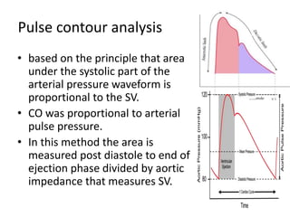
2. • Cardiac output (CO) is the volume of blood
ejected by each ventricle per minute and is
the product of stroke volume and heart rate.
• CO can thus be manipulated by alteration in
heart rate or rhythm, preload, contractility
and afterload.
• thermodilution method using pulmonary
artery catheter (PAC) is till date considered as
gold standard method.
3. • here are various methods of CO monitoring based on
a) Ficks principle,
b) thermodilution,
c) Doppler,
d) pulse contour analysis and
e) bioimpedance.
An ideal CO monitor should be
1. minimally or non-invasive,
2. continuous,
3. cost effective,
4. reproducible,
5. reliable during various physiological states and
6. have fast response time
4. • Methods of CO monitoring are broadly classified
as follows:
1. Invasive-Intermittent bolus pulmonary artery
thermodilution, Continuous pulmonary artery
thermodilution;
2. Minimally invasive-Lithium dilution CO (LiDCO),
Pulse contour analysis CO (PiCCO and FloTrac),
Esophgeal Doppler (ED), transesophgeal
echocardiography (TEE);
3. Non-invasive-Partial gas rebreathing, Thoracic
bioimpedance and bioreactance, endotracheal
cardiac output monitor (ECOM), Doppler
method and Photoelectric plethysmography
5. INVASIVE METHODS: Cardiac output
measurement by pulmonary artery
catheter
• still considered as gold standard monitor to
measure CO since 1970’s.
• its use has been associated with various
complications like pneumothorax, arrhythmia,
infection, pulmonary artery rupture, valve injury,
knotting and thrombosis leading to embolism.
• various technical errors may lead to false
readings , Moreover, intracardiac shunts,
mechanical ventilation or valvular dysfunction
may lead to incorrect readings.
6. CONTINUOUS CO MEASUREMENT BY
PAC
• Continuous CO (CCO, Edwards Lifesciences, Irvine,
California, United States) is a modification of PAC with
copper filament in the catheter that remains in the
right ventricle.
• There is intermittent heating of blood in the right heart
by the filament and the resultant signal is captured by
thermistor near the tip of the catheter. Average value
of CO measured over time is displayed on the monitor.
• Main advantages of CCO over conventional PAC are
avoidance of repeated boluses thus reducing the
infection risk and operator errors.
7. • continuous monitoring of stroke volume (SV), systemic
vascular resistance (SVR) and mixed venous saturation
can also be performed with this catheter.
• PAC use increased mortality after myocardial
infarctionand SUPPORT trial also showed increased
mortalityat 30 d.
• PAC-MAN trial failed to show any benefit or harm with
the use of PAC.
• ESCAPE trial demonstrated functional improvement
with PAC guided therapy used in patients with
congestive heart failure.
• Inspite of various arguments PAC is still considered as
the “Gold Standard” for monitoring of CO.
8. MINIMALLY INVASIVE METHODS
• LiDCO(Lithium dilution CO ): system combines pulse
contour analysis with lithium indicator dilution for
continuous monitoring of SV and SV variation (SVV).
• first described in 1993.It is a minimally invasive
technique and requires a venous (central or peripheral)
line and an arterial catheter.
• A bolus of lithium chloride is injected into venous line
and arterial concentration is measured by withdrawing
blood across disposable lithium sensitive sensor
containing an ionophor selectively permeable to Li.
• CO is calculated based on Li dose and area according
to the concentration time circulation.
9. • It requires calibration every 8 h and during major
hemodynamic changes.
• It is contraindicated in patients on Li therapy
and calibration is also affected by neuromuscular
blockers as quaternary ammonium residue
causes electrode to drift.
• Its accuracy is affected by aortic regurgitation,
intraaortic balloon pump (IABP), damped arterial
line, postaortic surgery, arrhythmia and intra or
extracardiac shunts.
• in relation with PAC, found good correlation with
PAC BY Linton et al.
10. Pulse contour analysis
• based on the principle that area
under the systolic part of the
arterial pressure waveform is
proportional to the SV.
• CO was proportional to arterial
pulse pressure.
• In this method the area is
measured post diastole to end of
ejection phase divided by aortic
impedance that measures SV.
11. • It also measures SVV and pulse pressure
variation (PVV) which is useful in predicting
fluid responsiveness.
• SVV is the difference between maximum and
minimum SV over the respiratory cycle and is
caused by changes in preload with alteration
in intrathoracic pressure.
12. PICCO system
• The PiCCO system (PULSION medical system, Munich,
Germany) was the first pulse contour device introduced
and was replaced with PiCCO2 in 2007.
• It requires both central venous (femoral or internal jugular)
and arterial cannulation (femoral/radial).
• Indicator solution injected via central venous cannula and
blood temperature changes are detected by a thermistor
tip catheter placed in the artery. Thus, it combines pulse
contour analysis with the transpulmonary thermodilution
CO to determine hemodynamic variables.
• It requires manual calibration every 8 h and hourly during
hemodynamic instability.
13. • In addition, thermodilution curve can be used to
measure intrathoracic blood volume (ITBV), global end
diastolic volume (GEDV) and extravascular lung water
(EVLW).
• GEDV and ITBV are a measure of cardiac preload and
EVLW (interstitial, intracellular or intra alveolar) is a
mean to quantify pulmonary edema.
• It also measures SVV/PVV which is marker of fluid
responsiveness.
• Its accuracy may be affected be vascular compliance,
aortic impedence and peripheral arterial resistance.
• Valvular regurgitation, aortic aneurysm, significant
arrhythmia and rapidly changing temperature may also
affect its accuracy.
• tudies have found good correlation with PAC during
coronary artery bypass grafting
14. FloTrac system
• FloTrac (Edwards LifeSciences. Irvine, United States) is a
pulse contour device introduced in 2005 and is a minimally
invasive method as it requires only an arterial line (femoral
or radial).
• The system does not need any external calibration, is
operator independent and easy to use.
• based on the principle that there is a linear relationship
between the pulse pressure and SV.
• Good arterial waveform quality is a prerequisite for
accurate reading of CO. Accuracy is affected in patients
with significant arrhythmias, IABP or morbid obesity.
• Various studies have validated the efficacy of FloTrac with
PAC and find good correlation.
• However, in patients with low SVR undergoing liver
transplantation or septicemia it is not found as accurate as
PAC
15. Pressure recording analytic
method(PRAM)
• measures the area under the curve of arterial
waveform.
• Major advantage is that it does not require
external calibration and internal calibration is
done by morphology of the arterial waveform.
• However the accuracy of this method is still
not proven.
16. Esophageal doppler
• the midthoracic level it measures flow as it is presumed to
be parallel to the descending aorta. Since aorta is
considered as a cylinder, the flow can be measured by
multiplying cross-sectional area (CSA) and velocity.
• Doppler ultrasound is used to measure the SV.
• Major limiting factor is that it measures flow only in
descending thoracic aorta which is 70% of total flow. A
correction factor needs to be added to compensate aortic
arch flow.
• Moreover discrepancies in flow may be seen in aortic
coarctation, aneurysm or crossclamp, IABP and various
metabolic states. Various factors like changes in pulse
pressure, vascular compliance, volume status or inotropes
may affect the CSA.
17. • Various studies have compared ED with PAC
and found good agreement.
• ED has also been used in GDT and shown
greater improvement in SV and CO with faster
recovery and shorter length of stay.
18. TEE
• now been a widely used monitor in perioperative
setting.
• important tool for the assessment of cardiac
structures, filling status and cardiac contractility.
• Doppler technique is used to measure CO by
Simpson’s rule measuring SV multiplied by HR.
Flow is measured by area under the Doppler
velocity waveform that gives VTI( VELOCITY TIME
INTERFERANCE)
19. • Measurement can be done at the level of
pulmonary artery, mitral or aortic valve. TEE
views used for measurement are
midesophageal aortic long axis view and deep
transgastric long axis view with pulsed and
continuous wave Doppler respectively.
• It is a useful tool in hemodynamically
unstable patient under mechanical ventilation.
20. NON INVASIVE METHODS
Partial gas rebreathing:
• known as the NICO system (Novametrix Medical
Systems, Wallingford, Conn, United States) or
partial gas re-breathing monitor and uses indirect
Fick’s principle to calculate CO.
• It is used in intubated patients under mechanical
ventilation.
• At steady state, the amount of CO2 entering the
lungs via the pulmonary artery is proportional to
the CO and equals the amount exiting the lungs
via expiration and pulmonary veins
21. • CO is calculated according to following formula: CO =
VCO2/CvCO2 - CaCO2.
• VCO2 is CO2 consumption, CaCO2 and CvCO2 is arterial and
venous CO2 content respectively.
• The diffusion rate of carbon dioxide is 22 times more rapid
than that of oxygen, it is assumed that no difference in
venous CO2 (CvCO2) will occur, whether under normal or
rebreathing conditions.
• Major limitation is that tracheal intubation with fixed
ventilator setting is required.
• It is also not very accurate in patients with severe chest
trauma, significant intrapulmonary shunt, high CO states
and low minute ventilation
• not found accuracy of this device with PAC.
• Studies have shown underestimation preoperatively and
overestimation postoperatively after cardiac surgery
22. Thoracic bioimpedance
• Thoracic bioimpedance (TEB) is a non-invasive method of
CO monitoring.
• It is based on the hypothesis by considering
• thorax as a cylinder perfused with fluid with specific
resistivity.
• Electrodes six in number are placed (two on either side of
neck and four in lower thorax) on the patient and the
resistance to current flowing from the outermost to
innermost electrodes is measured. The bioimpedance is
indirectly proportional to the content of thoracic fluid.
Tissue fluid volume, pulmonary and venous blood, and the
aortic blood volume all contribute to the TEB
measurement. Changes in CO will change the amount of
aortic blood and will be reflected in a change TEB.
23. SV= VEPT × VET × EPCI
VEPT = volume of electrically participating tissue
(gender, height, and weight). VET = ventricular
ejection time taken from the R-R interval. EPCI =
ejection phase contractility index which is indirectly
proportional to TEB.
• Major limitations like interference with
electrocautery, proper electrode placement,
patient’s movements and arrhythmia may affect
its accuracy.
• Results were also not encouraging in critically ill
patients. Moreover, it has been considered as
trend analysis monitor rather than a diagnostic
one.