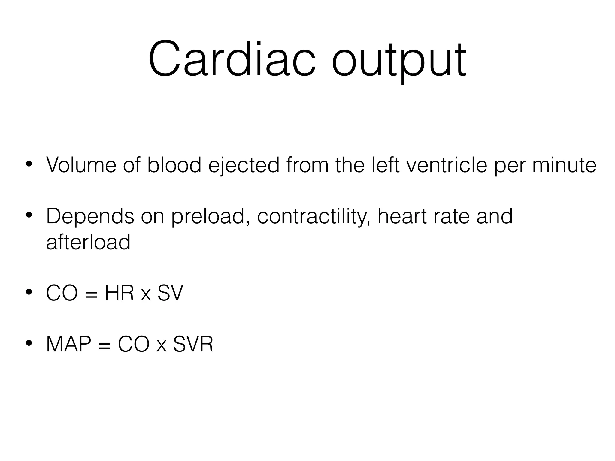
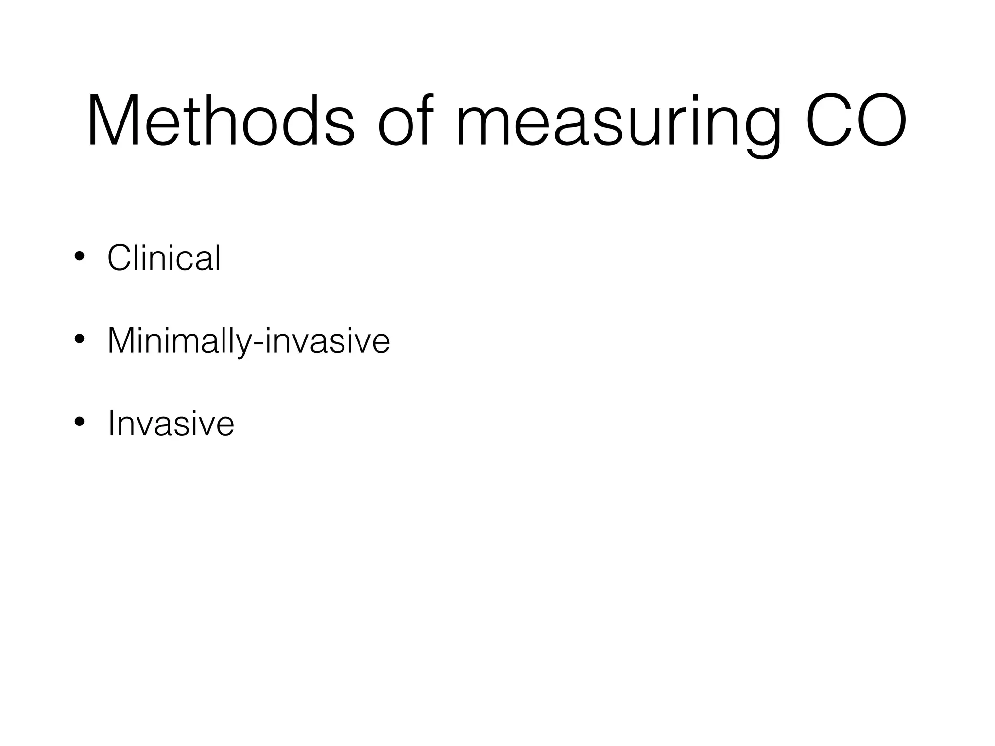
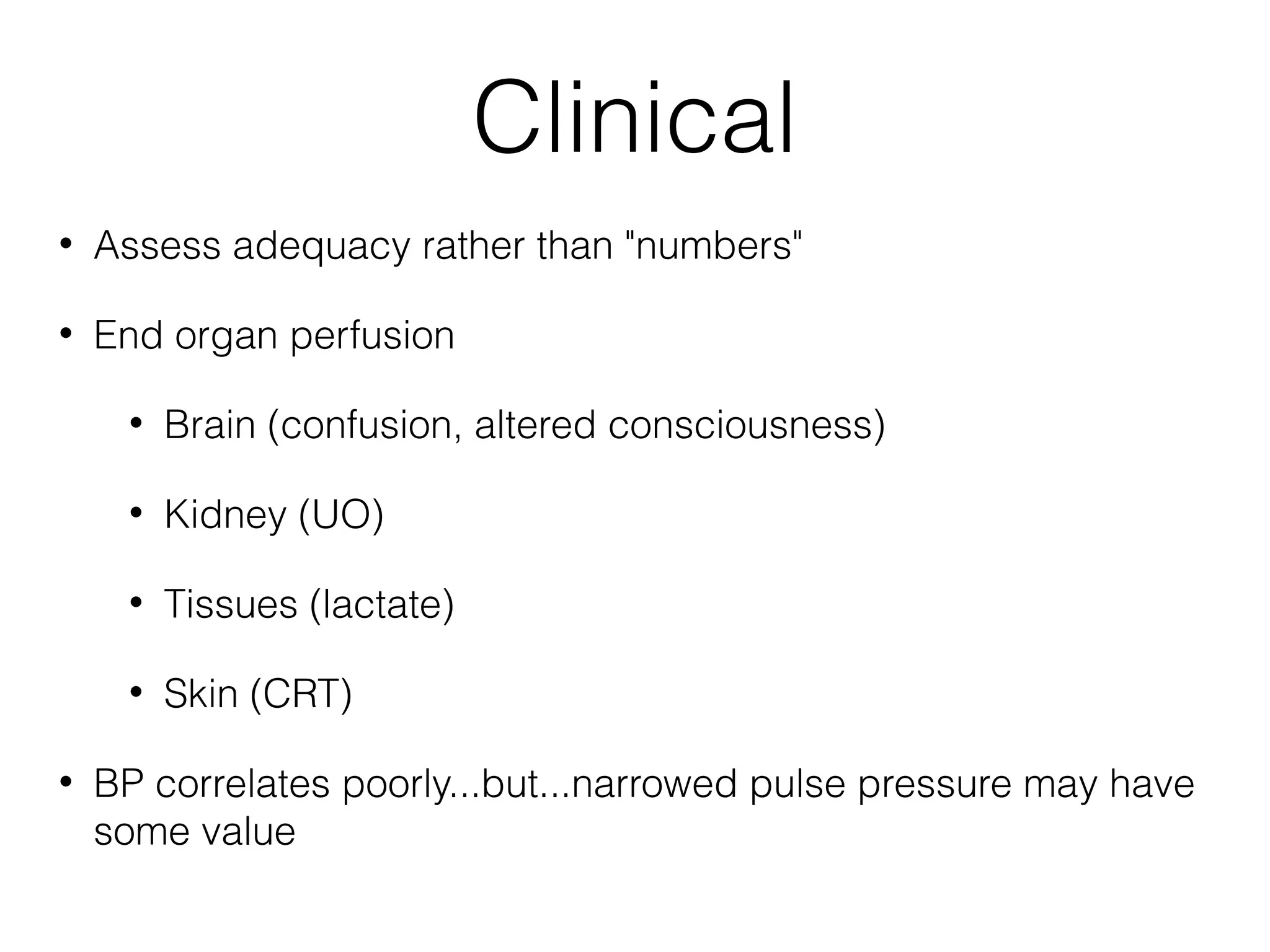
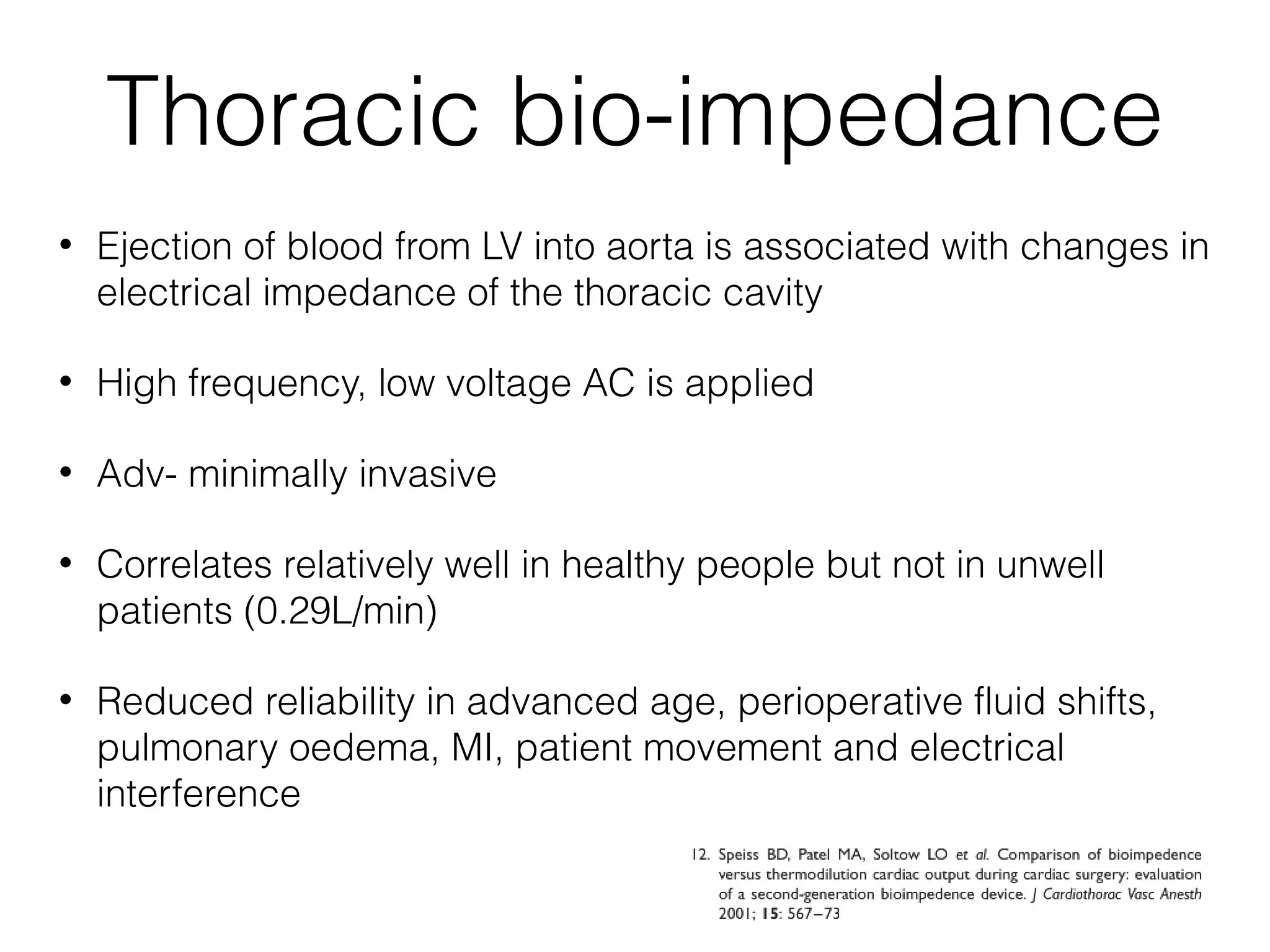
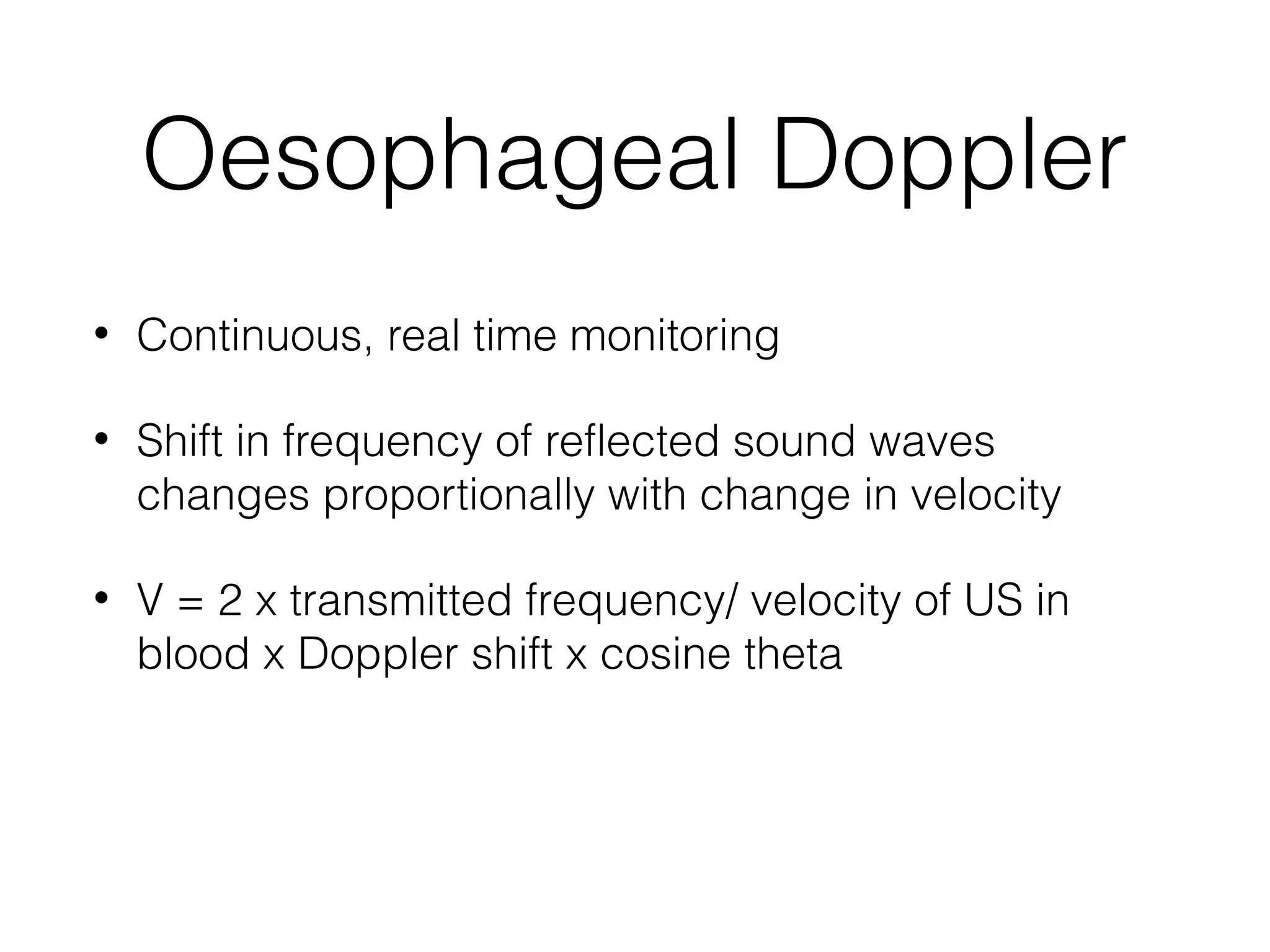
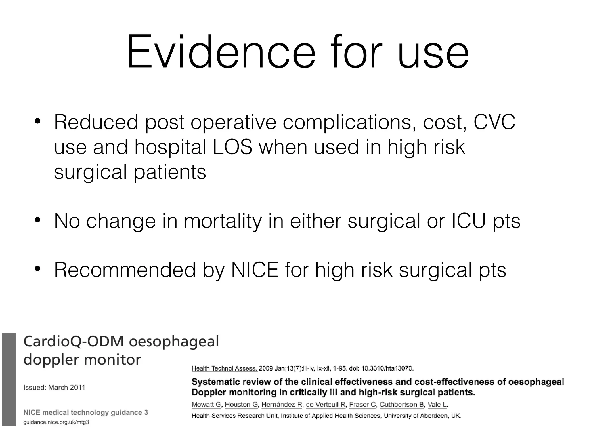
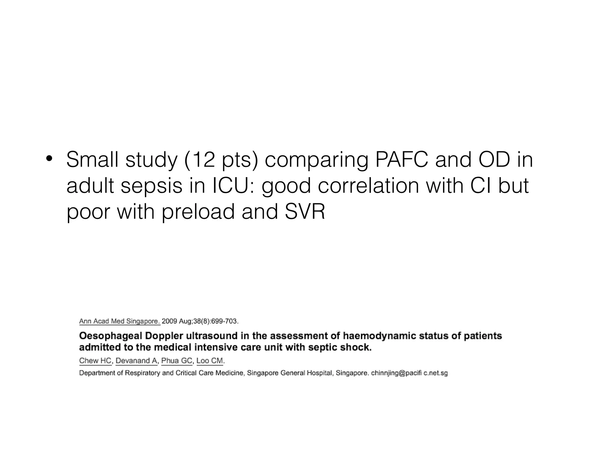
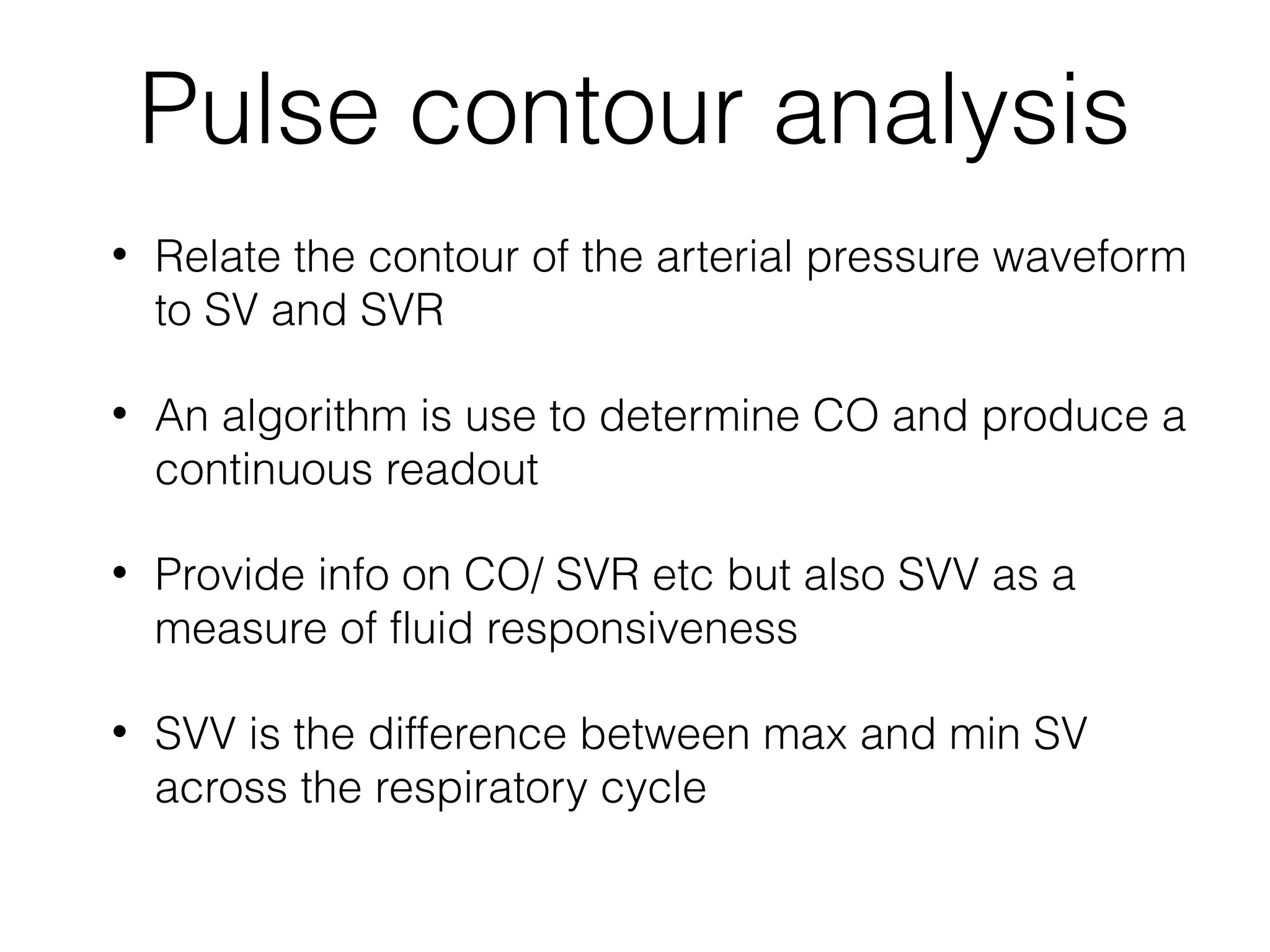
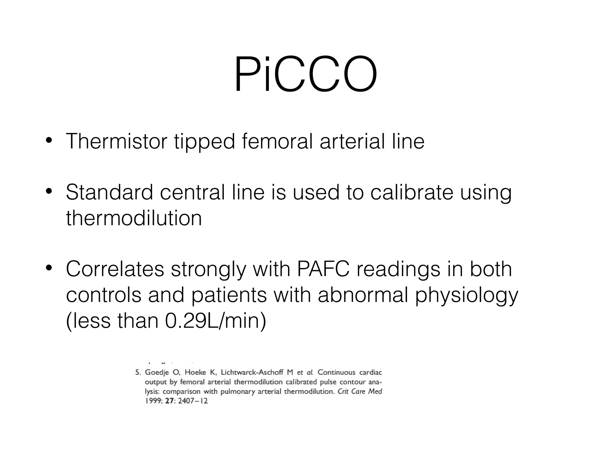
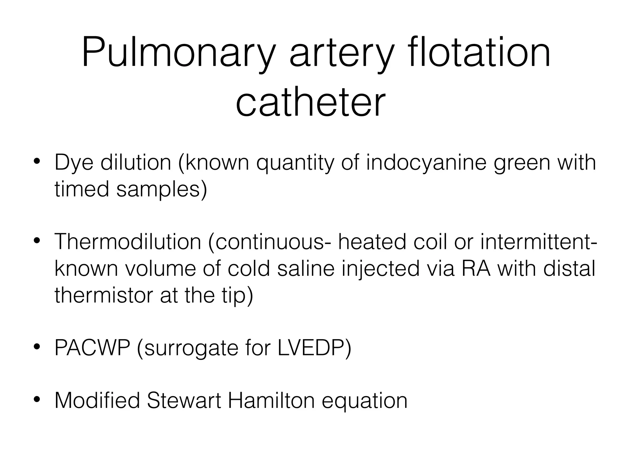
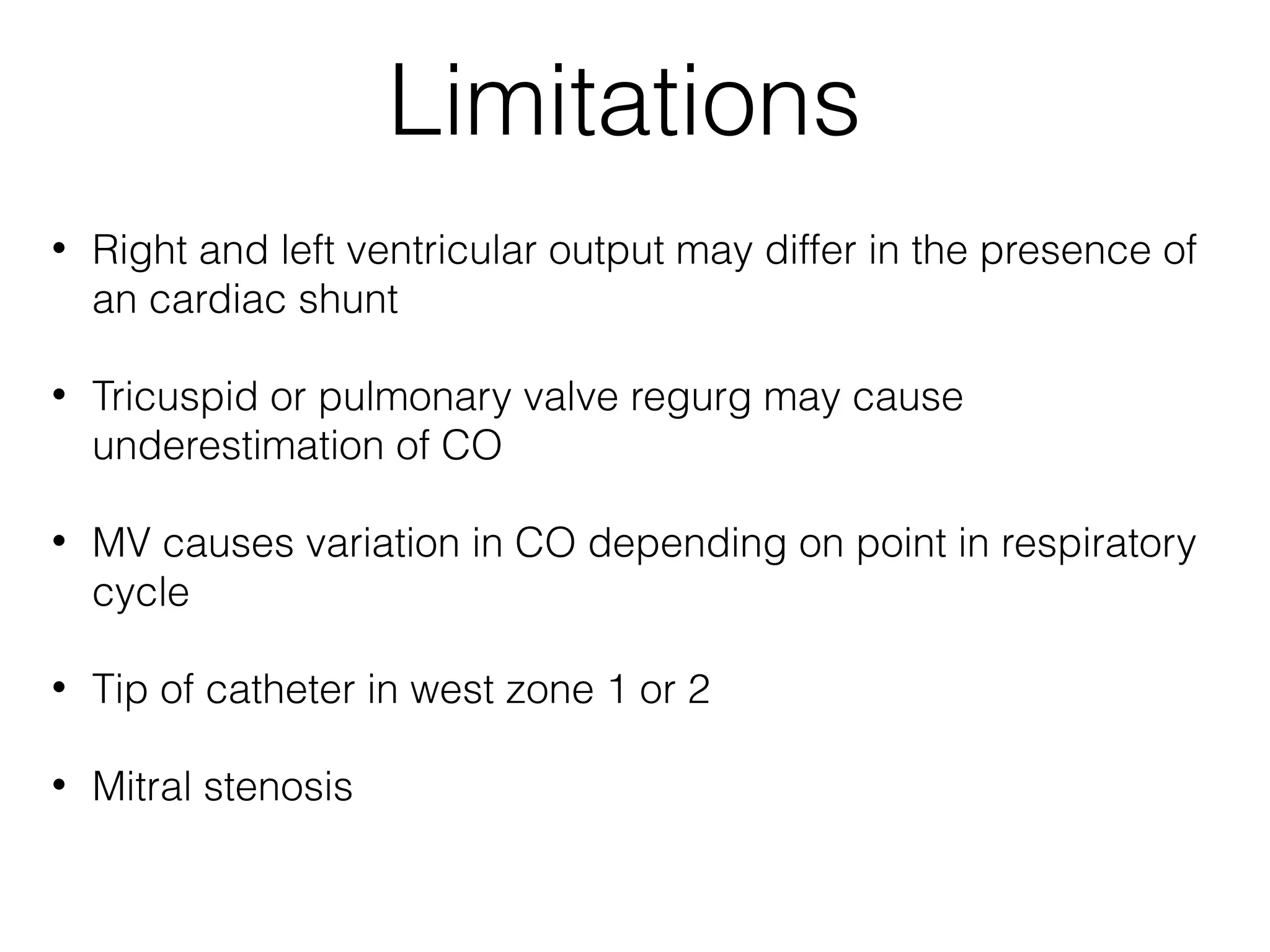
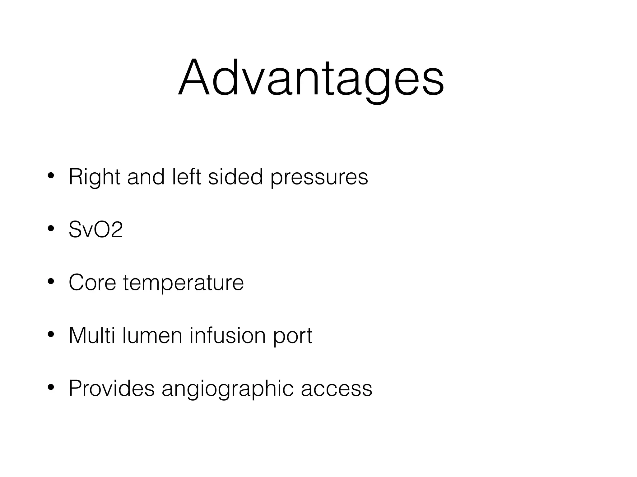
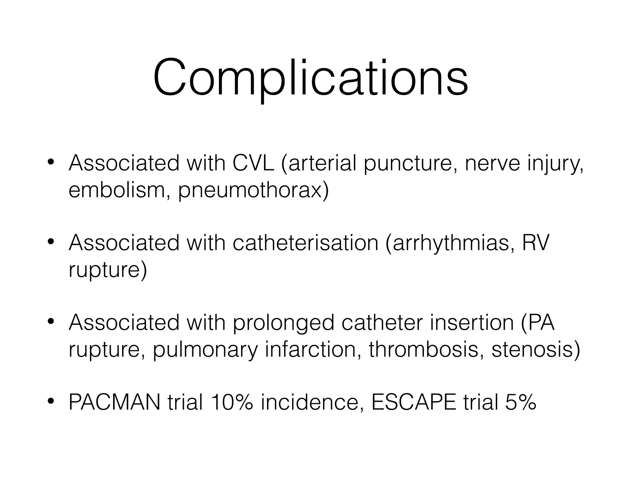
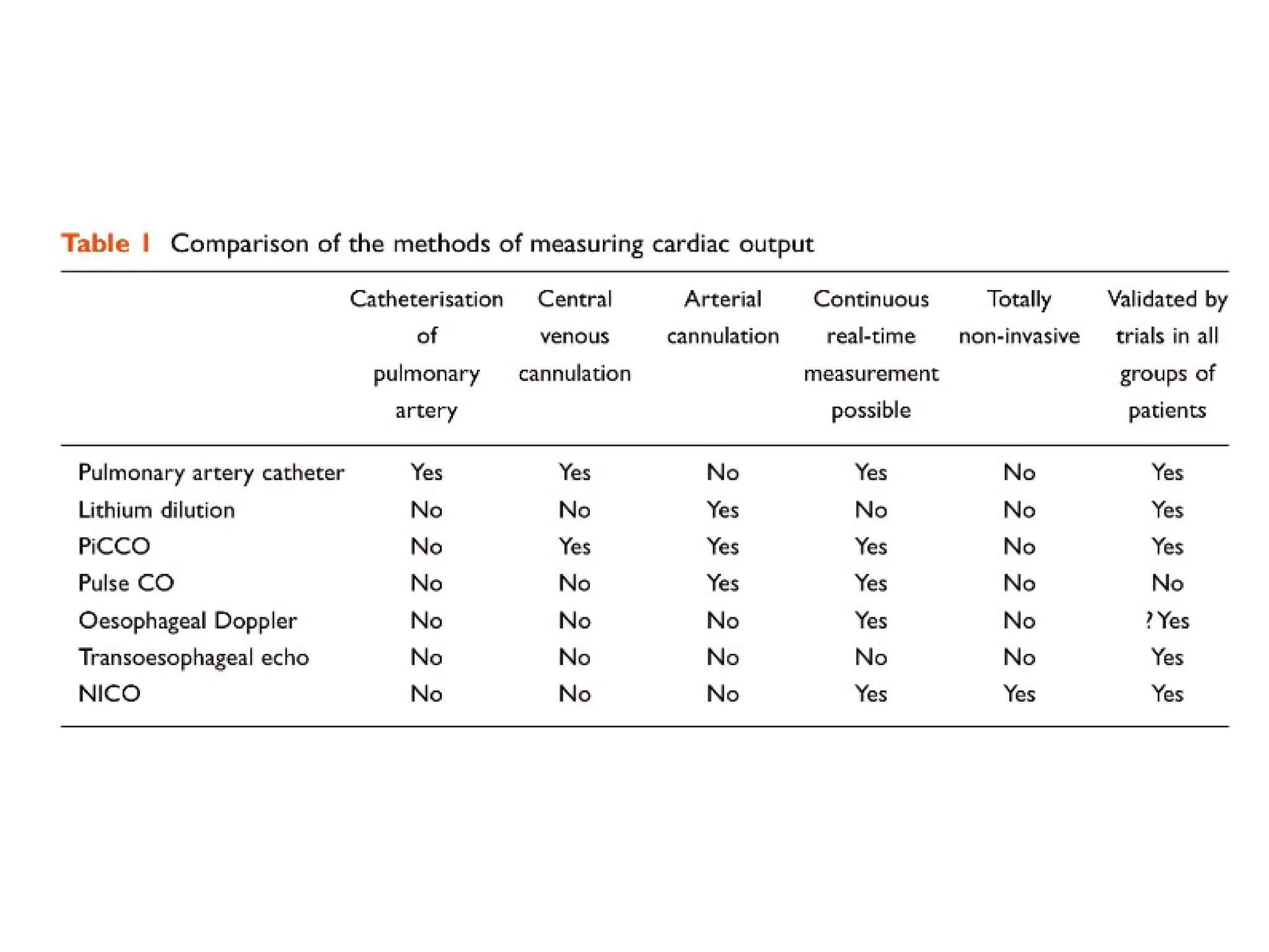
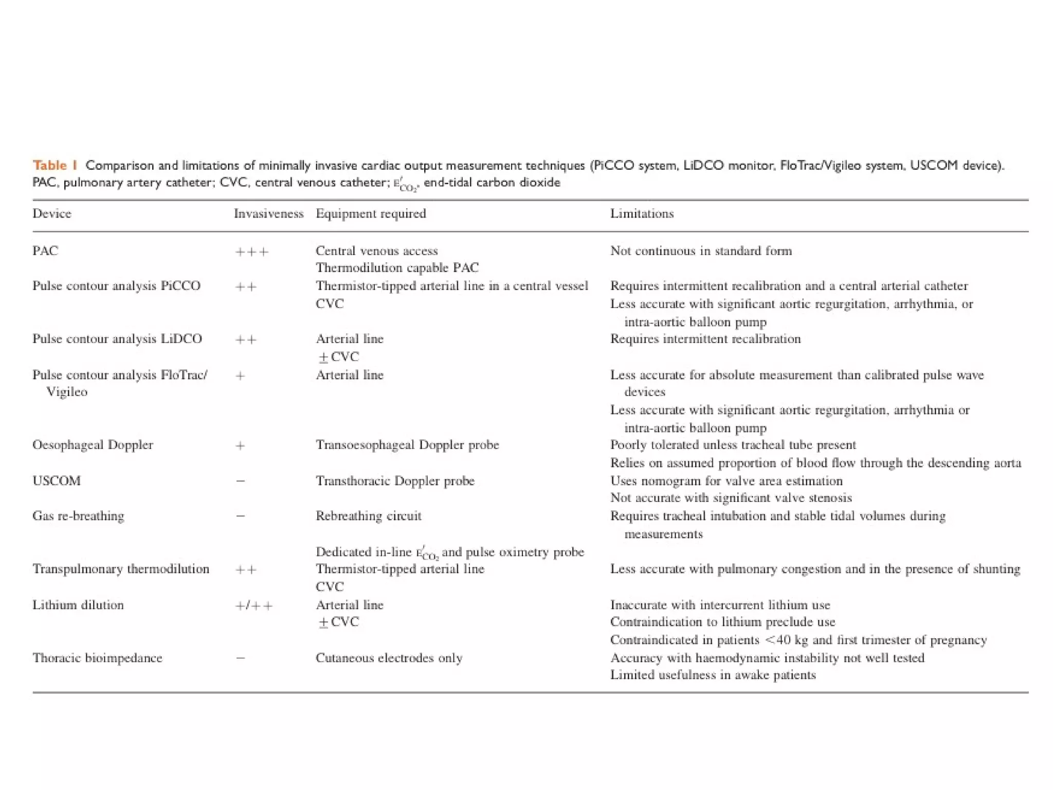
Cardiac output
monitoring
Dr Karen Orr
ST6 anaesthetics/ICM
Altnagelvin ICM study day 7/11/13
Cardiac output
•

Volume of blood ejected from the left ventricle per minute

•

Depends on preload, contractility, heart rate and
afterload

•

CO = HR x SV

•

MAP = CO x SVR
Methods of measuring CO
•

Clinical

•

Minimally-invasive

•

Invasive
Clinical
•

Assess adequacy rather than "numbers"

•

End organ perfusion
•
•

Kidney (UO)

•

Tissues (lactate)

•
•

Brain (confusion, altered consciousness)

Skin (CRT)

BP correlates poorly...but...narrowed pulse pressure may have
some value
•

Increased intrathoracic pressure during inspiration

•

Reduced venous return therefore reduced SV and BP

•

More pronounced during MV
Minimally invasive
Efficacy compared to PAFC?????
Fick principle
•

Amount of a substance taken up by an organ per unit time is
equal to the arterial minus the venous concentration multiplied
by blood flow

•

CO = VCO2/ CaCO2- CvCO2

•

CO2 production can be measured via sensors on breathing
circuit

•

CO2 content in mixed venous and arterial blood

•

Reduced accuracy in sicker patients, severe chest trauma, intra
pulmonary shunt, low MV and high CO
Thoracic bio-impedance
•

Ejection of blood from LV into aorta is associated with changes in
electrical impedance of the thoracic cavity

•

High frequency, low voltage AC is applied

•

Adv- minimally invasive

•

Correlates relatively well in healthy people but not in unwell
patients (0.29L/min)

•

Reduced reliability in advanced age, perioperative fluid shifts,
pulmonary oedema, MI, patient movement and electrical
interference
Oesophageal Doppler
•

Continuous, real time monitoring

•

Shift in frequency of reflected sound waves
changes proportionally with change in velocity

•

V = 2 x transmitted frequency/ velocity of US in
blood x Doppler shift x cosine theta
Assumptions
•

70% of blood enters descending aorta

•

Blood flow is uniform and maximal

•

Cross sectional area is constant (calculated using
formula dependent on age, sex and height)
Measured variables
•

CO

•

SV (stroke distance x aortic root diameter)
•

Stroke distance is AUC x HR

•

FTc (corrected flow time): indicates preload

•

Peak velocity: indicates contractility

•

HR
Contraindications
•

Oesophageal varices

•

IABP

•

Severe coarctation

•

Known oesophageal pathology
Limitations
•

Intubated patients

•

Probe must be as close as possible to parallel to aorta

•

Operator dependent

•

Learning curve for operator

•

Probe displacement
Evidence for use
•

Reduced post operative complications, cost, CVC
use and hospital LOS when used in high risk
surgical patients

•

No change in mortality in either surgical or ICU pts

•

Recommended by NICE for high risk surgical pts
•

Small study (12 pts) comparing PAFC and OD in
adult sepsis in ICU: good correlation with CI but
poor with preload and SVR
Pulse contour analysis
•

Relate the contour of the arterial pressure waveform
to SV and SVR

•

An algorithm is use to determine CO and produce a
continuous readout

•

Provide info on CO/ SVR etc but also SVV as a
measure of fluid responsiveness

•

SVV is the difference between max and min SV
across the respiratory cycle
PiCCO
•

Thermistor tipped femoral arterial line

•

Standard central line is used to calibrate using
thermodilution

•

Correlates strongly with PAFC readings in both
controls and patients with abnormal physiology
(less than 0.29L/min)
FloTrac
•

Standard arterial catheter

•

Algorithm is used based on age, height, gender, weight and
waveform characteristics

•

No external calibration

•

Conflicting evidence for accuracy compared with PiCCO and
PAFC

•

One study showed CO underestimated by up to 2L/min in 40%
of readings

•

Main advantage is ease of use
LiDCO
•

Pulse power analysis (based on law of conservation of mass)

•

Assumes that net power equates to net flow

•

Standard arterial line +/- CVL

•

Calibrated using lithium dilution

•

Good correlation with PAFC across a range of values (error of
<0.11L/min)

•

Contraindicated in chronic lithium use, recent NDNMB, early
pregnancy
Limitations
•

Rely on optimal arterial signal

•

Arrhythmias

•

IABP

•

Severe aortic regurg

•

Changes in SVR
Invasive
Pulmonary artery flotation
catheter
•

Dye dilution (known quantity of indocyanine green with
timed samples)

•

Thermodilution (continuous- heated coil or intermittentknown volume of cold saline injected via RA with distal
thermistor at the tip)

•

PACWP (surrogate for LVEDP)

•

Modified Stewart Hamilton equation
Limitations
•

Right and left ventricular output may differ in the presence of
an cardiac shunt

•

Tricuspid or pulmonary valve regurg may cause
underestimation of CO

•

MV causes variation in CO depending on point in respiratory
cycle

•

Tip of catheter in west zone 1 or 2

•

Mitral stenosis
Advantages
•

Right and left sided pressures

•

SvO2

•

Core temperature

•

Multi lumen infusion port

•

Provides angiographic access
Complications
•

Associated with CVL (arterial puncture, nerve injury,
embolism, pneumothorax)

•

Associated with catheterisation (arrhythmias, RV
rupture)

•

Associated with prolonged catheter insertion (PA
rupture, pulmonary infarction, thrombosis, stenosis)

•

PACMAN trial 10% incidence, ESCAPE trial 5%
The evidence
•

No effect on mortality, LOS, or cost of care in either
general ICU or high risk surgical patients

•

No effect on surgical outcomes when used preoperatively to optimise haemodynamics
What about cardiac surgery?
•

Increased mortality and end organ complications in
propensity matched obs study

•

Authors recognise need for RCT
Device comparison
Cardiac output monitoring

Cardiac output monitoring

  • 1.
    Cardiac output monitoring Dr KarenOrr ST6 anaesthetics/ICM Altnagelvin ICM study day 7/11/13
  • 2.
    Cardiac output • Volume ofblood ejected from the left ventricle per minute • Depends on preload, contractility, heart rate and afterload • CO = HR x SV • MAP = CO x SVR
  • 3.
    Methods of measuringCO • Clinical • Minimally-invasive • Invasive
  • 4.
    Clinical • Assess adequacy ratherthan "numbers" • End organ perfusion • • Kidney (UO) • Tissues (lactate) • • Brain (confusion, altered consciousness) Skin (CRT) BP correlates poorly...but...narrowed pulse pressure may have some value
  • 5.
    • Increased intrathoracic pressureduring inspiration • Reduced venous return therefore reduced SV and BP • More pronounced during MV
  • 6.
  • 7.
  • 8.
    Fick principle • Amount ofa substance taken up by an organ per unit time is equal to the arterial minus the venous concentration multiplied by blood flow • CO = VCO2/ CaCO2- CvCO2 • CO2 production can be measured via sensors on breathing circuit • CO2 content in mixed venous and arterial blood • Reduced accuracy in sicker patients, severe chest trauma, intra pulmonary shunt, low MV and high CO
  • 9.
    Thoracic bio-impedance • Ejection ofblood from LV into aorta is associated with changes in electrical impedance of the thoracic cavity • High frequency, low voltage AC is applied • Adv- minimally invasive • Correlates relatively well in healthy people but not in unwell patients (0.29L/min) • Reduced reliability in advanced age, perioperative fluid shifts, pulmonary oedema, MI, patient movement and electrical interference
  • 10.
    Oesophageal Doppler • Continuous, realtime monitoring • Shift in frequency of reflected sound waves changes proportionally with change in velocity • V = 2 x transmitted frequency/ velocity of US in blood x Doppler shift x cosine theta
  • 11.
    Assumptions • 70% of bloodenters descending aorta • Blood flow is uniform and maximal • Cross sectional area is constant (calculated using formula dependent on age, sex and height)
  • 12.
    Measured variables • CO • SV (strokedistance x aortic root diameter) • Stroke distance is AUC x HR • FTc (corrected flow time): indicates preload • Peak velocity: indicates contractility • HR
  • 14.
  • 15.
    Limitations • Intubated patients • Probe mustbe as close as possible to parallel to aorta • Operator dependent • Learning curve for operator • Probe displacement
  • 16.
    Evidence for use • Reducedpost operative complications, cost, CVC use and hospital LOS when used in high risk surgical patients • No change in mortality in either surgical or ICU pts • Recommended by NICE for high risk surgical pts
  • 17.
    • Small study (12pts) comparing PAFC and OD in adult sepsis in ICU: good correlation with CI but poor with preload and SVR
  • 18.
    Pulse contour analysis • Relatethe contour of the arterial pressure waveform to SV and SVR • An algorithm is use to determine CO and produce a continuous readout • Provide info on CO/ SVR etc but also SVV as a measure of fluid responsiveness • SVV is the difference between max and min SV across the respiratory cycle
  • 19.
    PiCCO • Thermistor tipped femoralarterial line • Standard central line is used to calibrate using thermodilution • Correlates strongly with PAFC readings in both controls and patients with abnormal physiology (less than 0.29L/min)
  • 20.
    FloTrac • Standard arterial catheter • Algorithmis used based on age, height, gender, weight and waveform characteristics • No external calibration • Conflicting evidence for accuracy compared with PiCCO and PAFC • One study showed CO underestimated by up to 2L/min in 40% of readings • Main advantage is ease of use
  • 21.
    LiDCO • Pulse power analysis(based on law of conservation of mass) • Assumes that net power equates to net flow • Standard arterial line +/- CVL • Calibrated using lithium dilution • Good correlation with PAFC across a range of values (error of <0.11L/min) • Contraindicated in chronic lithium use, recent NDNMB, early pregnancy
  • 22.
    Limitations • Rely on optimalarterial signal • Arrhythmias • IABP • Severe aortic regurg • Changes in SVR
  • 23.
  • 24.
    Pulmonary artery flotation catheter • Dyedilution (known quantity of indocyanine green with timed samples) • Thermodilution (continuous- heated coil or intermittentknown volume of cold saline injected via RA with distal thermistor at the tip) • PACWP (surrogate for LVEDP) • Modified Stewart Hamilton equation
  • 26.
    Limitations • Right and leftventricular output may differ in the presence of an cardiac shunt • Tricuspid or pulmonary valve regurg may cause underestimation of CO • MV causes variation in CO depending on point in respiratory cycle • Tip of catheter in west zone 1 or 2 • Mitral stenosis
  • 27.
    Advantages • Right and leftsided pressures • SvO2 • Core temperature • Multi lumen infusion port • Provides angiographic access
  • 28.
    Complications • Associated with CVL(arterial puncture, nerve injury, embolism, pneumothorax) • Associated with catheterisation (arrhythmias, RV rupture) • Associated with prolonged catheter insertion (PA rupture, pulmonary infarction, thrombosis, stenosis) • PACMAN trial 10% incidence, ESCAPE trial 5%
  • 29.
    The evidence • No effecton mortality, LOS, or cost of care in either general ICU or high risk surgical patients • No effect on surgical outcomes when used preoperatively to optimise haemodynamics
  • 30.
    What about cardiacsurgery? • Increased mortality and end organ complications in propensity matched obs study • Authors recognise need for RCT
  • 31.