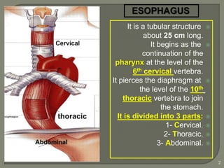
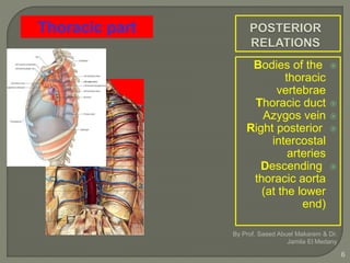
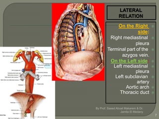
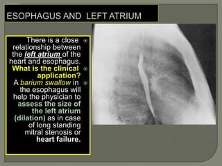
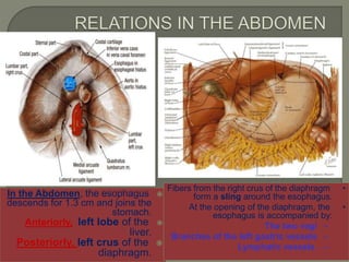
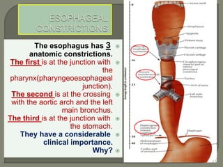
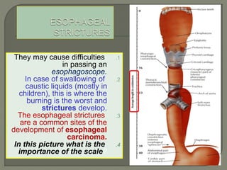
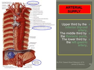
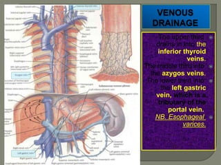
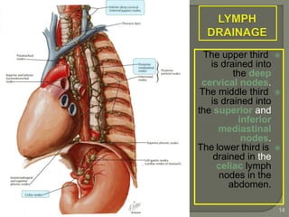
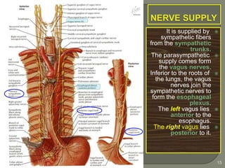
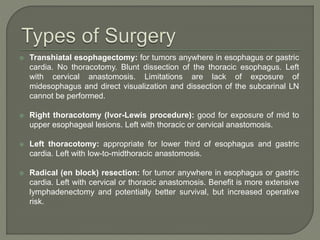
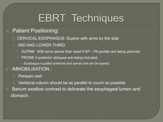
It is a tubular structure
about 25 cm long.
It begins as the
continuation of the
pharynx at the level of the
6th cervical vertebra.
It pierces the diaphragm at
the level of the 10th
thoracic vertebra to join
the stomach.
It is divided into 3 parts:
1- Cervical.
2- Thoracic.
3- Abdominal.
2
Abdominal
thoracic
Cervical
Posteriorly:
Vertebral column.
Laterally:
Lobes of the thyroid
gland.
Anteriorly:
Trachea and the
recurrent laryngeal
nerves.
By Prof. Saeed Abuel Makarem & Dr. Jamila El Medany
3
RELATIONS
In the thorax, it passes
downward and to the left
through superior then to
posterior mediastinum
At the level of the sternal
angle, the aortic arch
pushes the esophagus again
to the midline.
By Prof. Saeed Abuel Makarem & Dr.
Jamila El Medany4
Trachea
Left recurrent
laryngeal
nerve
Left principal
bronchus
Pericardium
Left atrium
By Prof. Saeed Abuel Makarem & Dr. Jamila El Medany
5
Thoracic part
Bodies of the
thoracic
vertebrae
Thoracic duct
Azygos vein
Right posterior
intercostal
arteries
Descending
thoracic aorta
(at the lower
end)
By Prof. Saeed Abuel Makarem & Dr.
Jamila El Medany
6
Thoracic part
On the Right
side:
Right mediastinal
pleura
Terminal part of the
azygos vein.
On the Left side:
Left mediastinal
pleura
Left subclavian
artery
Aortic arch
Thoracic duct
By Prof. Saeed Abuel Makarem & Dr.
Jamila El Medany
7
There is a close
relationship between
the left atrium of the
heart and esophagus.
What is the clinical
application?
A barium swallow in
the esophagus will
help the physician to
assess the size of
the left atrium
(dilation) as in case
of long standing
mitral stenosis or
heart failure.
In the Abdomen, the esophagus
descends for 1.3 cm and joins the
stomach.
Anteriorly, left lobe of the
liver.
Posteriorly, left crus of the
diaphragm. 9
•Fibers from the right crus of the diaphragm
form a sling around the esophagus.
•At the opening of the diaphragm, the
esophagus is accompanied by:
–The two vagi
–Branches of the left gastric vessels
–Lymphatic vessels.
The esophagus has 3
anatomic constrictions.
The first is at the junction with
the
pharynx(pharyngeoesophageal
junction).
The second is at the crossing
with the aortic arch and the left
main bronchus.
The third is at the junction with
the stomach.
They have a considerable
clinical importance.
Why?
.1They may cause difficulties
in passing an
esophagoscope.
.2In case of swallowing of
caustic liquids (mostly in
children), this is where the
burning is the worst and
strictures develop.
.3The esophageal strictures
are a common sites of the
development of esophageal
carcinoma.
.4In this picture what is the
importance of the scale?
Upper third by the
inferior thyroid
artery.
The middle third by
the thoracic aorta.
The lower third by
the left gastric
artery.
By Prof. Saeed Abuel Makarem & Dr.
Jamila El Medany 12
The upper third
drains in into the
inferior thyroid
veins.
The middle third into
the azygos veins.
The lower third into
the left gastric
vein, which is a
tributary of the
portal vein.
NB. Esophageal
varices.
The upper third
is drained into
the deep
cervical nodes.
The middle third
is drained into
the superior and
inferior
mediastinal
nodes.
The lower third is
drained in the
celiac lymph
nodes in the
abdomen.
By Prof. Saeed Abuel Makarem & Dr. Jamila El
Medany
14
It is supplied by
sympathetic fibers
from the sympathetic
trunks.
The parasympathetic
supply comes form
the vagus nerves.
Inferior to the roots of
the lungs, the vagus
nerves join the
sympathetic nerves to
form the esophageal
plexus.
The left vagus lies
anterior to the
esophagus.
The right vagus lies
posterior to it.
By Prof. Saeed Abuel Makarem & Dr. Jamila El
Medany
15
 Esophageal cancer is the 7th leading cause of cancer deaths.
 accounts for 1% of all malignancy & 6% of all GI malignancy.
 Most common in China, Iran, South Africa, India and the former Soviet
Union.
 The incidence rises steadily with age, reaching a peak in the 6th to 7th
decade of life.
 Male : Female = 3.5 : 1
 African-American males : White males = 5:1
 Worldwide SCC responsible for most of the cases.
 Adenocarcinoma now accounts for over 50% of esophageal cancer in
the USA, due to association with GERD , Barretts’s esophagus &
obesity.
 SCC usually occurs in the middle 3rd of the esophagus (the ratio of
upper : middle : lower is 15 : 50 : 35).
 Adenocarcinoma is most common in the lower 3rd of the esophagus,
accounting for over 65% of cases.
 Two most common forms of esophageal
cancer are named for the type of cells that
become malignant
 Squamous cell carcinoma forms
in squamous cells, the thin, flat cells lining the
esophagus. This cancer is most often found
in the upper and middle part of the
esophagus
 Adenocarcinoma begins
in glandular (secretory) cells. Glandular cells
in the lining of the esophagus produce and
release fluids such as mucus.
Adenocarcinomas usually form in the lower
part of the esophagus, near the stomach.
 Smoking and alcohol (80% - 90%)
 Dietary factors
• N-nitroso compounds (animal carcinogens)
• Pickled vegetables and other food-products
• Toxin-producing fungi
• Betel nut chewing
• Ingestion of very hot foods and beverages (such as tea)
 Underlying esophageal disease (such as achalasia and
caustic strictures, Tylosis)
 Genetic abnormalities:
• p53 mutation, loss of 3p and 9q alleli, amp. Cyclin D1 & amp.
EGFR
 Associated with Barretts’s esophagus, GERD
& hiatal hernia.
 Obesity (3 to 4 fold risk)
 Smoking (2 to 3 fold risk)
 Increased esophageal acid exposure such as
Zollinger-Ellison syndrome.
Barrett’s esophagus is a
metaplasia of the esophageal epithelial lining. The
squamous epithelium is replaced by columnar
epithelium,with 0.5% annual rate of neoplastic
transformation.
 No serosal covering, direct invasion of contiguous structures occurs
early.
 Commonly spread by lymphatics (70%)
 25% - 30% hematogenous metastases at time of presentation.
 Most common site of metastases are
• lung, liver, pleura, bone, kidney & adrenal gland
 Median survival with distant metastases – 6 to 12 months
• Esophageal intraepithelial neoplasia
• Glandular epithelial dysplasia/adenocarcinoma in situ
in Barrett's mucosa
Preinvasive
Neoplasia
• Squamous cell carcinoma
• Adenocarcinoma,
• Adenoid cystic carcinoma
• Mucoepidermoid carcinoma
• Adenosquamous carcinoma
• Small cell carcinoma
• Carcinoid tumor
• Malignant melanoma
• Sarcomas
Invasive Malignant
Neoplasia
95
%
 It is commonly associated with the
symptoms of dysphagia, wt. loss, pain,
anorexia, and vomiting
 Symptoms often start 3 to 4 months
before diagnosis
 Dysphagia - in more than 90% pt.
Odynophagia - in 50% of pt.
 Wt. loss – more than 5 % of total body
wt. in 40 – 70% pt. associated with
worst prognosis.
Complications:
 Cachexia, Malnutrition, dehydration, anaemia,.
 Aspiration pneumonia.
 Distant metastasis.
 Invasion of near by structures: e.g.
• Recurrent laryngeal nerve → Hoarseness of voice
• Trachea → Stridor & TOF→ cough, choking &
cyanosis
• Perforation into the pleural cavity → Empyema
• back pain in celiac axis node involvement
There are a plethora of modalities
available to diagnose and stage
esophageal cancer
Radiologic tests, endoscopic
procedures, and minimally invasive
surgical techniques all add value to a
solid staging workup in a patient with
esophageal cancer.
A barium esophagram is recommended
for any patient presenting with
dysphagia
is able to differentiate intraluminal from
intramural lesions and to discriminate
between intrinsic (from a mass
protruding into the lumen) and extrinsic
(from compression of a structures
outside the esophagus) compression
The classic finding of an apple-core
lesion in patients with esophageal
cancer is recognized easily
Although the esophagram will not be
specific for cancer, it is a good first test
to perform in patients presenting with
dysphagia and a suspicion of
esophageal cancer
The diagnosis of esophageal cancer is
made best from an endoscopic biopsy
any patient undergoing surgery for
esophageal cancer must have an
endoscopy performed by the operating
surgeon before entering the operating
room for a definitive resection
CT scan of the chest and abdomen is
important to assess the length of the
tumor, thickness of the esophagus and
stomach, regional lymph node status
and distant disease to the liver and
lungs
PET scan evaluates the primary mass,
regional lymph nodes, and distant
disease
 Its sensitivity and specificity slightly
exceed those of CT; however, they
remain low for definitive staging
EUS is the most critical component of
esophageal cancer staging.
The information obtained from EUS will
help guide both medical and surgical
therapy
biopsy samples can be obtained of the
mass and lymph nodes in the
paratracheal, subcarinal,
paraesophageal, celiac region
Chemotherpay
Radiation therap
Chemo-radiotherap
Surgical resection
 Site of disease
 Extent of disease involvement
 Co-morbid conditions
 Patient preference.
 Prerequisite for surgery
• disease should be 5 cm beyond cricophyrangeus.
 Surgery indications
• Lower 1/3 rd oesophageal ds involving GE junction.
• Tumor size <5 cm .
• palliative surgery
 5-Year OS for surgery alone is 20–25% (no significant difference
between surgical techniques according to results of 2 meta-analyses)
 Local failure rate around 19–57% when used alone
 surgical morbidity/mortality related to experience of the surgeons.
 Transhiatal esophagectomy: for tumors anywhere in esophagus or gastric
cardia. No thoracotomy. Blunt dissection of the thoracic esophagus. Left
with cervical anastomosis. Limitations are lack of exposure of
midesophagus and direct visualization and dissection of the subcarinal LN
cannot be performed.
 Right thoracotomy (Ivor-Lewis procedure): good for exposure of mid to
upper esophageal lesions. Left with thoracic or cervical anastomosis.
 Left thoracotomy: appropriate for lower third of esophagus and gastric
cardia. Left with low-to-midthoracic anastomosis.
 Radical (en block) resection: for tumor anywhere in esophagus or gastric
cardia. Left with cervical or thoracic anastomosis. Benefit is more extensive
lymphadenectomy and potentially better survival, but increased operative
risk.
 No data proving that chemotherapy alone provides improved
survival or palliation. Partial response, not long-term remission, is
the rule
 Indication
• Used in combination with radiation for locally advanced cancers
• Used as single treatment modality in stage IV disease
• Combination chemotherapy has been used preoperatively in a combined
modality approach to esophageal Ca in hopes of controlling occult metastatic
disease and improving the resectability rate.
 Platinum doublet is preferred over single agents
 Cisplatin plus 5-FU or docetaxel are commonly used combinations
Regimens:
 Paclitaxel and carboplatin
 Cisplatin and 5-FU or capecitabine
 Oxaliplatin and 5-FU or capecitabine
 Paclitaxel or docetaxel and cisplatin
 Carboplatin and 5-FU
 Irinotecan and cisplatin
 Oxaliplatin, docetaxel and capecitabine
 Epirubicin, cisplatin and 5-FU (Only for adenocarcinoma)
 Curative
• Radical RT
• Pre-Op RT
• Post Op RT
• Concurrent chemo-radiation
 Palliative
 EBRT
 Brachytherapy
 Patient Positioning:
• CERVICAL ESOPHAGUS: Supine with arms by the side
• MID AND LOWER THIRD:
 SUPINE With arms above their head if AP – PA portals are being planned
 PRONE if posterior obliques are being included.
 Esophagus is pulled anteriorly and spinal cord can be spared.
 IMMOBILISATION :
• Perspex cast
• Vertebral column should be as parallel to couch as possible.
 Barium swallow contrast to delineate the esophageal lumen and
stomach.
Field Portals:
 AP – PA foll. by opposed oblique pair.
 2 anterior obliques and 1 posterior field.
 2 posterior obliques and 1 anterior field
 4 field box with soft tissue compensators foll by obliques ( Univ of
Florida tech )
z SUPERIOR BORDER: At C 7
z INFERIOR BORDER : At T 4 ( carina )
z 2 cm lateral margins.
z SC nodes irradiated electively.
z SC nodes will be underdosed if oblique portals are used to treat
primary; can be boosted by a separate field if required.
 AP – PA followed by 1 Ant and 2 Post oblique pair
 4 FIELD : AP-PA & opposed laterals – for mid 1/3rd lesions with
patient in prone position.
 AP-PA upto 43 Gy foll by 2 Post obliques upto 50 Gy ( gross disease
boosted to 60 Gy )
z SUPERIOR BORDER: 5 cm proximal to superior extent of disease.
z INFERIOR BORDER:
y MID 1/3RD – AT GE jn. As visualised by Barium swallow
y LOWER 1/3RD - Coeliac plexus ( L 1 ) to be included.
 Energy
• 6 – 10 MV linac or Co60
 Chemoradiation:
• 50.4 Gy in 28 # at 1.8 Gy per #
• Boost to 60 – 66 Gy for residual disease
 Radical RT:
• 45 Gy / 25 # / 1.8 Gy per #
• boost with 2 cm margin to total dose of 60Gy
 Dose limitations
• Spinal cord Dmax:45 Gy at 1.8 Gy/fx
• Lung: Limit 70% of both lungs <20 Gy
• Heart: Limit 50% of ventricles <25 Gy
 As a boost after EBRT or as a palliative measure
 Local control of 25% - 35 in palliative setting
 In curative setting, addition of brachytherapy does not improve
results compared to Chemoradiation.
 Limit dose to critical structure
 Dose escalation to primary
 Relief bleeding, pain and improves swallowing status in palliative
setting.
Patient selection:
• Primary tumor length ≤ 10 cm length
• Tumor confined to esophageal wall
• Thoracic esophagus location
• No nodal / systemic metastasis.
Contraindications:
• T E fistula
• Cervical esophagous location
• Stenosis which cannot be bypassed
 EBRT 45 – 50 Gy in 1.8-2.0Gy /#,5#/wk
 HDR – 5 Gy x two # one week apart , 2 – 3weeks after EBRT.
 LDR – single 20 Gy # @ 0.4 – 1.0 Gy per hr, 2 -3 weeks after EBRT.
 Never concurrently with chemotherapy
 Ext diameter of applicator must be 6 – 10 mm.
 Active length : visible tumor by UGI scopy plus 1 – 2 cm proximal &
distal margin.
 Dose is prescribed 1 cm from mid source or mid dwell position.
 No randomized trials of RT Vs Sx
 5 yr survival with conventional RT : < 10%
 Tumors < 5 cm , 5 yr survival : 20%
 Stage wise 5 yr survival:
• Stage I – 20%
• Stage II – 10%
• Stage III – 3 %
• Stage IV – 0%
 For cervical esophagus, cure rates were similar with Radical RT or
Sx alone.
 RT or Sx alone DOES NOT alter the natural history of the disease.
 RTOG 8501: RT Vs Chemo RT
• Better LRC and improved OS with ChemoRT
 RT alone should be used for palliation or in medically unfit patients.
 Principle:
•  resectability,  likelihood of tumor dissemination during Sx , 
radioresponsiveness due to unaltered blood supply
• 5 randomised trials ,shows no apparent clinical benefit to use of
preop rt alone except,
• Only one trial ( Huang et al ) showed survival advantage of 46%
Vs 25% with 40 Gy RT
• Recent meta analysis Oesophageal Cancer Collaborative Group
study showed no clear survival advantage.
 No difference in resectability rates, LRC or survival with pre-op RT
 Advantages:
• Treat areas at risk for recurrences while minimizing
dose to OAR.
• Patients with node negative , completely resected T1 /
T2 tumors can be excluded.
 Disadvantage:
• Tolerance of stomach or bowel used for
interpositioning.
 2 randomised trials:
• Peniere et al :-
 221 pts, mid / low 1/3rd growth
 RT : 45- 55 Gy @ 1.8 Gy per #
 3 yrs -  local failure rate ( from 35% to 10%)
- no significant disease free survival.
• Fok et al :-
 130 pts , RT – 49 Gy @ 3.5 Gy per #
 Local failure rate  in patients who had palliative resection
( from 46% to 20% )
 No difference for completely resected patients
 Post op RT improves local control, but does not confer any survival
advantage.
ChemoRT Vs RT Alone
 RTOG 8501 INTERGROUP TRIAL:
• 121 pts: 60 pts RT alone – 64 Gy @ 2 Gy per #
61 pts chemoRT – 50 Gy RT +
5 FU + CDDP – on 1 , 5 , 8 & 11 weeks
• Median survival : 8.9 Vs 12.5 months
• 5 yr survival : 0% Vs 30 %
• Distant mets @ 5 yrs: 40% Vs 12 %
• Acute toxicity : 25% Vs 44 %
  Median & overall survival, LRR and Acute toxicity in Chemo RT
arm.
 Chemoradiation is a standard Non-surgical Tx.
RT dose escalation in Chemo RT
 Intergroup 0123 TRIAL – 218 pts
• Chemoradiation - either 50.4 Gy or 64.8 Gy
• No significant difference in median survival, 2 yr survival or loco-
regional failure.
 Intensification of RT dose beyond 50.4 Gy(in combination with
chemotherapy ) does not improve results.
PRE OP CHEMO RT Vs Sx ALONE
 44 Randomised trials
 2 studies showed  in local recurrence
• Urba et al – 19 % Vs 42 %
• Bosset et al ( EORTC ) – 28% Vs 40%
 One study showed significant survival benefit at 3 yrs (in pts who had a
pathologic CR )
• Urba et al – 64% Vs 19%
 One study (Walsh et al) showed benefit in median (16 Vs 11 months )
and overall survival at 3 yrs ( 32 Vs 6%)
 Results support TRIMODALITY approach.
The role of preoperative
chemotherapy alone is
controversial, according to
mixed results from clinical
trials.
Stage Recommended treatment
Stage I–III and IVA
resectable
medically-fit
definitive chemo-RT (preferred for cervical esophagus)
Or, Pre-op chemo-RT → surgery. Surgery preferred for adenocarcinoma
regardless of response to chemo-RT.
Or, surgery. (noncervical T1N0 and young T2N0 patients with primaries
of lower esophagus or gastroesophageal junction.
Indications for post-op chemo-RT include: unfavorable T2N0, T3/4,
LN+, and/or close/+ margin.
Stage I–III inoperable Definitive chemo-RT
Stage IV palliative Concurrent chemo-RT (5-FU + cisplatin, 50 Gy) or RT alone (e.g., 2.5
Gy × 14 fx) or chemo alone or best supportive care.
Pain: medications ± RT
Bleeding: endoscopic therapy, surgery, or RT
 EBRT using 3D-CRT to a total dose of 50.4 Gy (1.8 Gy per daily fraction) is
standard.
 IMRT is often utilized to minimize exposure to adjacent structures.
 Proton beam in combination with chemotherapy is being explored.
 Targeted biologic agents added to standard cytotoxic chemotherapy is being
explored
 Esophageal cancer is the 7th leading cause of cancer deaths.
 Adenocarcinoma now accounts for over 50% of esophageal cancer in the
USA, due to association with GERD & obesity.
 Dysphagia and weight loss are the two most common presentations in patients
with esophageal cancer.
 Endoscopic ultrasound (EUS) is necessary to accompany a complete workup
for proper staging and diagnosis of esophageal cancer.
 Surgery is the standard of care for early-stage esophageal cancer.
 Preoperative chemotherapy and radiation is the standard option for locally
advanced esophageal cancer in surgically eligible patients.

Ca esophagus

  • 2.
    It is atubular structure about 25 cm long. It begins as the continuation of the pharynx at the level of the 6th cervical vertebra. It pierces the diaphragm at the level of the 10th thoracic vertebra to join the stomach. It is divided into 3 parts: 1- Cervical. 2- Thoracic. 3- Abdominal. 2 Abdominal thoracic Cervical
  • 3.
    Posteriorly: Vertebral column. Laterally: Lobes ofthe thyroid gland. Anteriorly: Trachea and the recurrent laryngeal nerves. By Prof. Saeed Abuel Makarem & Dr. Jamila El Medany 3 RELATIONS
  • 4.
    In the thorax,it passes downward and to the left through superior then to posterior mediastinum At the level of the sternal angle, the aortic arch pushes the esophagus again to the midline. By Prof. Saeed Abuel Makarem & Dr. Jamila El Medany4
  • 5.
    Trachea Left recurrent laryngeal nerve Left principal bronchus Pericardium Leftatrium By Prof. Saeed Abuel Makarem & Dr. Jamila El Medany 5 Thoracic part
  • 6.
    Bodies of the thoracic vertebrae Thoracicduct Azygos vein Right posterior intercostal arteries Descending thoracic aorta (at the lower end) By Prof. Saeed Abuel Makarem & Dr. Jamila El Medany 6 Thoracic part
  • 7.
    On the Right side: Rightmediastinal pleura Terminal part of the azygos vein. On the Left side: Left mediastinal pleura Left subclavian artery Aortic arch Thoracic duct By Prof. Saeed Abuel Makarem & Dr. Jamila El Medany 7
  • 8.
    There is aclose relationship between the left atrium of the heart and esophagus. What is the clinical application? A barium swallow in the esophagus will help the physician to assess the size of the left atrium (dilation) as in case of long standing mitral stenosis or heart failure.
  • 9.
    In the Abdomen,the esophagus descends for 1.3 cm and joins the stomach. Anteriorly, left lobe of the liver. Posteriorly, left crus of the diaphragm. 9 •Fibers from the right crus of the diaphragm form a sling around the esophagus. •At the opening of the diaphragm, the esophagus is accompanied by: –The two vagi –Branches of the left gastric vessels –Lymphatic vessels.
  • 10.
    The esophagus has3 anatomic constrictions. The first is at the junction with the pharynx(pharyngeoesophageal junction). The second is at the crossing with the aortic arch and the left main bronchus. The third is at the junction with the stomach. They have a considerable clinical importance. Why?
  • 11.
    .1They may causedifficulties in passing an esophagoscope. .2In case of swallowing of caustic liquids (mostly in children), this is where the burning is the worst and strictures develop. .3The esophageal strictures are a common sites of the development of esophageal carcinoma. .4In this picture what is the importance of the scale?
  • 12.
    Upper third bythe inferior thyroid artery. The middle third by the thoracic aorta. The lower third by the left gastric artery. By Prof. Saeed Abuel Makarem & Dr. Jamila El Medany 12
  • 13.
    The upper third drainsin into the inferior thyroid veins. The middle third into the azygos veins. The lower third into the left gastric vein, which is a tributary of the portal vein. NB. Esophageal varices.
  • 14.
    The upper third isdrained into the deep cervical nodes. The middle third is drained into the superior and inferior mediastinal nodes. The lower third is drained in the celiac lymph nodes in the abdomen. By Prof. Saeed Abuel Makarem & Dr. Jamila El Medany 14
  • 15.
    It is suppliedby sympathetic fibers from the sympathetic trunks. The parasympathetic supply comes form the vagus nerves. Inferior to the roots of the lungs, the vagus nerves join the sympathetic nerves to form the esophageal plexus. The left vagus lies anterior to the esophagus. The right vagus lies posterior to it. By Prof. Saeed Abuel Makarem & Dr. Jamila El Medany 15
  • 16.
     Esophageal canceris the 7th leading cause of cancer deaths.  accounts for 1% of all malignancy & 6% of all GI malignancy.  Most common in China, Iran, South Africa, India and the former Soviet Union.  The incidence rises steadily with age, reaching a peak in the 6th to 7th decade of life.  Male : Female = 3.5 : 1  African-American males : White males = 5:1
  • 17.
     Worldwide SCCresponsible for most of the cases.  Adenocarcinoma now accounts for over 50% of esophageal cancer in the USA, due to association with GERD , Barretts’s esophagus & obesity.  SCC usually occurs in the middle 3rd of the esophagus (the ratio of upper : middle : lower is 15 : 50 : 35).  Adenocarcinoma is most common in the lower 3rd of the esophagus, accounting for over 65% of cases.
  • 18.
     Two mostcommon forms of esophageal cancer are named for the type of cells that become malignant  Squamous cell carcinoma forms in squamous cells, the thin, flat cells lining the esophagus. This cancer is most often found in the upper and middle part of the esophagus  Adenocarcinoma begins in glandular (secretory) cells. Glandular cells in the lining of the esophagus produce and release fluids such as mucus. Adenocarcinomas usually form in the lower part of the esophagus, near the stomach.
  • 19.
     Smoking andalcohol (80% - 90%)  Dietary factors • N-nitroso compounds (animal carcinogens) • Pickled vegetables and other food-products • Toxin-producing fungi • Betel nut chewing • Ingestion of very hot foods and beverages (such as tea)  Underlying esophageal disease (such as achalasia and caustic strictures, Tylosis)  Genetic abnormalities: • p53 mutation, loss of 3p and 9q alleli, amp. Cyclin D1 & amp. EGFR
  • 20.
     Associated withBarretts’s esophagus, GERD & hiatal hernia.  Obesity (3 to 4 fold risk)  Smoking (2 to 3 fold risk)  Increased esophageal acid exposure such as Zollinger-Ellison syndrome. Barrett’s esophagus is a metaplasia of the esophageal epithelial lining. The squamous epithelium is replaced by columnar epithelium,with 0.5% annual rate of neoplastic transformation.
  • 21.
     No serosalcovering, direct invasion of contiguous structures occurs early.  Commonly spread by lymphatics (70%)  25% - 30% hematogenous metastases at time of presentation.  Most common site of metastases are • lung, liver, pleura, bone, kidney & adrenal gland  Median survival with distant metastases – 6 to 12 months
  • 22.
    • Esophageal intraepithelialneoplasia • Glandular epithelial dysplasia/adenocarcinoma in situ in Barrett's mucosa Preinvasive Neoplasia • Squamous cell carcinoma • Adenocarcinoma, • Adenoid cystic carcinoma • Mucoepidermoid carcinoma • Adenosquamous carcinoma • Small cell carcinoma • Carcinoid tumor • Malignant melanoma • Sarcomas Invasive Malignant Neoplasia 95 %
  • 23.
     It iscommonly associated with the symptoms of dysphagia, wt. loss, pain, anorexia, and vomiting  Symptoms often start 3 to 4 months before diagnosis  Dysphagia - in more than 90% pt. Odynophagia - in 50% of pt.  Wt. loss – more than 5 % of total body wt. in 40 – 70% pt. associated with worst prognosis.
  • 24.
    Complications:  Cachexia, Malnutrition,dehydration, anaemia,.  Aspiration pneumonia.  Distant metastasis.  Invasion of near by structures: e.g. • Recurrent laryngeal nerve → Hoarseness of voice • Trachea → Stridor & TOF→ cough, choking & cyanosis • Perforation into the pleural cavity → Empyema • back pain in celiac axis node involvement
  • 25.
    There are aplethora of modalities available to diagnose and stage esophageal cancer Radiologic tests, endoscopic procedures, and minimally invasive surgical techniques all add value to a solid staging workup in a patient with esophageal cancer.
  • 26.
    A barium esophagramis recommended for any patient presenting with dysphagia is able to differentiate intraluminal from intramural lesions and to discriminate between intrinsic (from a mass protruding into the lumen) and extrinsic (from compression of a structures outside the esophagus) compression
  • 27.
    The classic findingof an apple-core lesion in patients with esophageal cancer is recognized easily Although the esophagram will not be specific for cancer, it is a good first test to perform in patients presenting with dysphagia and a suspicion of esophageal cancer
  • 29.
    The diagnosis ofesophageal cancer is made best from an endoscopic biopsy any patient undergoing surgery for esophageal cancer must have an endoscopy performed by the operating surgeon before entering the operating room for a definitive resection
  • 31.
    CT scan ofthe chest and abdomen is important to assess the length of the tumor, thickness of the esophagus and stomach, regional lymph node status and distant disease to the liver and lungs
  • 35.
    PET scan evaluatesthe primary mass, regional lymph nodes, and distant disease  Its sensitivity and specificity slightly exceed those of CT; however, they remain low for definitive staging
  • 37.
    EUS is themost critical component of esophageal cancer staging. The information obtained from EUS will help guide both medical and surgical therapy biopsy samples can be obtained of the mass and lymph nodes in the paratracheal, subcarinal, paraesophageal, celiac region
  • 39.
  • 40.
     Site ofdisease  Extent of disease involvement  Co-morbid conditions  Patient preference.
  • 41.
     Prerequisite forsurgery • disease should be 5 cm beyond cricophyrangeus.  Surgery indications • Lower 1/3 rd oesophageal ds involving GE junction. • Tumor size <5 cm . • palliative surgery  5-Year OS for surgery alone is 20–25% (no significant difference between surgical techniques according to results of 2 meta-analyses)  Local failure rate around 19–57% when used alone  surgical morbidity/mortality related to experience of the surgeons.
  • 42.
     Transhiatal esophagectomy:for tumors anywhere in esophagus or gastric cardia. No thoracotomy. Blunt dissection of the thoracic esophagus. Left with cervical anastomosis. Limitations are lack of exposure of midesophagus and direct visualization and dissection of the subcarinal LN cannot be performed.  Right thoracotomy (Ivor-Lewis procedure): good for exposure of mid to upper esophageal lesions. Left with thoracic or cervical anastomosis.  Left thoracotomy: appropriate for lower third of esophagus and gastric cardia. Left with low-to-midthoracic anastomosis.  Radical (en block) resection: for tumor anywhere in esophagus or gastric cardia. Left with cervical or thoracic anastomosis. Benefit is more extensive lymphadenectomy and potentially better survival, but increased operative risk.
  • 43.
     No dataproving that chemotherapy alone provides improved survival or palliation. Partial response, not long-term remission, is the rule  Indication • Used in combination with radiation for locally advanced cancers • Used as single treatment modality in stage IV disease • Combination chemotherapy has been used preoperatively in a combined modality approach to esophageal Ca in hopes of controlling occult metastatic disease and improving the resectability rate.
  • 44.
     Platinum doubletis preferred over single agents  Cisplatin plus 5-FU or docetaxel are commonly used combinations Regimens:  Paclitaxel and carboplatin  Cisplatin and 5-FU or capecitabine  Oxaliplatin and 5-FU or capecitabine  Paclitaxel or docetaxel and cisplatin  Carboplatin and 5-FU  Irinotecan and cisplatin  Oxaliplatin, docetaxel and capecitabine  Epirubicin, cisplatin and 5-FU (Only for adenocarcinoma)
  • 45.
     Curative • RadicalRT • Pre-Op RT • Post Op RT • Concurrent chemo-radiation  Palliative  EBRT  Brachytherapy
  • 46.
     Patient Positioning: •CERVICAL ESOPHAGUS: Supine with arms by the side • MID AND LOWER THIRD:  SUPINE With arms above their head if AP – PA portals are being planned  PRONE if posterior obliques are being included.  Esophagus is pulled anteriorly and spinal cord can be spared.  IMMOBILISATION : • Perspex cast • Vertebral column should be as parallel to couch as possible.  Barium swallow contrast to delineate the esophageal lumen and stomach.
  • 47.
    Field Portals:  AP– PA foll. by opposed oblique pair.  2 anterior obliques and 1 posterior field.  2 posterior obliques and 1 anterior field  4 field box with soft tissue compensators foll by obliques ( Univ of Florida tech ) z SUPERIOR BORDER: At C 7 z INFERIOR BORDER : At T 4 ( carina ) z 2 cm lateral margins. z SC nodes irradiated electively. z SC nodes will be underdosed if oblique portals are used to treat primary; can be boosted by a separate field if required.
  • 48.
     AP –PA followed by 1 Ant and 2 Post oblique pair  4 FIELD : AP-PA & opposed laterals – for mid 1/3rd lesions with patient in prone position.  AP-PA upto 43 Gy foll by 2 Post obliques upto 50 Gy ( gross disease boosted to 60 Gy ) z SUPERIOR BORDER: 5 cm proximal to superior extent of disease. z INFERIOR BORDER: y MID 1/3RD – AT GE jn. As visualised by Barium swallow y LOWER 1/3RD - Coeliac plexus ( L 1 ) to be included.
  • 50.
     Energy • 6– 10 MV linac or Co60  Chemoradiation: • 50.4 Gy in 28 # at 1.8 Gy per # • Boost to 60 – 66 Gy for residual disease  Radical RT: • 45 Gy / 25 # / 1.8 Gy per # • boost with 2 cm margin to total dose of 60Gy  Dose limitations • Spinal cord Dmax:45 Gy at 1.8 Gy/fx • Lung: Limit 70% of both lungs <20 Gy • Heart: Limit 50% of ventricles <25 Gy
  • 51.
     As aboost after EBRT or as a palliative measure  Local control of 25% - 35 in palliative setting  In curative setting, addition of brachytherapy does not improve results compared to Chemoradiation.  Limit dose to critical structure  Dose escalation to primary  Relief bleeding, pain and improves swallowing status in palliative setting.
  • 52.
    Patient selection: • Primarytumor length ≤ 10 cm length • Tumor confined to esophageal wall • Thoracic esophagus location • No nodal / systemic metastasis. Contraindications: • T E fistula • Cervical esophagous location • Stenosis which cannot be bypassed
  • 53.
     EBRT 45– 50 Gy in 1.8-2.0Gy /#,5#/wk  HDR – 5 Gy x two # one week apart , 2 – 3weeks after EBRT.  LDR – single 20 Gy # @ 0.4 – 1.0 Gy per hr, 2 -3 weeks after EBRT.  Never concurrently with chemotherapy  Ext diameter of applicator must be 6 – 10 mm.  Active length : visible tumor by UGI scopy plus 1 – 2 cm proximal & distal margin.  Dose is prescribed 1 cm from mid source or mid dwell position.
  • 56.
     No randomizedtrials of RT Vs Sx  5 yr survival with conventional RT : < 10%  Tumors < 5 cm , 5 yr survival : 20%  Stage wise 5 yr survival: • Stage I – 20% • Stage II – 10% • Stage III – 3 % • Stage IV – 0%
  • 57.
     For cervicalesophagus, cure rates were similar with Radical RT or Sx alone.  RT or Sx alone DOES NOT alter the natural history of the disease.  RTOG 8501: RT Vs Chemo RT • Better LRC and improved OS with ChemoRT  RT alone should be used for palliation or in medically unfit patients.
  • 58.
     Principle: • resectability,  likelihood of tumor dissemination during Sx ,  radioresponsiveness due to unaltered blood supply • 5 randomised trials ,shows no apparent clinical benefit to use of preop rt alone except, • Only one trial ( Huang et al ) showed survival advantage of 46% Vs 25% with 40 Gy RT • Recent meta analysis Oesophageal Cancer Collaborative Group study showed no clear survival advantage.  No difference in resectability rates, LRC or survival with pre-op RT
  • 59.
     Advantages: • Treatareas at risk for recurrences while minimizing dose to OAR. • Patients with node negative , completely resected T1 / T2 tumors can be excluded.  Disadvantage: • Tolerance of stomach or bowel used for interpositioning.
  • 60.
     2 randomisedtrials: • Peniere et al :-  221 pts, mid / low 1/3rd growth  RT : 45- 55 Gy @ 1.8 Gy per #  3 yrs -  local failure rate ( from 35% to 10%) - no significant disease free survival. • Fok et al :-  130 pts , RT – 49 Gy @ 3.5 Gy per #  Local failure rate  in patients who had palliative resection ( from 46% to 20% )  No difference for completely resected patients  Post op RT improves local control, but does not confer any survival advantage.
  • 61.
    ChemoRT Vs RTAlone  RTOG 8501 INTERGROUP TRIAL: • 121 pts: 60 pts RT alone – 64 Gy @ 2 Gy per # 61 pts chemoRT – 50 Gy RT + 5 FU + CDDP – on 1 , 5 , 8 & 11 weeks • Median survival : 8.9 Vs 12.5 months • 5 yr survival : 0% Vs 30 % • Distant mets @ 5 yrs: 40% Vs 12 % • Acute toxicity : 25% Vs 44 %   Median & overall survival, LRR and Acute toxicity in Chemo RT arm.  Chemoradiation is a standard Non-surgical Tx.
  • 62.
    RT dose escalationin Chemo RT  Intergroup 0123 TRIAL – 218 pts • Chemoradiation - either 50.4 Gy or 64.8 Gy • No significant difference in median survival, 2 yr survival or loco- regional failure.  Intensification of RT dose beyond 50.4 Gy(in combination with chemotherapy ) does not improve results.
  • 63.
    PRE OP CHEMORT Vs Sx ALONE  44 Randomised trials  2 studies showed  in local recurrence • Urba et al – 19 % Vs 42 % • Bosset et al ( EORTC ) – 28% Vs 40%  One study showed significant survival benefit at 3 yrs (in pts who had a pathologic CR ) • Urba et al – 64% Vs 19%  One study (Walsh et al) showed benefit in median (16 Vs 11 months ) and overall survival at 3 yrs ( 32 Vs 6%)  Results support TRIMODALITY approach.
  • 64.
    The role ofpreoperative chemotherapy alone is controversial, according to mixed results from clinical trials.
  • 65.
    Stage Recommended treatment StageI–III and IVA resectable medically-fit definitive chemo-RT (preferred for cervical esophagus) Or, Pre-op chemo-RT → surgery. Surgery preferred for adenocarcinoma regardless of response to chemo-RT. Or, surgery. (noncervical T1N0 and young T2N0 patients with primaries of lower esophagus or gastroesophageal junction. Indications for post-op chemo-RT include: unfavorable T2N0, T3/4, LN+, and/or close/+ margin. Stage I–III inoperable Definitive chemo-RT Stage IV palliative Concurrent chemo-RT (5-FU + cisplatin, 50 Gy) or RT alone (e.g., 2.5 Gy × 14 fx) or chemo alone or best supportive care. Pain: medications ± RT Bleeding: endoscopic therapy, surgery, or RT
  • 66.
     EBRT using3D-CRT to a total dose of 50.4 Gy (1.8 Gy per daily fraction) is standard.  IMRT is often utilized to minimize exposure to adjacent structures.  Proton beam in combination with chemotherapy is being explored.  Targeted biologic agents added to standard cytotoxic chemotherapy is being explored
  • 67.
     Esophageal canceris the 7th leading cause of cancer deaths.  Adenocarcinoma now accounts for over 50% of esophageal cancer in the USA, due to association with GERD & obesity.  Dysphagia and weight loss are the two most common presentations in patients with esophageal cancer.  Endoscopic ultrasound (EUS) is necessary to accompany a complete workup for proper staging and diagnosis of esophageal cancer.  Surgery is the standard of care for early-stage esophageal cancer.  Preoperative chemotherapy and radiation is the standard option for locally advanced esophageal cancer in surgically eligible patients.