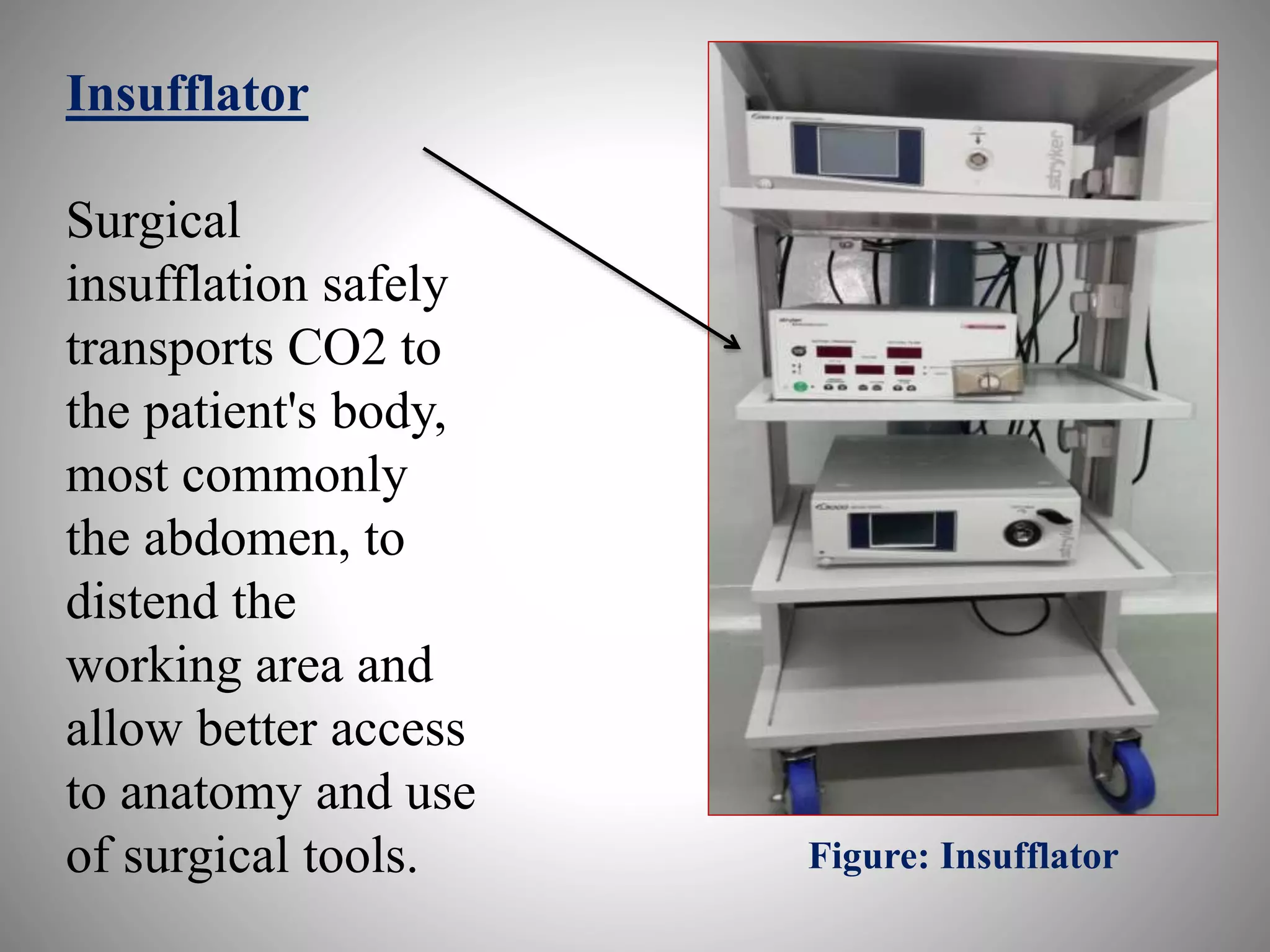
laparoscopic instruments
Prepared by
Dr Waseem Ahmad
Junior resident-3.
Dept. of Surgery (Ilmul Jarahat)
National Institute of Unani Medicine
Bangalore
ContentsContents
1. General description
2. Principals of laparoscopy
3. Composition of laparoscope
I. Equipment cart
II. Insufflating system
III. Imaging system
IV. Instrumentations
V. Electro-surgical-cautery
system
Laparoscopy
Laparoscopy is a technologically dependent
surgery comprising of computerized designed
instruments with microprocessor controlled safety
features.
Principal of laparoscopy
Identification of pathology and surgical
intervention is the solely principal of laparoscopy.
COMPOSITION OF A LAPAROSCOPE
1. Equipment cart
2. Insufflating system
3. Imaging system
4. Instrumentations
5. Electro-surgical-cautery system
Equipment cart
Multiple shelves for different equipments
1. Monitor shelf
2. Light source shelf
3. Co2 insufflators shelf
4. Electro-surgical shelf
Monitor shelf
Camera reader shelf
Co2 insufflators shelf
Light source shelf
Electro-surgical shelf
Figure: Equipment cart
Insufflating system
Insufflation system allows gas to fill the
abdominopelvic cavity to maximize visualization.
Insufflation system are designed to deliver the gas at
low as well as high pressure to ensure the constant set
intra-abdomino-pelvic pressure during surgery.
Insufflations tube with 0.3 micron filter is
recommended to prevent contamination with bacteria,
micro-particles and debris from insufflators and gas
tank.
CO2 Cylinders
Available sizes are
2.5 lb CO2 tanks
• 5 lb CO2 cylinders
• 7 lb CO2 tanks
• 10 lb CO2 cylinders
• 15 lb CO2 tanks
• 20 lb CO2 tanks
• 35 lb CO2 tanks
• 50 lb CO2 tanks
Figure: CO2 Cylinders
CO2 Cable:
It is flexible,
friendly use and
highly tensile in
strength to ensure
the proper
delivery of gas
from insufflators
to intra-peritoneal
cavity without
leakage.
Figure: CO2 Cable
Insufflator
Surgical
insufflation safely
transports CO2 to
the patient's body,
most commonly
the abdomen, to
distend the
working area and
allow better access
to anatomy and use
of surgical tools. Figure: Insufflator
Imaging system
1. Light source
2. Fiber-optic cable
3. Camera console
4. Endoscope/Telescope
5. Monitor
Light source
Three types are there
1. Halide
2. Halogen
3. Xenon
Halogen: 250 watts inbuilt
lamp and this is economical
Xenon: 175-300 watts lamp
and this is expensive Figure: Light source
LIGHT CABLES
There are two types of
light cables
Fluid filled cables and
fiber-optic cables
Fiber-optic cables
They conduct less heat
They are soft and sound to handle
They don’t produce hindrance
while handling camera console
They are easily autoclaved
Fluid filled cables
They conduct more heat
Stiffer and tough in nature
They produce hindrance while
handling camera console
They are difficult to be autoclaved
Figure: Fiber-optic cable
Fiber-optic cable
Connected to “light source”
Connected to “telescope”
Camera console
The camera head consists of a goal lens, a prism assembly
and three sensors for acquiring the primary colors of the
image.
Optical zoom is advantageous because it doesn't have
negative effect on the look resolution.
White balancing and contrast
Zoom options
Digital enhancement options to create sharper images.
Camera console
Connected to “Camera reader” Connected to “Telescope”
Endoscope/Telescope:
British physicist Hopkins invented in 1952.
Normally used telescope is the Hopkins Forward
Oblique Telescope 30°, diameter 10mm length 33cm,
and is autoclavable.
At the distal end is a front lens complex (inverting real-
image lens system, IRILS) which creates an inverted
and real image of the subject.
Forward viewing, 300 angled and 450 angled.
Endoscope/Telescope
10 mm telescope 5 mm telescope
300 beveled
tip
Metallic shaft
Fiber-optic cable is connected to this end
Connected to
camera
console
Figure: Endoscope/Telescope
Monitor
Surgical monitors are no different from the T.V. We watch at home.
The existing television systems in use differ according to the country.
The U.S.A uses the NTSC (National Television System Committee)
system.
In European countries the PAL (Phase Alternation by Line) system is in
use.
There is also a French system called SECAM (Sequential color and
memory).
The broadcasting standards for each are summarized below:
Standard resolution monitors have 400 lines
High definition monitors have 1200 lines.
Monitor
Handling devices
It contains following parts
i. Tip
ii. Insulated outer tube
iii. Rotator
iv. Attachment for electro-surgical
cord
v. Rachet for locking
Handling device
Tip
TROCAR
A trocar is a laparoscopic device that is made up of
an obturator (which may be a metal or plastic
sharpened or non-bladed tip) and a cannula (basically a
hollow tube).
Trocars are placed through the abdomen
during laparoscopic surgery.
The trocar functions as a portal for the subsequent
placement of other instruments, such as graspers,
scissors, staplers, etc
TROCAR
5 mm Trocar 10 mm Trocar
Obturator
Trocar cannula
What should be the good qualities of a
handling device?
It is usually 33 cm long
Jaws to be adequately elastic to perform atraumatic
handling
Easy to disassemble and reassemble
Parts should be interchangeable between similar
instruments
Easy cleaning and sterilization
Simple design with minimum number of hinges and bolts
Scissors
Reposable scissors: They combine reusable handle with
disposable scissor. They can be straight or curved scissors.
Reusable scissor: They combine reusable handle and reusable
scissors. They can be straight or curved scissors.
These scissors are called Metzenbaum scissors
Types
Straight scissor
Micro curved scissor
Hook scissor
Micro curved scissor
Slide locking grasper
They are designed to provide highest level of tissue
grasping properties.
Types
Straight fenestrated grasper
Allis grasper
Maxi grip grasper
Toothed grasper
Babcock grasper
Cobra grasper
Bowel grasper
Maxi grip grasper
Cobra grasper
Babcock grasper
Dissectors
Most commonly used is Maryland dissector.
Maryland dissector:
· Maryland dissector instrument is inserted through 5 mm trocar.
· Dissector comes with long, curved jaws and fine-tapered tips
· Dissectors used mainly for separation of sac from cord structures
·Also finding use for other blunt dissection purposes
· Rotating shaft for allowing easy dissection with lesser strain to surgeon's
wrist
· Sharp and insulated scissor finish
· As a monopolar electrosurgical instrument it finds use for dissection and
hemostasis using flexible endoscopes with working channel
Maryland dissector
Needle holders
Most commonly
used are-
Storz needle holder
And
Ethicon type
needle holder.
Figure: Storz needle holder
Clip applicator
This is used to ligate the vessels and ducts. The
applicator is first loaded with metallic clip and then
clip is applied by applying pressure to handle of the
instrument.
Clip
applicator
Veress needle
A Veress needle is a spring-loaded needle used to
create pneumoperitoneum for laparoscopic surgery.
The tool was first developed in 1932 by Janos Veres,
a Hungarian internist.
Modern needles are 12 to 15 cm long, with an external diameter of
2 mm.
The outer cannula consists of a beveled needle point for cutting
through tissues of the abdominal wall. A spring-loaded, inner stylet is
positioned within the outer cannula. This inner stylet has a dull tip to
protect any viscera from injury by the sharp, outer cannula.
Veress needle
Suction and irrigation cannula
This tube is essentially used in case of antra-
abdominal leakage in order to was out the
affected area.
This is three way cannula in which one end is used
for suction, another for irrigation and third one for
being placed in abdominal cavity.
Suction and
irrigation
cannula
This end is placed
in abdominal cavity
Thumb control
Port for irrigation
Part for drainage
Figure: Suction thumb control
Thank you for watching

Laparoscopic instruments

  • 1.
    laparoscopic instruments Prepared by DrWaseem Ahmad Junior resident-3. Dept. of Surgery (Ilmul Jarahat) National Institute of Unani Medicine Bangalore
  • 2.
    ContentsContents 1. General description 2.Principals of laparoscopy 3. Composition of laparoscope I. Equipment cart II. Insufflating system III. Imaging system IV. Instrumentations V. Electro-surgical-cautery system
  • 3.
    Laparoscopy Laparoscopy is atechnologically dependent surgery comprising of computerized designed instruments with microprocessor controlled safety features. Principal of laparoscopy Identification of pathology and surgical intervention is the solely principal of laparoscopy.
  • 4.
    COMPOSITION OF ALAPAROSCOPE 1. Equipment cart 2. Insufflating system 3. Imaging system 4. Instrumentations 5. Electro-surgical-cautery system
  • 5.
    Equipment cart Multiple shelvesfor different equipments 1. Monitor shelf 2. Light source shelf 3. Co2 insufflators shelf 4. Electro-surgical shelf
  • 6.
    Monitor shelf Camera readershelf Co2 insufflators shelf Light source shelf Electro-surgical shelf Figure: Equipment cart
  • 7.
    Insufflating system Insufflation systemallows gas to fill the abdominopelvic cavity to maximize visualization. Insufflation system are designed to deliver the gas at low as well as high pressure to ensure the constant set intra-abdomino-pelvic pressure during surgery. Insufflations tube with 0.3 micron filter is recommended to prevent contamination with bacteria, micro-particles and debris from insufflators and gas tank.
  • 8.
    CO2 Cylinders Available sizesare 2.5 lb CO2 tanks • 5 lb CO2 cylinders • 7 lb CO2 tanks • 10 lb CO2 cylinders • 15 lb CO2 tanks • 20 lb CO2 tanks • 35 lb CO2 tanks • 50 lb CO2 tanks Figure: CO2 Cylinders
  • 9.
    CO2 Cable: It isflexible, friendly use and highly tensile in strength to ensure the proper delivery of gas from insufflators to intra-peritoneal cavity without leakage. Figure: CO2 Cable
  • 10.
    Insufflator Surgical insufflation safely transports CO2to the patient's body, most commonly the abdomen, to distend the working area and allow better access to anatomy and use of surgical tools. Figure: Insufflator
  • 11.
    Imaging system 1. Lightsource 2. Fiber-optic cable 3. Camera console 4. Endoscope/Telescope 5. Monitor
  • 12.
    Light source Three typesare there 1. Halide 2. Halogen 3. Xenon Halogen: 250 watts inbuilt lamp and this is economical Xenon: 175-300 watts lamp and this is expensive Figure: Light source
  • 13.
    LIGHT CABLES There aretwo types of light cables Fluid filled cables and fiber-optic cables Fiber-optic cables They conduct less heat They are soft and sound to handle They don’t produce hindrance while handling camera console They are easily autoclaved Fluid filled cables They conduct more heat Stiffer and tough in nature They produce hindrance while handling camera console They are difficult to be autoclaved
  • 14.
    Figure: Fiber-optic cable Fiber-opticcable Connected to “light source” Connected to “telescope”
  • 15.
    Camera console The camerahead consists of a goal lens, a prism assembly and three sensors for acquiring the primary colors of the image. Optical zoom is advantageous because it doesn't have negative effect on the look resolution. White balancing and contrast Zoom options Digital enhancement options to create sharper images.
  • 16.
    Camera console Connected to“Camera reader” Connected to “Telescope”
  • 17.
    Endoscope/Telescope: British physicist Hopkinsinvented in 1952. Normally used telescope is the Hopkins Forward Oblique Telescope 30°, diameter 10mm length 33cm, and is autoclavable. At the distal end is a front lens complex (inverting real- image lens system, IRILS) which creates an inverted and real image of the subject. Forward viewing, 300 angled and 450 angled.
  • 18.
  • 19.
    300 beveled tip Metallic shaft Fiber-opticcable is connected to this end Connected to camera console Figure: Endoscope/Telescope
  • 20.
    Monitor Surgical monitors areno different from the T.V. We watch at home. The existing television systems in use differ according to the country. The U.S.A uses the NTSC (National Television System Committee) system. In European countries the PAL (Phase Alternation by Line) system is in use. There is also a French system called SECAM (Sequential color and memory). The broadcasting standards for each are summarized below: Standard resolution monitors have 400 lines High definition monitors have 1200 lines.
  • 21.
  • 22.
    Handling devices It containsfollowing parts i. Tip ii. Insulated outer tube iii. Rotator iv. Attachment for electro-surgical cord v. Rachet for locking
  • 23.
  • 24.
    TROCAR A trocar isa laparoscopic device that is made up of an obturator (which may be a metal or plastic sharpened or non-bladed tip) and a cannula (basically a hollow tube). Trocars are placed through the abdomen during laparoscopic surgery. The trocar functions as a portal for the subsequent placement of other instruments, such as graspers, scissors, staplers, etc
  • 25.
    TROCAR 5 mm Trocar10 mm Trocar Obturator Trocar cannula
  • 26.
    What should bethe good qualities of a handling device? It is usually 33 cm long Jaws to be adequately elastic to perform atraumatic handling Easy to disassemble and reassemble Parts should be interchangeable between similar instruments Easy cleaning and sterilization Simple design with minimum number of hinges and bolts
  • 27.
    Scissors Reposable scissors: Theycombine reusable handle with disposable scissor. They can be straight or curved scissors. Reusable scissor: They combine reusable handle and reusable scissors. They can be straight or curved scissors. These scissors are called Metzenbaum scissors Types Straight scissor Micro curved scissor Hook scissor
  • 28.
  • 29.
    Slide locking grasper Theyare designed to provide highest level of tissue grasping properties. Types Straight fenestrated grasper Allis grasper Maxi grip grasper Toothed grasper Babcock grasper Cobra grasper Bowel grasper
  • 30.
  • 31.
  • 32.
  • 33.
    Dissectors Most commonly usedis Maryland dissector. Maryland dissector: · Maryland dissector instrument is inserted through 5 mm trocar. · Dissector comes with long, curved jaws and fine-tapered tips · Dissectors used mainly for separation of sac from cord structures ·Also finding use for other blunt dissection purposes · Rotating shaft for allowing easy dissection with lesser strain to surgeon's wrist · Sharp and insulated scissor finish · As a monopolar electrosurgical instrument it finds use for dissection and hemostasis using flexible endoscopes with working channel
  • 34.
  • 35.
    Needle holders Most commonly usedare- Storz needle holder And Ethicon type needle holder. Figure: Storz needle holder
  • 36.
    Clip applicator This isused to ligate the vessels and ducts. The applicator is first loaded with metallic clip and then clip is applied by applying pressure to handle of the instrument.
  • 37.
  • 38.
    Veress needle A Veressneedle is a spring-loaded needle used to create pneumoperitoneum for laparoscopic surgery. The tool was first developed in 1932 by Janos Veres, a Hungarian internist. Modern needles are 12 to 15 cm long, with an external diameter of 2 mm. The outer cannula consists of a beveled needle point for cutting through tissues of the abdominal wall. A spring-loaded, inner stylet is positioned within the outer cannula. This inner stylet has a dull tip to protect any viscera from injury by the sharp, outer cannula.
  • 39.
  • 40.
    Suction and irrigationcannula This tube is essentially used in case of antra- abdominal leakage in order to was out the affected area. This is three way cannula in which one end is used for suction, another for irrigation and third one for being placed in abdominal cavity.
  • 41.
    Suction and irrigation cannula This endis placed in abdominal cavity Thumb control Port for irrigation Part for drainage Figure: Suction thumb control
  • 42.
    Thank you forwatching

Editor's Notes

  • #19 5 mm telescope5 mm telescope