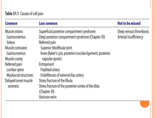
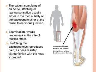
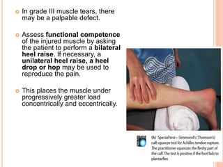
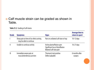
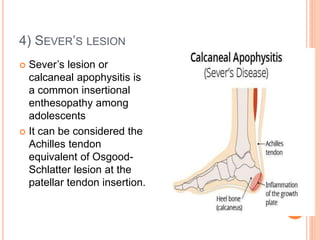
1. The document discusses various injuries to the calf and ankle, including gastrocnemius muscle strains, soleus muscle strains, Achilles tendon ruptures, Sever's lesion, and lateral and medial ligament injuries.
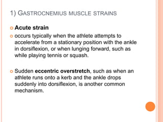
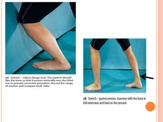
2. It describes the mechanisms, symptoms, and physical exam findings for each injury. Gastrocnemius strains typically occur during plantarflexion while soleus strains have a history of increasing tightness.
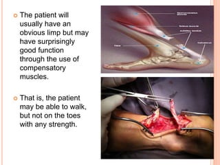
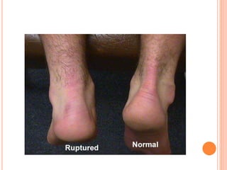
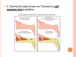
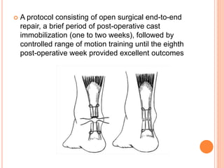
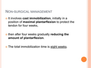
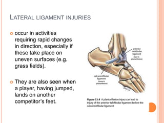
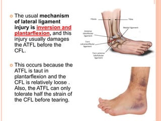
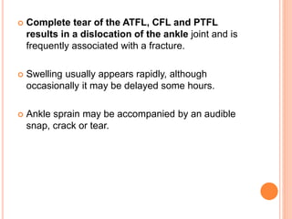
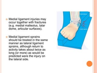
3. Achilles tendon ruptures result in a palpable gap and reduced plantarflexion strength. Ligament injuries involve inversion or eversion strains and medial injuries have a longer recovery time.
3. 1) GASTROCNEMIUS MUSCLE STRAINS
Acute strain
occurs typically when the athlete attempts to
accelerate from a stationary position with the ankle
in dorsiflexion, or when lunging forward, such as
while playing tennis or squash.
Sudden eccentric overstretch, such as when an
athlete runs onto a kerb and the ankle drops
suddenly into dorsiflexion, is another common
mechanism.
4. The patient complains of
an acute, stabbing or
tearing sensation usually
either in the medial belly of
the gastrocnemius or at the
musculotendinous junction.
Examination reveals
tenderness at the site of
muscle strain.
Stretching the
gastrocnemius reproduces
pain, as does resisted
plantarflexion with the knee
extended.
5. In grade III muscle tears, there
may be a palpable defect.
Assess functional competence
of the injured muscle by asking
the patient to perform a bilateral
heel raise. If necessary, a
unilateral heel raise, a heel
drop or hop may be used to
reproduce the pain.
This places the muscle under
progressively greater load
concentrically and eccentrically.
6. Calf muscle strain can be graded as shown in
Table.
7. CHRONIC STRAIN
Chronic gastrocnemius muscle strain may occur as
an overuse injury or following inadequate
rehabilitation of an acute injury.
Inadequate rehabilitation results in disorganized,
weak scar tissue that is susceptible to further injury.
8. 2) SOLEUS MUSCLE STRAINS
Strains of the soleus muscle are a relatively common
sports injury.
sudden onset pain,
history of increasing calf tightness over a period of
days or weeks.
Examination reveals tenderness deep to the
gastrocnemius, usually in the lateral aspect of the
soleus muscle.
Both the soleus stretch and resisted soleus contraction
provoke pain. This can be differentiated from the stretch
and contraction that provoke pain in gastrocnemius
strains.
9.
10. 3) ACHILLES TENDON RUPTURE (COMPLETE)
Complete rupture of the Achilles tendon classically
occurs in athletes in their 30s or 40s.
male:female ratio is 10:1.
The patient describes feeling ‘as if I was hit or kicked in
the back of the leg’;
pain is not always the strongest sensation.
This is immediately followed by grossly diminished
function.
A snap or tear may be audible.
11. The patient will
usually have an
obvious limp but may
have surprisingly
good function
through the use of
compensatory
muscles.
That is, the patient
may be able to walk,
but not on the toes
with any strength.
12. FOUR CLINICAL TESTS CAN GREATLY SIMPLIFY
EXAMINATION OF COMPLETE ACHILLES TENDON
RUPTURE:
1. On careful inspection with the patient prone and
both ankles fully relaxed, the foot on the side with
the ruptured tendon hangs straight down (because
of the absence of tendon tone); the foot on the non-
ruptured side maintains a little plantarflexion.
2. Acutely, there may be a palpable gap in the
tendon, approximately 3–6 cm proximal to the
insertion into the calcaneus.
3. The strength of plantarflexion is markedly
reduced.
13.
14. 4. Simmond’s (also known as Thomson’s) calf
squeeze test is positive.
15. SURGICAL MANAGEMENT
Open surgical treatment of Achilles tendon rupture is
associated with a 27% lower risk of re-rupture compared
with non-surgical treatment.
Complications including infection, adhesions and
disturbed skin sensitivity.
Another approach to reduce these complications is to
perform surgery ‘percutaneously’.
Early post-operative mobilization with a functional
brace reduced the complication rate compared with in
those who had been managed with post-operative cast
immobilization for eight weeks.
16. A protocol consisting of open surgical end-to-end
repair, a brief period of post-operative cast
immobilization (one to two weeks), followed by
controlled range of motion training until the eighth
post-operative week provided excellent outcomes
17. NON-SURGICAL MANAGEMENT
It involves cast immobilization, initially in a
position of maximal plantarflexion to protect the
tendon for four weeks,
then after four weeks gradually reducing the
amount of plantarflexion.
The total immobilization time is eight weeks.
18. 4) SEVER’S LESION
Sever’s lesion or
calcaneal apophysitis is
a common insertional
enthesopathy among
adolescents
It can be considered the
Achilles tendon
equivalent of Osgood-
Schlatter lesion at the
patellar tendon insertion.
19. LATERAL LIGAMENT INJURIES
occur in activities
requiring rapid changes
in direction, especially if
these take place on
uneven surfaces (e.g.
grass fields).
They are also seen when
a player, having jumped,
lands on another
competitor’s feet.
20. The usual mechanism
of lateral ligament
injury is inversion and
plantarflexion, and this
injury usually damages
the ATFL before the
CFL.
This occurs because the
ATFL is taut in
plantarflexion and the
CFL is relatively loose .
Also, the ATFL can only
tolerate half the strain of
the CFL before tearing.
21. Complete tear of the ATFL, CFL and PTFL
results in a dislocation of the ankle joint and is
frequently associated with a fracture.
Swelling usually appears rapidly, although
occasionally it may be delayed some hours.
Ankle sprain may be accompanied by an audible
snap, crack or tear.
22. MEDIAL (DELTOID) LIGAMENT INJURIES
Because the deltoid ligament is stronger than the
lateral ligament, and probably
because eversion is a less common mechanism
of ankle sprain, medial ankle ligament injuries are
less common than lateral ligament injuries.
Occasionally, medial and lateral ligament injuries
occur in the same ankle sprain.
23. Medial ligament injuries may
occur together with fractures
(e.g. medial malleolus, talar
dome, articular surfaces).
Medial ligament sprains
should be treated in the same
manner as lateral ligament
sprains, although return to
activity takes about twice as
long (or more) as would be
predicted were the injury on
the lateral side.