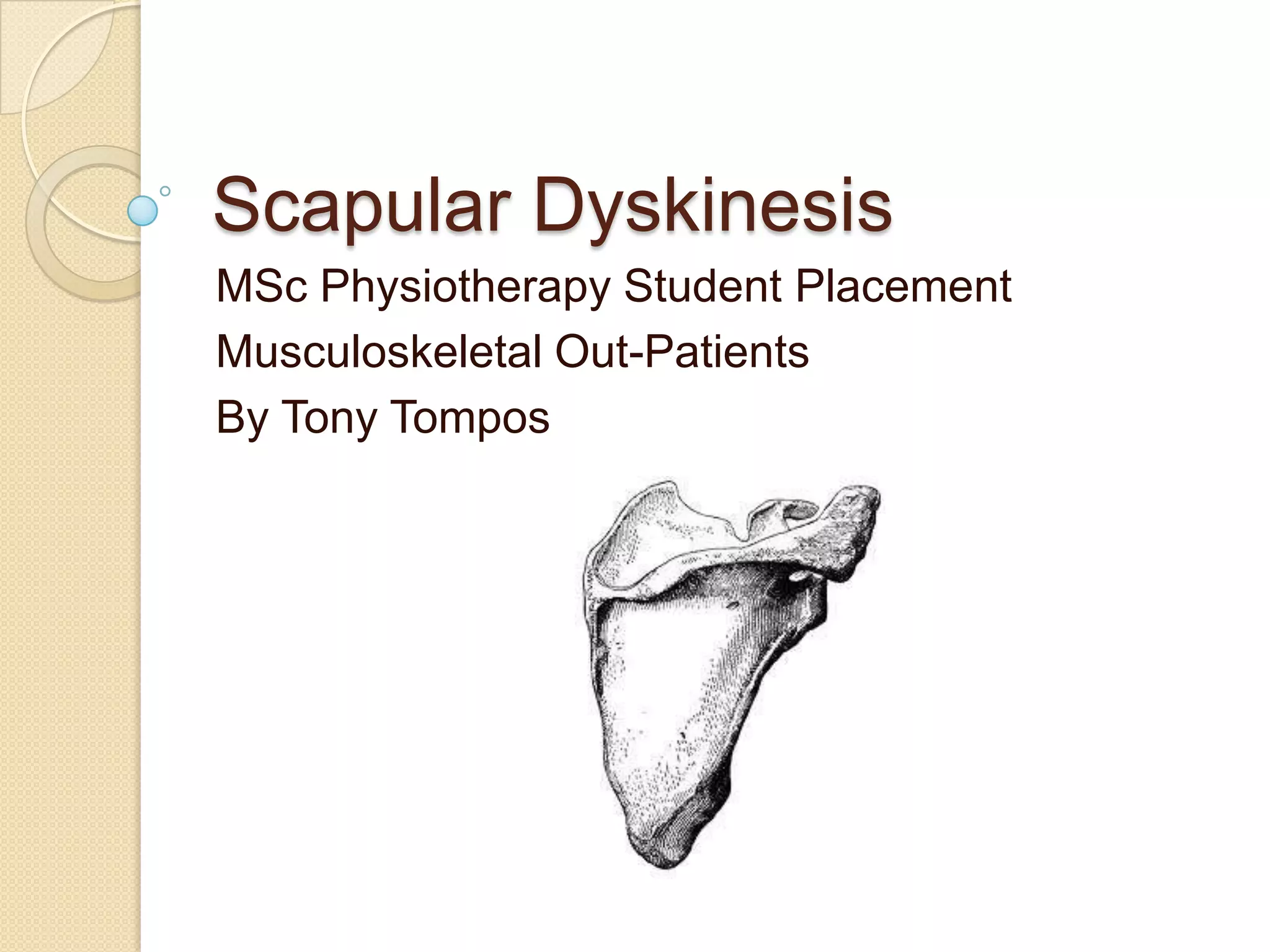
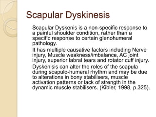
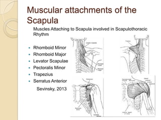
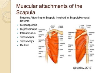
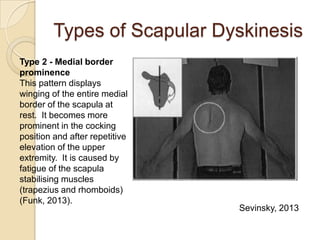
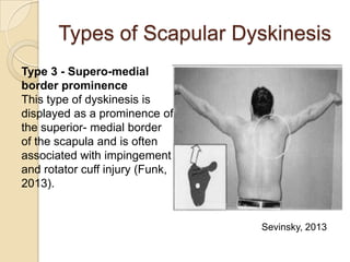
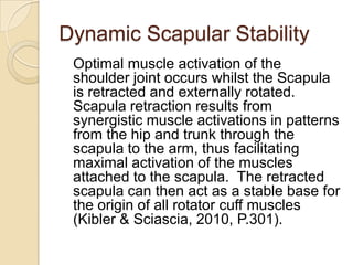
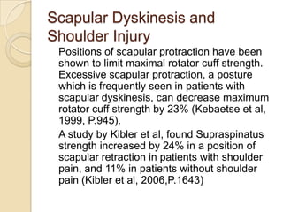
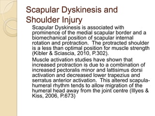
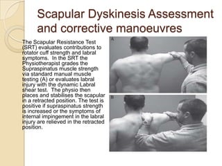
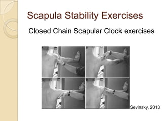
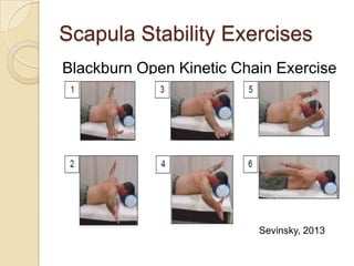
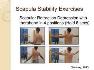
Scapular Dyskinesis
MSc Physiotherapy Student Placement
Musculoskeletal Out-Patients
By Tony Tompos
Contents
 Introduction to Scapular Dyskinesis
 Muscular attachments of the scapula
 Types of Scapular Dyskinesis
 Dynamic scapular stability
 Scapular Dyskinesis and shoulder
injury
 Scapular Assessment and Corrective
manoeuvres
 Treatment
Scapular Dyskinesis
Scapular dyskinesis has been identified
as (1)‘abnormal static scapular position
and/or dynamic scapular motion
characterised by medial border
prominence or
(2) ‘inferior angle prominence and/or
early scapular elevation or shrugging on
arm elevation’ and/or
(3) ‘rapid downward rotation during arm
lowering’ (Kibler & Sciascia, 2010 p.300)
Scapular Dyskinesis
However due to the difference in
dynamic scapular motion and static
position of the scapula; when
describing the static position of the
scapula, if an asymmetry is observed,
then this should be referred to as
‘altered scapular resting position’
rather than scapular dyskinesis (Kibler
& Sciascia, 2010 p.301)
Scapular Dyskinesis
Scapular Dyskenis is a non-specific response to
a painful shoulder condition, rather than a
specific response to certain glenohumeral
pathology.
It has multiple causative factors including Nerve
injury, Muscle weakness/imbalance, AC joint
injury, superior labral tears and rotator cuff injury.
Dyskenisis can alter the roles of the scapula
during scapulo-humeral rhythm and may be due
to alterations in bony stabilisers, muscle
activation patterns or lack of strength in the
dynamic muscle stabilisers. (Kibler, 1998, p.325).
Muscular attachments of the
Scapula
Muscles Attaching to Scapula involved in Scapulothoracic
Rhythm
 Rhomboid Minor
 Rhomboid Major
 Levator Scapulae
 Pectoralis Minor
 Trapezius
 Serratus Anterior
Sevinsky, 2013
Muscular attachments of the
Scapula
Muscles Attaching to Scapula involved in ScapuloHumeral
Rhythm
 Subscapularis
 Supraspinatus
 Infraspinatus
 Teres Minor
 Teres Major
 Deltoid
Sevinsky, 2013
Types of Scapular Dyskinesis
Type 1 - Infero-medial scapula
border prominence.
This becomes more evident in
the cocking position of overhead
sports. It is often associated
with tightness anteriorly
(inflexibility of the pectoralis
major/ minor muscles) with
weakness of the lower trapezius
and serratus anterior
muscles. This is often noticed
in the early stages of shoulder
disorders (Funk, 2013).
Sevinsky, 2013
Types of Scapular Dyskinesis
Type 2 - Medial border
prominence
This pattern displays
winging of the entire medial
border of the scapula at
rest. It becomes more
prominent in the cocking
position and after repetitive
elevation of the upper
extremity. It is caused by
fatigue of the scapula
stabilising muscles
(trapezius and rhomboids)
(Funk, 2013).
Sevinsky, 2013
Types of Scapular Dyskinesis
Type 3 - Supero-medial
border prominence
This type of dyskinesis is
displayed as a prominence of
the superior- medial border
of the scapula and is often
associated with impingement
and rotator cuff injury (Funk,
2013).
Sevinsky, 2013
Dynamic Scapular Stability
Stabilisation of the scapula and motion on the
thorax is provided by the coupling of the
upper and lower Trapezius with the Serratus
anterior and Rhomboid muscles. Scapular
elevation during arm elevation is
accomplished through coupling of the
Serratus anterior and lower Trapezius with
the upper Trapezius and Rhomboids.
Returning from upwards elevation, the
appropriately positioned Trapezius, when
working efficiently, helps maintain the
Scapula against the Thorax (Kibler &
Sciascia, 2010 p.301)
Dynamic Scapular Stability
Serratus anterior is a key stabiliser of the
Scapula as it contributes to all components of
three-dimensional motion of the scapula
during arm elevation. It helps produce
scapular upward rotation, posterior tilt, and
external rotation whilst stabilising the medial
border and inferior angle, thus preventing
scapular winging (Ludewig, 1996, P.57).
The primary role of the Serratus Anterior
during activities such as throwing, and
shoulder elevation is to act as an external
rotator/stabiliser of the scapular during arm
motion (Kibler & Sciascia, 2010, P.301)
Dynamic Scapular Stability
Optimal muscle activation of the
shoulder joint occurs whilst the Scapula
is retracted and externally rotated.
Scapula retraction results from
synergistic muscle activations in patterns
from the hip and trunk through the
scapula to the arm, thus facilitating
maximal activation of the muscles
attached to the scapula. The retracted
scapula can then act as a stable base for
the origin of all rotator cuff muscles
(Kibler & Sciascia, 2010, P.301).
Scapular Dyskinesis and
Shoulder Injury
Positions of scapular protraction have been
shown to limit maximal rotator cuff strength.
Excessive scapular protraction, a posture
which is frequently seen in patients with
scapular dyskinesis, can decrease maximum
rotator cuff strength by 23% (Kebaetse et al,
1999, P.945).
A study by Kibler et al, found Supraspinatus
strength increased by 24% in a position of
scapular retraction in patients with shoulder
pain, and 11% in patients without shoulder
pain (Kibler et al, 2006,P.1643)
Scapular Dyskinesis and
Shoulder Injury
Scapular Dyskinesis is associated with
prominence of the medial scapular border and a
biomechanical position of scapular internal
rotation and protraction. The protracted shoulder
is a less than optimal position for muscle strength
(Kibler & Sciascia, 2010, P.302).
Muscle activation studies have shown that
increased protraction is due to a combination of
increased pectoralis minor and lattisimus dorsi
activation and decreased lower trapezius and
serratus anterior activation. This altered scapula-
humeral rhythm tends to allow migration of the
humeral head away from the joint centre (Illyes &
Kiss, 2006, P.673)
Scapular Dyskinesis Assessment
and corrective manoeuvres
The Scapular Assistance
Test (SAT) helps evaluate
scapular contributions to
impingement and rotator
cuff strength. In the SAT,
gentle pressure is applied
to assist scapular upward
rotation and posterior tilt as
the patient elevates the
arm. The test is positive
when the painful arc of
impingement symptoms is
is relieved and the arc of
motion is increased.
Scapular Dyskinesis Assessment
and corrective manoeuvres
The Scapular Resistance Test
(SRT) evaluates contributions to
rotator cuff strength and labral
symptoms. In the SRT the
Physiotherapist grades the
Supraspinatus muscle strength
via standard manual muscle
testing (A) or evaluates labral
injury with the dynamic Labral
shear test. The physio then
places and stabilises the scapular
in a retracted position. The test is
positive if supraspinatus strength
is increased or the symptoms of
internal impingement in the labral
injury are relieved in the retracted
position.
Scapular Dyskinesis Assessment
and corrective manoeuvres
Neither the SRT or the SAT are capable
of diagnosing a specific form of shoulder
pathology, however a positive SAT or
SRT shows that Scapular Dyskinesis is
directly involved in producing the
patients symptoms and would indicate
the need for the inclusion of early
scapular rehabilitation exercises to
improve scapular control (Kibler &
Sciascia, 2010, P.303).
Treatment
Bony injury and/or tissue derangement such
as AC joint separation, clavicle fractures,
labral tears or rotator cuff disease may
require surgical intervention before treatment
of scapular dyskinesis can be addressed.
However, the large majority of cases of
dyskinesis are caused due to muscle
weakness, inhibition or inflexibility and can be
managed with conservative rehabilition. The
goal of initial treatment is to achieve the
position of optimal scapular function –
Posterior tilt, external rotation and upward
elevation (Kibler & Sciascia, 2010, P.303).
Treatment
Rehabilitation emphasis should start proximally and end
distally. Proximal control of core stability, leading to an
increased control of three dimensional control scapular
motion is achieved through a rehab regimen in which larger
muscles of the lower extremity and trunk are utilised during
the treatment of the scapula and shoulder.
Hip and trunk flexion
facilitate scapular
protraction, whilst hip
and trunk extension
along with trunk
rotation, aid in
facilitating scapular
retraction.
Treatment
Scapular stabilisation protocols
should focus on re-educating the
muscles to act as dynamic scapular
stabilisers as maximal rotator cuff
strength is achieved via a stabilised,
retracted scapula. Scapular control
should be achieved prior to
emphasising rotator cuff strength
and should emphasise closed chain,
humeral head co-contractions.
A logical progression of exercises
(isometric to dynamic) should be
focussed on strengthening the lower
trapezius and serratus anterior
whilst minimising upper trapezius
activation.
Scapula Stability Exercises
Closed Chain Scapular Clock exercises
Sevinsky, 2013
Scapula Stability Exercises
Blackburn Open Kinetic Chain Exercise
Sevinsky, 2013
Scapula Stability Exercises
Scapular Retraction Depression with
theraband in 4 positions (Hold 6 secs)
Sevinsky, 2013
Questions?
References
Funk L (2013) Available at: http://www.shoulderdoc.co.uk/article.asp?article=930
Illyes A & Kiss RM (2003) Kinematic and muscle activity characteristics of
multidirectional shoulder joint instability during elevation. Knee Surgery, Sports
Traumatology, Arthroscopy 14:673-85
Kibler BW & Sciascia A, (2010) Current Concepts: Scapular Dyskinesis. British Journal
of Sports Medicine, 2010; 44: 300-305.
Kibler BW (1998) The role of the scapula in function. American journal of sports
medicine 26:325-37.
Kebbatse M, Mclure P, Pratt NA (1999) Thoracic position effect on shoulder range of
motion, strength, and three-dimensional kinematics. Archives of physical medicine and
rehabilitation 80:945-50
Ludewig PM, Cook TM, Nawoczenski D (1996) Three dimensional scapular orientation
and muscle activity at selected positions of humeral elevation. Journal of orthopaedic
sports and physical therapy. 24:57-65
Sevinsky S (2013) Scapular Dyskinesis. Available at:
http://www.smogshoulder.com/images/Scapular_dyskinesis.pdf

Scapular dyskinesis

  • 1.
    Scapular Dyskinesis MSc PhysiotherapyStudent Placement Musculoskeletal Out-Patients By Tony Tompos
  • 2.
    Contents  Introduction toScapular Dyskinesis  Muscular attachments of the scapula  Types of Scapular Dyskinesis  Dynamic scapular stability  Scapular Dyskinesis and shoulder injury  Scapular Assessment and Corrective manoeuvres  Treatment
  • 3.
    Scapular Dyskinesis Scapular dyskinesishas been identified as (1)‘abnormal static scapular position and/or dynamic scapular motion characterised by medial border prominence or (2) ‘inferior angle prominence and/or early scapular elevation or shrugging on arm elevation’ and/or (3) ‘rapid downward rotation during arm lowering’ (Kibler & Sciascia, 2010 p.300)
  • 4.
    Scapular Dyskinesis However dueto the difference in dynamic scapular motion and static position of the scapula; when describing the static position of the scapula, if an asymmetry is observed, then this should be referred to as ‘altered scapular resting position’ rather than scapular dyskinesis (Kibler & Sciascia, 2010 p.301)
  • 5.
    Scapular Dyskinesis Scapular Dyskenisis a non-specific response to a painful shoulder condition, rather than a specific response to certain glenohumeral pathology. It has multiple causative factors including Nerve injury, Muscle weakness/imbalance, AC joint injury, superior labral tears and rotator cuff injury. Dyskenisis can alter the roles of the scapula during scapulo-humeral rhythm and may be due to alterations in bony stabilisers, muscle activation patterns or lack of strength in the dynamic muscle stabilisers. (Kibler, 1998, p.325).
  • 6.
    Muscular attachments ofthe Scapula Muscles Attaching to Scapula involved in Scapulothoracic Rhythm  Rhomboid Minor  Rhomboid Major  Levator Scapulae  Pectoralis Minor  Trapezius  Serratus Anterior Sevinsky, 2013
  • 7.
    Muscular attachments ofthe Scapula Muscles Attaching to Scapula involved in ScapuloHumeral Rhythm  Subscapularis  Supraspinatus  Infraspinatus  Teres Minor  Teres Major  Deltoid Sevinsky, 2013
  • 8.
    Types of ScapularDyskinesis Type 1 - Infero-medial scapula border prominence. This becomes more evident in the cocking position of overhead sports. It is often associated with tightness anteriorly (inflexibility of the pectoralis major/ minor muscles) with weakness of the lower trapezius and serratus anterior muscles. This is often noticed in the early stages of shoulder disorders (Funk, 2013). Sevinsky, 2013
  • 9.
    Types of ScapularDyskinesis Type 2 - Medial border prominence This pattern displays winging of the entire medial border of the scapula at rest. It becomes more prominent in the cocking position and after repetitive elevation of the upper extremity. It is caused by fatigue of the scapula stabilising muscles (trapezius and rhomboids) (Funk, 2013). Sevinsky, 2013
  • 10.
    Types of ScapularDyskinesis Type 3 - Supero-medial border prominence This type of dyskinesis is displayed as a prominence of the superior- medial border of the scapula and is often associated with impingement and rotator cuff injury (Funk, 2013). Sevinsky, 2013
  • 11.
    Dynamic Scapular Stability Stabilisationof the scapula and motion on the thorax is provided by the coupling of the upper and lower Trapezius with the Serratus anterior and Rhomboid muscles. Scapular elevation during arm elevation is accomplished through coupling of the Serratus anterior and lower Trapezius with the upper Trapezius and Rhomboids. Returning from upwards elevation, the appropriately positioned Trapezius, when working efficiently, helps maintain the Scapula against the Thorax (Kibler & Sciascia, 2010 p.301)
  • 12.
    Dynamic Scapular Stability Serratusanterior is a key stabiliser of the Scapula as it contributes to all components of three-dimensional motion of the scapula during arm elevation. It helps produce scapular upward rotation, posterior tilt, and external rotation whilst stabilising the medial border and inferior angle, thus preventing scapular winging (Ludewig, 1996, P.57). The primary role of the Serratus Anterior during activities such as throwing, and shoulder elevation is to act as an external rotator/stabiliser of the scapular during arm motion (Kibler & Sciascia, 2010, P.301)
  • 13.
    Dynamic Scapular Stability Optimalmuscle activation of the shoulder joint occurs whilst the Scapula is retracted and externally rotated. Scapula retraction results from synergistic muscle activations in patterns from the hip and trunk through the scapula to the arm, thus facilitating maximal activation of the muscles attached to the scapula. The retracted scapula can then act as a stable base for the origin of all rotator cuff muscles (Kibler & Sciascia, 2010, P.301).
  • 14.
    Scapular Dyskinesis and ShoulderInjury Positions of scapular protraction have been shown to limit maximal rotator cuff strength. Excessive scapular protraction, a posture which is frequently seen in patients with scapular dyskinesis, can decrease maximum rotator cuff strength by 23% (Kebaetse et al, 1999, P.945). A study by Kibler et al, found Supraspinatus strength increased by 24% in a position of scapular retraction in patients with shoulder pain, and 11% in patients without shoulder pain (Kibler et al, 2006,P.1643)
  • 15.
    Scapular Dyskinesis and ShoulderInjury Scapular Dyskinesis is associated with prominence of the medial scapular border and a biomechanical position of scapular internal rotation and protraction. The protracted shoulder is a less than optimal position for muscle strength (Kibler & Sciascia, 2010, P.302). Muscle activation studies have shown that increased protraction is due to a combination of increased pectoralis minor and lattisimus dorsi activation and decreased lower trapezius and serratus anterior activation. This altered scapula- humeral rhythm tends to allow migration of the humeral head away from the joint centre (Illyes & Kiss, 2006, P.673)
  • 16.
    Scapular Dyskinesis Assessment andcorrective manoeuvres The Scapular Assistance Test (SAT) helps evaluate scapular contributions to impingement and rotator cuff strength. In the SAT, gentle pressure is applied to assist scapular upward rotation and posterior tilt as the patient elevates the arm. The test is positive when the painful arc of impingement symptoms is is relieved and the arc of motion is increased.
  • 17.
    Scapular Dyskinesis Assessment andcorrective manoeuvres The Scapular Resistance Test (SRT) evaluates contributions to rotator cuff strength and labral symptoms. In the SRT the Physiotherapist grades the Supraspinatus muscle strength via standard manual muscle testing (A) or evaluates labral injury with the dynamic Labral shear test. The physio then places and stabilises the scapular in a retracted position. The test is positive if supraspinatus strength is increased or the symptoms of internal impingement in the labral injury are relieved in the retracted position.
  • 18.
    Scapular Dyskinesis Assessment andcorrective manoeuvres Neither the SRT or the SAT are capable of diagnosing a specific form of shoulder pathology, however a positive SAT or SRT shows that Scapular Dyskinesis is directly involved in producing the patients symptoms and would indicate the need for the inclusion of early scapular rehabilitation exercises to improve scapular control (Kibler & Sciascia, 2010, P.303).
  • 19.
    Treatment Bony injury and/ortissue derangement such as AC joint separation, clavicle fractures, labral tears or rotator cuff disease may require surgical intervention before treatment of scapular dyskinesis can be addressed. However, the large majority of cases of dyskinesis are caused due to muscle weakness, inhibition or inflexibility and can be managed with conservative rehabilition. The goal of initial treatment is to achieve the position of optimal scapular function – Posterior tilt, external rotation and upward elevation (Kibler & Sciascia, 2010, P.303).
  • 20.
    Treatment Rehabilitation emphasis shouldstart proximally and end distally. Proximal control of core stability, leading to an increased control of three dimensional control scapular motion is achieved through a rehab regimen in which larger muscles of the lower extremity and trunk are utilised during the treatment of the scapula and shoulder. Hip and trunk flexion facilitate scapular protraction, whilst hip and trunk extension along with trunk rotation, aid in facilitating scapular retraction.
  • 21.
    Treatment Scapular stabilisation protocols shouldfocus on re-educating the muscles to act as dynamic scapular stabilisers as maximal rotator cuff strength is achieved via a stabilised, retracted scapula. Scapular control should be achieved prior to emphasising rotator cuff strength and should emphasise closed chain, humeral head co-contractions. A logical progression of exercises (isometric to dynamic) should be focussed on strengthening the lower trapezius and serratus anterior whilst minimising upper trapezius activation.
  • 22.
    Scapula Stability Exercises ClosedChain Scapular Clock exercises Sevinsky, 2013
  • 23.
    Scapula Stability Exercises BlackburnOpen Kinetic Chain Exercise Sevinsky, 2013
  • 24.
    Scapula Stability Exercises ScapularRetraction Depression with theraband in 4 positions (Hold 6 secs) Sevinsky, 2013
  • 25.
  • 26.
    References Funk L (2013)Available at: http://www.shoulderdoc.co.uk/article.asp?article=930 Illyes A & Kiss RM (2003) Kinematic and muscle activity characteristics of multidirectional shoulder joint instability during elevation. Knee Surgery, Sports Traumatology, Arthroscopy 14:673-85 Kibler BW & Sciascia A, (2010) Current Concepts: Scapular Dyskinesis. British Journal of Sports Medicine, 2010; 44: 300-305. Kibler BW (1998) The role of the scapula in function. American journal of sports medicine 26:325-37. Kebbatse M, Mclure P, Pratt NA (1999) Thoracic position effect on shoulder range of motion, strength, and three-dimensional kinematics. Archives of physical medicine and rehabilitation 80:945-50 Ludewig PM, Cook TM, Nawoczenski D (1996) Three dimensional scapular orientation and muscle activity at selected positions of humeral elevation. Journal of orthopaedic sports and physical therapy. 24:57-65 Sevinsky S (2013) Scapular Dyskinesis. Available at: http://www.smogshoulder.com/images/Scapular_dyskinesis.pdf