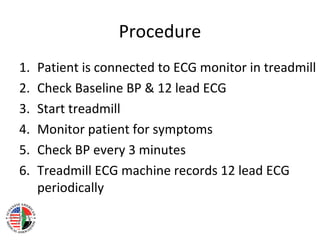
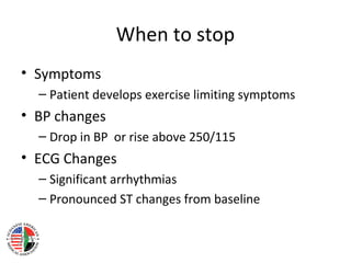
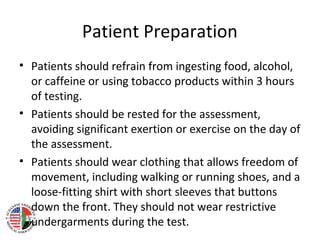
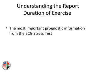
Exercise ECG Stress Testing for
Internists
Salah Abusin, MD, MRCP, ABIM, ABIM (Card)
Interventional Cardiologist
Dubuque, IA, USA
Stress Testing for Internists
• Principles & Purpose
• Patient Preparation
• Understanding the report
• When NOT to do it – Contraindications
• When to do it - Indications
Exercise ECG Stress Testing
Principles
• Elicit cardiovascular & ECG abnormalities not
present at rest and to determine the adequacy
of cardiac function.
Exercise ECG Stress Testing
Purpose
• estimate prognosis/determine functional
capacity
• the likelihood and extent of coronary artery
disease (CAD)
Procedure
1. Patient is connected to ECG monitor in treadmill
2. Check Baseline BP & 12 lead ECG
3. Start treadmill
4. Monitor patient for symptoms
5. Check BP every 3 minutes
6. Treadmill ECG machine records 12 lead ECG
periodically
When to stop
• Symptoms
– Patient develops exercise limiting symptoms
• BP changes
– Drop in BP or rise above 250/115
• ECG Changes
– Significant arrhythmias
– Pronounced ST changes from baseline
Patient Preparation
• Patients should refrain from ingesting food, alcohol,
or caffeine or using tobacco products within 3 hours
of testing.
• Patients should be rested for the assessment,
avoiding significant exertion or exercise on the day of
the assessment.
• Patients should wear clothing that allows freedom of
movement, including walking or running shoes, and a
loose-fitting shirt with short sleeves that buttons
down the front. They should not wear restrictive
undergarments during the test.
Understanding the Report
Duration of Exercise
• The most important prognostic information
from the ECG Stress Test
Understanding the Report
Target HR achieved?
• The target HR is measured as follows
– 220 – Age
• HR with exercise should rise to > 85% of target HR
to consider this sufficient exercise
• If the test is stopped early (in the absence of
evidence of ischemia) before >85% of target HR is
achieved then the test is considered inconclusive
Understanding the Report
Did the patient develop any
symptoms?
• Development of chest pain (with
hemodynamic/ECG changes) suggested an
increased likelihood of CAD
• Chest pain at low workload is considered a
poor prognostic sign
Understanding the Report
BP changes during exercise
• SBP should rise with exercise
• A drop in SBP with exercise is considered a
poor prognostic sign (indication to stop the
test)
Understanding the Report
ST segment changes with Exercise
• Development of significant ST depression
during Exercise increases the likelihood of
significant CAD
• Development of ST Elevation is considered a
poor prognostic sign (indication to stop the
test)
Understanding the Report
Arrhythmias during Exercise
• Development of VT is considered a poor
prognostic sign (indication to stop the test)
When not to do it?
When it is not safe
• ACS – Acute Phase
• Uncontrolled arrhythmia
• Uncontrolled HF, uncontrolled HTN
• Symptomatic Severe Ao stenosis
• Acute PE
• Suspected or known dissecting aneurysm
• Active or suspected myocarditis, pericarditis, or endocarditis
• Acute noncardiac disorder that may affect exercise
performance or be aggravated by exercise
When not to do it?
When the ECG cannot be interpreted
• ACS
• Uncontrolled arrhythmia
• Uncontrolled HF, uncontrolled HTN
• Symptomatic Severe Ao stenosis
• Acute PE
• Suspected or known dissecting aneurysm
• Active or suspected myocarditis, pericarditis, or endocarditis
• Acute noncardiac disorder that may affect exercise
performance or be aggravated by exercise
Indications - Stable Angina
• Diagnosis of Obstructive CAD
– Highest Yield in patients with intermediate
probability
– When used in patients with low probability,
increases likelihood of false positive (especially in
women)
• Risk Stratification in patients high probability
of CAD
Indications – Unstable Angina
• Risk Stratification for Low risk Unstable Angina
(negative troponin) after 8-12 hours
Indications – STEMI
• Before discharge for patients who underwent
successful thrombolysis and did NOT undergo
diagnostic coronary angiography
• Before discharge in patients who came with
late presenting STEMI and did NOT undergo
diagnostic coronary angiography
• Thanks

CAD 2014 - Introduction to Stress testing

  • 1.
    Exercise ECG StressTesting for Internists Salah Abusin, MD, MRCP, ABIM, ABIM (Card) Interventional Cardiologist Dubuque, IA, USA
  • 2.
    Stress Testing forInternists • Principles & Purpose • Patient Preparation • Understanding the report • When NOT to do it – Contraindications • When to do it - Indications
  • 3.
    Exercise ECG StressTesting Principles • Elicit cardiovascular & ECG abnormalities not present at rest and to determine the adequacy of cardiac function.
  • 4.
    Exercise ECG StressTesting Purpose • estimate prognosis/determine functional capacity • the likelihood and extent of coronary artery disease (CAD)
  • 5.
    Procedure 1. Patient isconnected to ECG monitor in treadmill 2. Check Baseline BP & 12 lead ECG 3. Start treadmill 4. Monitor patient for symptoms 5. Check BP every 3 minutes 6. Treadmill ECG machine records 12 lead ECG periodically
  • 6.
    When to stop •Symptoms – Patient develops exercise limiting symptoms • BP changes – Drop in BP or rise above 250/115 • ECG Changes – Significant arrhythmias – Pronounced ST changes from baseline
  • 7.
    Patient Preparation • Patientsshould refrain from ingesting food, alcohol, or caffeine or using tobacco products within 3 hours of testing. • Patients should be rested for the assessment, avoiding significant exertion or exercise on the day of the assessment. • Patients should wear clothing that allows freedom of movement, including walking or running shoes, and a loose-fitting shirt with short sleeves that buttons down the front. They should not wear restrictive undergarments during the test.
  • 8.
    Understanding the Report Durationof Exercise • The most important prognostic information from the ECG Stress Test
  • 9.
    Understanding the Report TargetHR achieved? • The target HR is measured as follows – 220 – Age • HR with exercise should rise to > 85% of target HR to consider this sufficient exercise • If the test is stopped early (in the absence of evidence of ischemia) before >85% of target HR is achieved then the test is considered inconclusive
  • 10.
    Understanding the Report Didthe patient develop any symptoms? • Development of chest pain (with hemodynamic/ECG changes) suggested an increased likelihood of CAD • Chest pain at low workload is considered a poor prognostic sign
  • 11.
    Understanding the Report BPchanges during exercise • SBP should rise with exercise • A drop in SBP with exercise is considered a poor prognostic sign (indication to stop the test)
  • 12.
    Understanding the Report STsegment changes with Exercise • Development of significant ST depression during Exercise increases the likelihood of significant CAD • Development of ST Elevation is considered a poor prognostic sign (indication to stop the test)
  • 13.
    Understanding the Report Arrhythmiasduring Exercise • Development of VT is considered a poor prognostic sign (indication to stop the test)
  • 16.
    When not todo it? When it is not safe • ACS – Acute Phase • Uncontrolled arrhythmia • Uncontrolled HF, uncontrolled HTN • Symptomatic Severe Ao stenosis • Acute PE • Suspected or known dissecting aneurysm • Active or suspected myocarditis, pericarditis, or endocarditis • Acute noncardiac disorder that may affect exercise performance or be aggravated by exercise
  • 17.
    When not todo it? When the ECG cannot be interpreted • ACS • Uncontrolled arrhythmia • Uncontrolled HF, uncontrolled HTN • Symptomatic Severe Ao stenosis • Acute PE • Suspected or known dissecting aneurysm • Active or suspected myocarditis, pericarditis, or endocarditis • Acute noncardiac disorder that may affect exercise performance or be aggravated by exercise
  • 21.
    Indications - StableAngina • Diagnosis of Obstructive CAD – Highest Yield in patients with intermediate probability – When used in patients with low probability, increases likelihood of false positive (especially in women) • Risk Stratification in patients high probability of CAD
  • 24.
    Indications – UnstableAngina • Risk Stratification for Low risk Unstable Angina (negative troponin) after 8-12 hours
  • 25.
    Indications – STEMI •Before discharge for patients who underwent successful thrombolysis and did NOT undergo diagnostic coronary angiography • Before discharge in patients who came with late presenting STEMI and did NOT undergo diagnostic coronary angiography
  • 26.