This document provides information about stress tests, including:
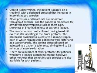
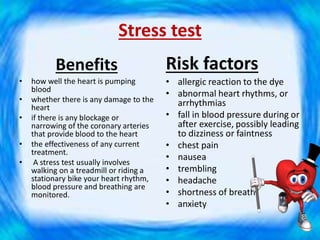
- Stress tests evaluate how the heart functions under physical stress by monitoring heart rate, blood pressure, and other vital signs during exercise or pharmaceutical stress.
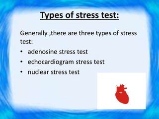
- There are three main types of stress tests: exercise stress tests, adenosine stress tests which use drugs, and nuclear stress tests which use radioactive tracers.
- Stress tests can indicate problems with blood flow, identify areas of reduced flow, and evaluate the effectiveness of heart treatments. They are used to investigate symptoms, assess cardiac function, and follow up on conditions like heart disease.


![Indications:
• Symptoms suggesting myocardial ischemia
• Acute chest pain in patients excluded for acute
coronary syndrome (ACS)
• Recent ACS treated without coronary angiography
• Known CAD with worsening symptoms
• Prior coronary revascularization (patients 5 years or
longer after Coronary artery bypass grafting [CABG] or
2 years or less after percutaneous coronary
intervention [PCI])
• Certain cardiac arrhythmias to assess chronotropic
competence
• Newly diagnosed heart failure or cardiomyopathy
Your doctor may recommend a test with imaging, such
as a nuclear stress test or echocardiographic stress test, if
an exercise stress test doesn't pinpoint the cause of your
symptoms.](https://image.slidesharecdn.com/stresstest-220707152514-4e21d845/85/stress-test-10-320.jpg)


![Stresstesting housestaffdidactic_10092014[1]](https://cdn.slidesharecdn.com/ss_thumbnails/stress20testinghousestaff20didactic100920141-141013101956-conversion-gate02-thumbnail.jpg?width=640&height=640&fit=bounds)


