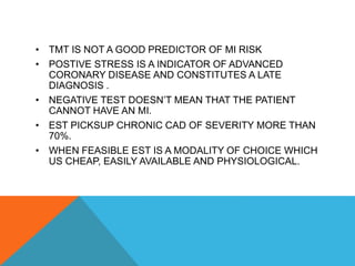
TREADMILL EXERCISE STRESS
TEST
PRESENTATION BY DR RAHUL METRI
UNIT HEAD – DR MAHESH DAVE SIR
ASSESSMENT OF CORONARY ARTERY DISEASE
1)Functional assessment - stress testing .
2)Anatomical assessment-
a) Non invasive - CT coronary angiography, Cardiac MRI
b) Invasive – Coronary Angiogram (gold std)
INTRODUCTION
• Exercise stress test is a noninvasive tool to evaluate the
cardiovascular system response to exercise.
• Exercise is the body’s most common physiologic stress, and
it places major demand on the cardiopulmonary system. Thus
exercise can be considered as the most practical test of
cardiac perfusion and function.
• The body increases its resting metabolic rate up to 20 times
and cardiac output about 6times during exercise.
In exercise stress test , we increase demand and
see if supply is proportionately increasing.
• Demand – o2 consumption –exercise
• Supply – measure blood flow – ecg changes
TYPES OF STRESS TESTING.
1.Exercise
a Treadmill
b Bicycle
(Treadmill testing provides a more common form of
physiologic stress (i.e., walking) in which patients are more
likely to attain a higher oxygen uptake and peak heart rate
than during stationary Cycling)
2.Pharmacological
a) Dobutamine ( chronotropic /inotropic)
b) adenosine (vasodilator)
c) Regadenosine
INDICATION FOR EXERCISE STRESS TESTING
• Diagnosis of IHD in intermediate CAD
• Assess functional capacity of patient
• Markedly abnormal calcium score on EBCT
• Post ACS risk stratification
• Exercise prescription
GENERAL CONCERNS PRIOR TO EXERCISE TEST.
• Patient preparation. Choosing a test type/ protocol. Patient
monitoring.
• Reasons to terminate test and Post test monitoring.
• The treadmill should have front and side rails for subjects to
steady themselves.
• It should be calibrated monthly. An emergency stop button
should be readily available to the staff only.
• Exercise test should be performed under the supervision of a
physician who has been trained to conduct exercise test.
PATIENT'S PREPARATION
• The Subject should be instructed have light breakfast and not to smoke or
take caffeinated beverages atleast 2-4 hrs prior to the test. Unusual physical
exertion should be avoided before testing. Specific questions should determine
which drugs are being taken. Medications should be brought along.
• Wear comfortable shoes and loose-fitting clothes
• Adequate skin preparation is essential for high quality recordings.
• The areas of electrode application are rubbed with alcohol saturated pad to
remove oil and it is rubbed with free sand paper or rough material to reduce
skin resistance .
• Advise patient about the risk and benefits of the procedure .A written informed
consent form is usually . A standard 12-lead ECG is usually obtained required
The ECG should be obtained and blood pressures recorded in both positions,
and patients should be instructed on how to perform the test
• Room temperature should be between 18 and 22 degrees and humidity less
than 60%
PATIENT'S PREPARATION
• Patients skin preparation for electrodes placement also very
important. Hyperventilation should be avoided before testing
.Subjects with or without disease can exhibit ST segments
changes with hyperventilation. The heart rate , blood pressure
and ECG should be recorded at the end of each stage of the
exercise , immediately before and immediately after stopping
the exercise , at the onset of an ischemic response.
EXERCISE PROTOCOLS
The exercise protocol should be progressive with even increments in speed
whenever possible.
• Smaller, even and more frequent work increments are preferable to larger,
uneven , and less frequent increases, because the former yield a more accurate
estimations of exercise capacity.
• The protocol should include a suitable recovery or cool down period
• In general, 6 to 12 minutes of continuous progressive exercise during which
the myocardial oxygen demand is elevated to the patient's maximal level is
optimal for diagnostic and prognostic purposes .Dynamic protocols most
frequently are used to asses to cardiovascular reserve, and those suitable for
clinical testing should include a low intensity warm-up phase
VARIOUS TREADMILL PROTOCOLS
• BRUCE
• MODIFIED BRUCE
• ASYMPTOMATIC CARDIAC ISCHAEMIA PILOT(ACIP)
• MODIFIED ACIP
• CORNELLBALKE WARE
• NAUGHTON
• WEBER
Most common and widely
adopted . Large diagnostic and
prognostic data base has been
published.
• The Bruce multistage maximal
treadmill protocol has 3-minute
periods to allow achievement of
a steady state before workload
is increased for next stage
• 6 stages , each stage has
3min period
• Maximum energy
expenditure – 13 mets
Starts at a lower workload
than the standard test .
Typically used for elderly
or sedentary patients
• The first two stages of the
modified Bruce test are
performed at a 1.7mph and
0% grade and 1.7mph 5%
grade.
• The third stage correspond
the first stage of the
standard Bruce test
protocol.
BRUCE PROTOCOL MODIFIED BRUCE
ACIP PROTOCOL.
• The asymptomatic Cardiac Ischemia Pilot protocol use 2-minute
stages, with 1.5-MET increments between stages after 1min
warm up stages with 1 MET increments.
• Developed to test patients with established CAD.
• Result in a linear increase in heart rate and, distributing the time
to occurrence of ST segment depression over a wider range
heart rate and exercise time than protocols with more abrupt
increments in workload between stages.
• The modified ACIP protocol produces a similar aerobic demand
as the standard ACIP protocol for each minute of the exercise .
• Well suited for short or older individuals who cannot keep up with
a working speed of 3mph
NAUGHTON AND WEBER PROTOCOLS
Use 1 to 2 minute stages with 1-MET increments between
stages.
These protocol is more suitable for patients with limited exercise
tolerance , such as patients with compensated heart failure
RECOVERY AFTER EXERCISE
• If maximal sensitivity is to be achieved with an exercise test,
patients should be supine as soon as possible during the post
exercise period (maximal wall stress.
• A cool-down walk can be helpful in performing test on patients
with an established diagnosis undergoing testing for other
diagnostic reasons-MI,CCF,valvular heart dx.
• A cool down walk after the test can delay or eliminate the
appearance of ST segment depression.
• Monitoring should continue for at least 5minutes after exercise
or until changes stabilize
MEASUREMENTS
• ECG
• BLOOD PRESSURE
• SYMPTOMS
• HEART RATE RESPONSE AND RECOVERY
• EXERCISE CAPACITY
ECG CHANGES
1. Exercise induced st depression
• fast up sloping =normal
• Slow up sloping = abnormal
• Horizontal = abnormal
• Down sloping = abnormal
2. Exercise induced ST elevation (usually rare ) = always
abnormal
3. Exercise induced U wave inversion
4. Exercise induced ST elevation in AVR
ST DEPRESSION
• ST depression has no localizing value
• PQ is the isoelectric point
• ST depression >1mm and horizontal or slow downsloping are
abnormal
• Usually measured 80 msec post J point
• Lateral leads are most specific and inferior leads least
specific.
ST ELEVATION
• Usually rare , if occurs its abnormal
• Can manifest with or without Q wave
• With Q wave can be in peri infarctional ischemia , dyskinetic ,
aneurysmal segment . And it has no localizing value .
• Without Q wave in critical lesion , coronary vasospasm .And it
has localizing value.
U WAVE INVERSION
• Chest pain with u wave inversion indicates significant CAD,
usually LAD
• U wave after exercise i.e recovery – indicates CAD . And it
has localizing value , V1 to V6 = anterior wall MI ,
lead 2,3,avf = RCA or Circumflex
ST ELEVATION IN AVR
• NO ST ELEVATION in AVR has high negative predictive value
i.e it rules out LMCA / triple vessel disease
NON ECG CHANGES
• Functional Capacity
• Exercise induced symptoms and signs
• HR response
• BP response
• Duke treadmill score
TERMINOLOGY
• Whole body peak O2 consumption –Vo2 max = CO * (A
– V o2 diff) ---METS
• 1 Metabolic equivalent (MET) , is defined as the amount
of oxygen consumed while sitting at rest and is equal to
3.5 ml O2 per kg body weight * minute .
• Differs with thyroid status , disease state , obesity ,
muscle mass .
• MET is useful for giving preop fitness. If a person is
able to perform > 5 met of physical activity , most of
the surgery can be uncomplicated.
MYOCARDIAL O2 CONSUMPTION
• Myocardial Peak O2 consumption –MVO2 max -
Coronary flow * coronary (A – V ) o2 difference ---
double product
• Accurate measurement – cardiac catheterization
• Indirectly measured by Double product = HR*SBP
• Normal >20,000
• < 20,000 indicates low heart work load .
• >29,000 indicates high work load .
FUNCTIONAL CAPACITY
• Functional capacity is a strong predictor of mortality and
nonfatal cardiovascular outcomes in both men and women
with and without CAD.
• Men predicted METs=18-(0.15× Age)
• Women predicted METs = 14-(0.13 × Age
ECG INDUCED SYMPTOMS AND SIGNS
• Chest pain quality
• Limiting vs non limiting angina
• Dysnea as a angina equivalent
• S3 , basal crepts – exercise induced LV failure
• S4 –CAD
• Wheeze – exercise induced asthma
HR AND BP RESPONSE
• Maximum HR = 220-age
• Our target HR = 85% of maximum HR
• Patient on beta blocker , max HR = 164-(0.7* age )
• Chronotropic incompetence and HR recovery are independent
predictor of mortality
• SBP – Increases to 160-200mmhg
• DBP –unchanged or minimal changes
• Hypertensive systolic response = 210-190; risk for future cardiac
event
• Exercise induced hypotension= indicates hypovolemia, LMCA,
LVD,
DUKE TREADMILL SCORE
• Duke treadmill score = Exercise duration(min) -5 (ST
deviation mm)-4 (angina index)
• Angina index = 0- none
1- typical anginal pain
2- angina causing test cessation .
TMT REPORTING
• TMT is positive for inducible ischemia at --- mets at a heart
rate --- bpm, patient experienced ----chest pain associate ecg
changes ---, which recovered within ---min. of rest.
• Double product of -----
• TMT IS NOT A GOOD PREDICTOR OF MI RISK
• POSTIVE STRESS IS A INDICATOR OF ADVANCED
CORONARY DISEASE AND CONSTITUTES A LATE
DIAGNOSIS .
• NEGATIVE TEST DOESN’T MEAN THAT THE PATIENT
CANNOT HAVE AN MI.
• EST PICKSUP CHRONIC CAD OF SEVERITY MORE THAN
70%.
• WHEN FEASIBLE EST IS A MODALITY OF CHOICE WHICH
US CHEAP, EASILY AVAILABLE AND PHYSIOLOGICAL.

Treadmill stress testing

  • 1.
    TREADMILL EXERCISE STRESS TEST PRESENTATIONBY DR RAHUL METRI UNIT HEAD – DR MAHESH DAVE SIR
  • 4.
    ASSESSMENT OF CORONARYARTERY DISEASE 1)Functional assessment - stress testing . 2)Anatomical assessment- a) Non invasive - CT coronary angiography, Cardiac MRI b) Invasive – Coronary Angiogram (gold std)
  • 5.
    INTRODUCTION • Exercise stresstest is a noninvasive tool to evaluate the cardiovascular system response to exercise. • Exercise is the body’s most common physiologic stress, and it places major demand on the cardiopulmonary system. Thus exercise can be considered as the most practical test of cardiac perfusion and function. • The body increases its resting metabolic rate up to 20 times and cardiac output about 6times during exercise.
  • 6.
    In exercise stresstest , we increase demand and see if supply is proportionately increasing. • Demand – o2 consumption –exercise • Supply – measure blood flow – ecg changes
  • 7.
    TYPES OF STRESSTESTING. 1.Exercise a Treadmill b Bicycle (Treadmill testing provides a more common form of physiologic stress (i.e., walking) in which patients are more likely to attain a higher oxygen uptake and peak heart rate than during stationary Cycling) 2.Pharmacological a) Dobutamine ( chronotropic /inotropic) b) adenosine (vasodilator) c) Regadenosine
  • 9.
    INDICATION FOR EXERCISESTRESS TESTING • Diagnosis of IHD in intermediate CAD • Assess functional capacity of patient • Markedly abnormal calcium score on EBCT • Post ACS risk stratification • Exercise prescription
  • 11.
    GENERAL CONCERNS PRIORTO EXERCISE TEST. • Patient preparation. Choosing a test type/ protocol. Patient monitoring. • Reasons to terminate test and Post test monitoring. • The treadmill should have front and side rails for subjects to steady themselves. • It should be calibrated monthly. An emergency stop button should be readily available to the staff only. • Exercise test should be performed under the supervision of a physician who has been trained to conduct exercise test.
  • 13.
    PATIENT'S PREPARATION • TheSubject should be instructed have light breakfast and not to smoke or take caffeinated beverages atleast 2-4 hrs prior to the test. Unusual physical exertion should be avoided before testing. Specific questions should determine which drugs are being taken. Medications should be brought along. • Wear comfortable shoes and loose-fitting clothes • Adequate skin preparation is essential for high quality recordings. • The areas of electrode application are rubbed with alcohol saturated pad to remove oil and it is rubbed with free sand paper or rough material to reduce skin resistance . • Advise patient about the risk and benefits of the procedure .A written informed consent form is usually . A standard 12-lead ECG is usually obtained required The ECG should be obtained and blood pressures recorded in both positions, and patients should be instructed on how to perform the test • Room temperature should be between 18 and 22 degrees and humidity less than 60%
  • 14.
    PATIENT'S PREPARATION • Patientsskin preparation for electrodes placement also very important. Hyperventilation should be avoided before testing .Subjects with or without disease can exhibit ST segments changes with hyperventilation. The heart rate , blood pressure and ECG should be recorded at the end of each stage of the exercise , immediately before and immediately after stopping the exercise , at the onset of an ischemic response.
  • 15.
    EXERCISE PROTOCOLS The exerciseprotocol should be progressive with even increments in speed whenever possible. • Smaller, even and more frequent work increments are preferable to larger, uneven , and less frequent increases, because the former yield a more accurate estimations of exercise capacity. • The protocol should include a suitable recovery or cool down period • In general, 6 to 12 minutes of continuous progressive exercise during which the myocardial oxygen demand is elevated to the patient's maximal level is optimal for diagnostic and prognostic purposes .Dynamic protocols most frequently are used to asses to cardiovascular reserve, and those suitable for clinical testing should include a low intensity warm-up phase
  • 16.
    VARIOUS TREADMILL PROTOCOLS •BRUCE • MODIFIED BRUCE • ASYMPTOMATIC CARDIAC ISCHAEMIA PILOT(ACIP) • MODIFIED ACIP • CORNELLBALKE WARE • NAUGHTON • WEBER
  • 17.
    Most common andwidely adopted . Large diagnostic and prognostic data base has been published. • The Bruce multistage maximal treadmill protocol has 3-minute periods to allow achievement of a steady state before workload is increased for next stage • 6 stages , each stage has 3min period • Maximum energy expenditure – 13 mets Starts at a lower workload than the standard test . Typically used for elderly or sedentary patients • The first two stages of the modified Bruce test are performed at a 1.7mph and 0% grade and 1.7mph 5% grade. • The third stage correspond the first stage of the standard Bruce test protocol. BRUCE PROTOCOL MODIFIED BRUCE
  • 19.
    ACIP PROTOCOL. • Theasymptomatic Cardiac Ischemia Pilot protocol use 2-minute stages, with 1.5-MET increments between stages after 1min warm up stages with 1 MET increments. • Developed to test patients with established CAD. • Result in a linear increase in heart rate and, distributing the time to occurrence of ST segment depression over a wider range heart rate and exercise time than protocols with more abrupt increments in workload between stages. • The modified ACIP protocol produces a similar aerobic demand as the standard ACIP protocol for each minute of the exercise . • Well suited for short or older individuals who cannot keep up with a working speed of 3mph
  • 20.
    NAUGHTON AND WEBERPROTOCOLS Use 1 to 2 minute stages with 1-MET increments between stages. These protocol is more suitable for patients with limited exercise tolerance , such as patients with compensated heart failure
  • 21.
    RECOVERY AFTER EXERCISE •If maximal sensitivity is to be achieved with an exercise test, patients should be supine as soon as possible during the post exercise period (maximal wall stress. • A cool-down walk can be helpful in performing test on patients with an established diagnosis undergoing testing for other diagnostic reasons-MI,CCF,valvular heart dx. • A cool down walk after the test can delay or eliminate the appearance of ST segment depression. • Monitoring should continue for at least 5minutes after exercise or until changes stabilize
  • 24.
    MEASUREMENTS • ECG • BLOODPRESSURE • SYMPTOMS • HEART RATE RESPONSE AND RECOVERY • EXERCISE CAPACITY
  • 26.
    ECG CHANGES 1. Exerciseinduced st depression • fast up sloping =normal • Slow up sloping = abnormal • Horizontal = abnormal • Down sloping = abnormal 2. Exercise induced ST elevation (usually rare ) = always abnormal 3. Exercise induced U wave inversion 4. Exercise induced ST elevation in AVR
  • 28.
    ST DEPRESSION • STdepression has no localizing value • PQ is the isoelectric point • ST depression >1mm and horizontal or slow downsloping are abnormal • Usually measured 80 msec post J point • Lateral leads are most specific and inferior leads least specific.
  • 29.
    ST ELEVATION • Usuallyrare , if occurs its abnormal • Can manifest with or without Q wave • With Q wave can be in peri infarctional ischemia , dyskinetic , aneurysmal segment . And it has no localizing value . • Without Q wave in critical lesion , coronary vasospasm .And it has localizing value.
  • 30.
    U WAVE INVERSION •Chest pain with u wave inversion indicates significant CAD, usually LAD • U wave after exercise i.e recovery – indicates CAD . And it has localizing value , V1 to V6 = anterior wall MI , lead 2,3,avf = RCA or Circumflex ST ELEVATION IN AVR • NO ST ELEVATION in AVR has high negative predictive value i.e it rules out LMCA / triple vessel disease
  • 31.
    NON ECG CHANGES •Functional Capacity • Exercise induced symptoms and signs • HR response • BP response • Duke treadmill score
  • 32.
    TERMINOLOGY • Whole bodypeak O2 consumption –Vo2 max = CO * (A – V o2 diff) ---METS • 1 Metabolic equivalent (MET) , is defined as the amount of oxygen consumed while sitting at rest and is equal to 3.5 ml O2 per kg body weight * minute . • Differs with thyroid status , disease state , obesity , muscle mass . • MET is useful for giving preop fitness. If a person is able to perform > 5 met of physical activity , most of the surgery can be uncomplicated.
  • 35.
    MYOCARDIAL O2 CONSUMPTION •Myocardial Peak O2 consumption –MVO2 max - Coronary flow * coronary (A – V ) o2 difference --- double product • Accurate measurement – cardiac catheterization • Indirectly measured by Double product = HR*SBP • Normal >20,000 • < 20,000 indicates low heart work load . • >29,000 indicates high work load .
  • 36.
    FUNCTIONAL CAPACITY • Functionalcapacity is a strong predictor of mortality and nonfatal cardiovascular outcomes in both men and women with and without CAD. • Men predicted METs=18-(0.15× Age) • Women predicted METs = 14-(0.13 × Age
  • 37.
    ECG INDUCED SYMPTOMSAND SIGNS • Chest pain quality • Limiting vs non limiting angina • Dysnea as a angina equivalent • S3 , basal crepts – exercise induced LV failure • S4 –CAD • Wheeze – exercise induced asthma
  • 38.
    HR AND BPRESPONSE • Maximum HR = 220-age • Our target HR = 85% of maximum HR • Patient on beta blocker , max HR = 164-(0.7* age ) • Chronotropic incompetence and HR recovery are independent predictor of mortality • SBP – Increases to 160-200mmhg • DBP –unchanged or minimal changes • Hypertensive systolic response = 210-190; risk for future cardiac event • Exercise induced hypotension= indicates hypovolemia, LMCA, LVD,
  • 39.
    DUKE TREADMILL SCORE •Duke treadmill score = Exercise duration(min) -5 (ST deviation mm)-4 (angina index) • Angina index = 0- none 1- typical anginal pain 2- angina causing test cessation .
  • 40.
    TMT REPORTING • TMTis positive for inducible ischemia at --- mets at a heart rate --- bpm, patient experienced ----chest pain associate ecg changes ---, which recovered within ---min. of rest. • Double product of -----
  • 41.
    • TMT ISNOT A GOOD PREDICTOR OF MI RISK • POSTIVE STRESS IS A INDICATOR OF ADVANCED CORONARY DISEASE AND CONSTITUTES A LATE DIAGNOSIS . • NEGATIVE TEST DOESN’T MEAN THAT THE PATIENT CANNOT HAVE AN MI. • EST PICKSUP CHRONIC CAD OF SEVERITY MORE THAN 70%. • WHEN FEASIBLE EST IS A MODALITY OF CHOICE WHICH US CHEAP, EASILY AVAILABLE AND PHYSIOLOGICAL.