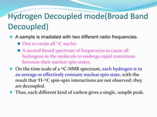
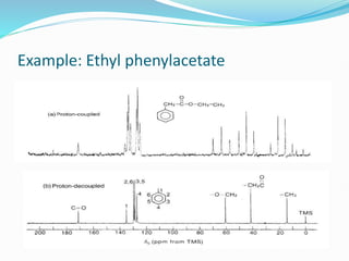
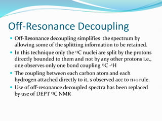
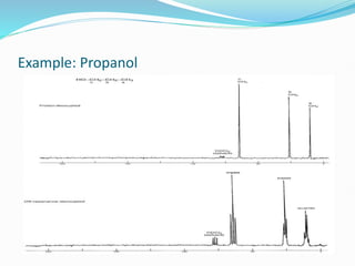
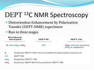
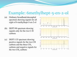
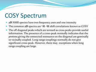
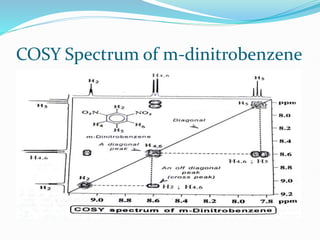
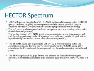
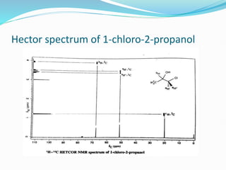
Presented By: Guided By:
Madhavanand Ingalageri Prof. Dr. A R Saundane
Introduction
 Nuclear magnetic resonance concern the magnetic properties of
certain atomic nuclei. It concerns the atoms having spin quantum
number.
 12C nucleus is not magnetically active the spin number I being zero. But
13C Have I = ½
 13C account for only 1.1% of naturally occurring carbon 13C- 13C coupling
is negligible and not observed.
 The gyromagnetic ratio of 13C is one-fourth of that of 1H.
 Each nonequivalent 13C gives a different signal.
 A 13C signal is split by the 1H bonded to it according to the (n + 1) rule.
 The most common mode of operation of a 13C-NMR spectrometer is a
hydrogen-decoupled mode.
Chemical Shift
 Carbon-13 chemical shifts are most affected by
 Hybridiasation state of carbon & Electronegative group attached to carbon
Hydrogen Decoupled mode(Broad Band
Decoupled)
 A sample is irradiated with two different radio frequencies.
 One to excite all 13C nuclei.
 A second broad spectrum of frequencies to cause all
hydrogens in the molecule to undergo rapid transitions
between their nuclear spin states.
 On the time scale of a 13C-NMR spectrum, each hydrogen is in
an average or effectively constant nuclear spin state, with the
result that 1H-13C spin-spin interactions are not observed; they
are decoupled.
 Thus, each different kind of carbon gives a single, unsplit peak.
Example: Ethyl phenylacetate
Off-Resonance Decoupling
 Off-Resonance decoupling simplifies the spectrum by
allowing some of the splitting information to be retained.
 In this technique only the 13C nuclei are split by the protons
directly bounded to them and not by any other protons i.e.,
one observes only one bond coupling 13C -1H
 The coupling between each carbon atom and each
hydrogen attached directly to it, s observed acc to n+1 rule.
 Use of off-resonance decoupled spectra has been replaced
by use of DEPT 13C NMR
Example: Propanol
DEPT 13C NMR Spectroscopy
 Distortionless Enhancement by Polarization
Transfer (DEPT-NMR) experiment
 Run in three stages
Example: 6methylhept-5-en-2-ol
(a) Ordinary broadband-decoupled
spectrum showing signals for all
eight of 6-methylhept-5-en-2-ol
(b) DEPT-90 spectrum showing
signals only for the two C-H
carbons
(c) DEPT-135 spectrum showing
positive signals for the two CH
carbons and the three CH3
carbons and negative signals for
the two CH2 carbons
COSY Spectrum
 2D NMR spectra have two frequency axes and one intensity
 The common 2D spectra are 1 H -1H shift correlations known as COSY
 The off diagonal peaks which are termed as cross peaks provide useful
information. The presence of a cross-peak normally indicates that the
protons giving the connected resonance on the diagonal are geminally
or vicinally coupled. Long range couplings normally do not give
significant cross-peak. However, there may exceptions when long
range coupling are large.
COSY Spectrum of m-dinitrobenzene
HECTOR Spectrum
 2D-NMR spectra that displays 13C – 1H-NMR shift correlations are called HETCOR
spectra. It shows coupling between protons and the carbon to which they are
attached. The HETCOR spectrum of 1-chloro-2propanol is shown in the fig.
 The 2-D spectrum is composed only of cross-peaks, each one relating carbon to its
directly bonded proton(s).
 The methyl doublet of 1H-NMR spectrum appears at δ 1.2 when drawn cross-peak
and then dropped down to the 13C spectrum axis indicates that the 13C peak at δ 20
is produced by the methyl carbon of 1-chloro-2-propanol(C-3)
 The 1H -NMR signal at δ 3.9 is due to CH-OH (C-2 proton) tracing out to the
correlation peak and down to the 13C spectrum shows the 13C NMR signal at 67
arises from the C-2 carbon of the compound i.e., the carbon carrying the hydroxyl
group.
 The 1H-NMR peaks at δ 3.4-3.5 for the two protons on the carbon bearing the
chlorine, the interpretation leads us to the cross-peak and down to the 13C peak at δ
51
Hector spectrum of 1-chloro-2-propanol
Applications of 13C NMR
 CMR is a noninvasive and nondestructive method,i.e,especially used in
repetitive In-vivo analysis of the sample without harming the tissues .
 CMR, chemical shift range(0-240 ppm) is wider compared to H-
NMR(0-14 ppm), which permits easy separation and identification of
chemically closely related metabolites.
 C-13 enrichment, which the signal intensities and helps in tracing the
cellular metabolism.
 CMR technique is used for quantification of drugs purity to
determination of the composition of high molecular weight synthetic
polymers.

c13ppt-150515121301-lva1-app6892 (1).pdf

  • 1.
    Presented By: GuidedBy: Madhavanand Ingalageri Prof. Dr. A R Saundane
  • 2.
    Introduction  Nuclear magneticresonance concern the magnetic properties of certain atomic nuclei. It concerns the atoms having spin quantum number.  12C nucleus is not magnetically active the spin number I being zero. But 13C Have I = ½  13C account for only 1.1% of naturally occurring carbon 13C- 13C coupling is negligible and not observed.  The gyromagnetic ratio of 13C is one-fourth of that of 1H.  Each nonequivalent 13C gives a different signal.  A 13C signal is split by the 1H bonded to it according to the (n + 1) rule.  The most common mode of operation of a 13C-NMR spectrometer is a hydrogen-decoupled mode.
  • 3.
    Chemical Shift  Carbon-13chemical shifts are most affected by  Hybridiasation state of carbon & Electronegative group attached to carbon
  • 4.
    Hydrogen Decoupled mode(BroadBand Decoupled)  A sample is irradiated with two different radio frequencies.  One to excite all 13C nuclei.  A second broad spectrum of frequencies to cause all hydrogens in the molecule to undergo rapid transitions between their nuclear spin states.  On the time scale of a 13C-NMR spectrum, each hydrogen is in an average or effectively constant nuclear spin state, with the result that 1H-13C spin-spin interactions are not observed; they are decoupled.  Thus, each different kind of carbon gives a single, unsplit peak.
  • 5.
  • 6.
    Off-Resonance Decoupling  Off-Resonancedecoupling simplifies the spectrum by allowing some of the splitting information to be retained.  In this technique only the 13C nuclei are split by the protons directly bounded to them and not by any other protons i.e., one observes only one bond coupling 13C -1H  The coupling between each carbon atom and each hydrogen attached directly to it, s observed acc to n+1 rule.  Use of off-resonance decoupled spectra has been replaced by use of DEPT 13C NMR
  • 7.
  • 8.
    DEPT 13C NMRSpectroscopy  Distortionless Enhancement by Polarization Transfer (DEPT-NMR) experiment  Run in three stages
  • 9.
    Example: 6methylhept-5-en-2-ol (a) Ordinarybroadband-decoupled spectrum showing signals for all eight of 6-methylhept-5-en-2-ol (b) DEPT-90 spectrum showing signals only for the two C-H carbons (c) DEPT-135 spectrum showing positive signals for the two CH carbons and the three CH3 carbons and negative signals for the two CH2 carbons
  • 10.
    COSY Spectrum  2DNMR spectra have two frequency axes and one intensity  The common 2D spectra are 1 H -1H shift correlations known as COSY  The off diagonal peaks which are termed as cross peaks provide useful information. The presence of a cross-peak normally indicates that the protons giving the connected resonance on the diagonal are geminally or vicinally coupled. Long range couplings normally do not give significant cross-peak. However, there may exceptions when long range coupling are large.
  • 11.
    COSY Spectrum ofm-dinitrobenzene
  • 12.
    HECTOR Spectrum  2D-NMRspectra that displays 13C – 1H-NMR shift correlations are called HETCOR spectra. It shows coupling between protons and the carbon to which they are attached. The HETCOR spectrum of 1-chloro-2propanol is shown in the fig.  The 2-D spectrum is composed only of cross-peaks, each one relating carbon to its directly bonded proton(s).  The methyl doublet of 1H-NMR spectrum appears at δ 1.2 when drawn cross-peak and then dropped down to the 13C spectrum axis indicates that the 13C peak at δ 20 is produced by the methyl carbon of 1-chloro-2-propanol(C-3)  The 1H -NMR signal at δ 3.9 is due to CH-OH (C-2 proton) tracing out to the correlation peak and down to the 13C spectrum shows the 13C NMR signal at 67 arises from the C-2 carbon of the compound i.e., the carbon carrying the hydroxyl group.  The 1H-NMR peaks at δ 3.4-3.5 for the two protons on the carbon bearing the chlorine, the interpretation leads us to the cross-peak and down to the 13C peak at δ 51
  • 13.
    Hector spectrum of1-chloro-2-propanol
  • 14.
    Applications of 13CNMR  CMR is a noninvasive and nondestructive method,i.e,especially used in repetitive In-vivo analysis of the sample without harming the tissues .  CMR, chemical shift range(0-240 ppm) is wider compared to H- NMR(0-14 ppm), which permits easy separation and identification of chemically closely related metabolites.  C-13 enrichment, which the signal intensities and helps in tracing the cellular metabolism.  CMR technique is used for quantification of drugs purity to determination of the composition of high molecular weight synthetic polymers.